- Causes of pulmonary edema
- What diseases are there a risk of edema?
- Types of pathology
- Symptoms of pulmonary edema
- Diagnostics
- Treatment of pulmonary edema
- Predictions and prevention
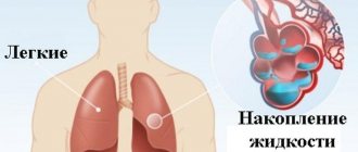
Pulmonary edema is a condition in which gas exchange is disrupted and respiratory failure develops. Swelling occurs due to an imbalance of pressure in the pulmonary capillaries and the release of a large amount of edematous fluid into the lung tissue without its reabsorption.
Causes of pulmonary edema
A mixture of edematous fluid (transudate) and a liquid substance that “lines” the alveoli of the lungs from the inside (it is called “pulmonary surfactant”) enters the lumen of the alveoli, where it combines with air. This leads to the formation of foam, which creates an obstacle to the flow of oxygen. Lack of oxygen provokes the development of shortness of breath, due to which the pressure inside the chest decreases and blood flow to the right side of the heart increases.
Pulmonary edema causes infiltration of the alveoli and is manifested by symptoms that indicate oxygen starvation, which is dangerous for tissues. The patient requires emergency care due to the high risk of death due to suffocation.
Possible complications
Due to cardiogenic pulmonary edema, oxygen starvation of all organs develops. Hypoxia leads to inevitable death of brain cells, which without timely help causes death. In addition, the consequences may be as follows:
- ischemic disorders. Oxygen starvation leads to disruptions in the functioning of the kidneys and liver;
- scarring of lung tissue called pneumosclerosis;
- cardiogenic shock;
- pulmonary edema of a recurrent nature;
- deformation of the structure of the alveoli and bronchioles;
- Secondary pneumonia develops against the background of circulatory disorders.
Important! Complications can begin within 4–5 minutes after the development of cardiogenic pulmonary edema, so if signs of pathology appear, you should act immediately.
What diseases are there a risk of edema?
Pathology indicates a complication of the underlying disease, increases its duration and worsens the prognosis of treatment. The most dangerous from the point of view of pulmonary edema include:
- Heart diseases. Respiratory failure often occurs against the background of pathologies of the heart and blood vessels. Cardiogenic pulmonary edema is a life-threatening complication that develops with myocardial infarction, aneurysm, endocarditis and myocarditis, severe hypertension, and pathological conditions caused by congenital or acquired heart defects.
- Pulmonary diseases. At risk are patients with injuries and severe diseases of the respiratory tract. Respiratory edema develops against the background of exacerbation of chronic bronchitis and bronchial asthma, tuberculosis, and lobar pneumonia. The risk is increased by tumor lesions of the lung tissue, pleurisy, and chest injuries, which are accompanied by pneumothorax. If a person is connected to a ventilator for a long time and receives a mixture with a high concentration of oxygen, this also leads to an imbalance of pressure in the pulmonary capillaries and the release of exudate.
- Infectious diseases. In severe cases or inadequate treatment of whooping cough, scarlet fever, influenza, diphtheria and other infectious diseases, edema develops, followed by acute pulmonary failure. In young children, this clinical syndrome can cause acute laryngitis and the proliferation of adenoids.
- Pathologies of intrauterine development and difficult childbirth. Severe pregnancy, prematurity, oxygen hypoxia, fetal bronchopulmonary dysplasia - these factors increase the risk of developing pulmonary failure in newborns. In pregnant women, pulmonary failure can develop due to eclampsia. The same phenomenon occurs with ovarian hyperstimulation syndrome against the background of stimulation of ovulation by hormonal drugs.
- Foreign bodies entering the respiratory tract. Mechanical asphyxia is one of the possible causes of acute respiratory failure. Pathology occurs when foreign objects enter the respiratory tract, after drowning or attempted hanging, or blockage of the respiratory tract with vomit.
- Diseases of the urinary system and gastrointestinal tract. Nephrogenic pulmonary edema can be complicated by acute inflammatory diseases, which are accompanied by renal dysfunction and the development of severe renal failure. In gastroenterology, patients with liver cirrhosis, severe pancreatitis, and intestinal obstruction have to deal with respiratory failure.
- Neurological diseases. Traumatic brain injuries, meningitis, encephalitis, neoplasms, cerebral hemorrhages, complications after brain surgery - all this leads to neurogenic pulmonary edema.
- Allergies, injuries, poisoning. Acute respiratory failure can be caused by the ingestion of various toxic substances into the body. Pulmonary edema is encountered by patients with alcohol and drug intoxication, poisoning with drugs, salts of heavy metals, and toxic gases. Pulmonary insufficiency is diagnosed in case of blood poisoning, burns with a large affected area, acute allergic reactions.
Development mechanism
The development of cardiogenic pulmonary edema is associated with impaired pumping function of the heart muscle. This occurs under the influence of various pathologies. These include heart failure, acquired or congenital heart defects, myocardial infarction, and arrhythmias. Due to insufficient blood circulation, stagnation occurs in the body. As a result, fluid from the lymphatic vessels and capillaries begins to pass through the walls of the vessels into the pulmonary alveoli. This process is medically called fluid effusion. The liquid gradually displaces air from the lung tissue, which causes difficulty breathing. As the lungs fill with transudate, breathing becomes impossible, severe oxygen starvation of all tissues and organs develops, and death occurs.
Types of pathology
Depending on the cause of development, pulmonary edema can be cardiogenic or non-cardiogenic. In the first case, the cause of respiratory failure is cardiovascular disease. The second category includes all other pulmonary edema caused by diseases of the gastrointestinal tract, nervous system, and injuries.
Pulmonary edema is also classified according to the nature of its course. There are four types of pathology development:
- With fulminant development, severe respiratory failure occurs within a few minutes and leaves no chance of survival.
- Acute swelling can take up to four hours to develop. This form is considered less dangerous than the previous one, but in the absence of timely assistance, the risk of death is extremely high. Most often, acute pulmonary edema develops with myocardial infarction, traumatic brain injury, and acute allergic reactions.
- In case of intoxication, renal and liver failure, subacute pulmonary edema may occur. The intensity of symptoms is constantly changing, so continuous medical monitoring of the patient's condition is required.
- Prolonged swelling may take several days to develop. Symptoms are mild compared to the acute and subacute forms. Prolonged pulmonary edema usually occurs against the background of various chronic diseases, including heart failure.
Who is at risk
In some patients, the risk of developing a dangerous condition increases due to certain provoking factors. These include:
- all heart diseases;
- frequent stress;
- age over 45 years;
- bad habits, namely drinking alcohol, smoking.
The conditions described above do not directly cause the development of edema, but they can increase the risk of its occurrence.
Elderly patients with cardiac dysfunction are at risk for developing cardiac pulmonary edema.
Symptoms of pulmonary edema
Before the main symptoms of pulmonary failure appear, patients experience weakness and dizziness, a feeling of tightness in the chest, headache, and dry cough. A few hours after the first symptoms of the pathology, an attack of cardiac asthma develops - the first stage of pulmonary edema. It manifests itself:
- severe lack of air;
- severe dry cough;
- increased heart rate;
- hoarse breathing;
- profuse sweating;
- pale skin, blueness of nails and lips;
- feelings of fear and anxiety.
As a rule, the attack occurs at night or before dawn. In patients with cardiac diseases, cardiac asthma can be caused by physical or emotional stress and hypothermia.
As edema develops, the alveoli become involved in the pathological process. This leads to increased suffocation. The patient experiences severe shortness of breath, the face becomes burgundy-blue and puffy, the veins in the neck swell, and mental retardation appears. Breathing becomes hoarse, foam mixed with blood comes out of the mouth.
In the absence of medical care, the patient loses consciousness and falls into a coma. In this state, blood pressure decreases critically, breathing becomes shallow, and the pulse weakens. The cause of human death is asphyxia, in which the body experiences an acute lack of oxygen with an excess of carbon dioxide.
Clinical manifestations in adults
Pathology does not always develop rapidly. In some cases, it is preceded by weakness, dizziness, chest tightness, dry cough, headache and tachypnea. These symptoms can appear a few minutes or several hours before swelling develops.
Primary manifestations: compressive chest pain, increasing tachycardia, dry wheezing turning into wet wheezing, increased breathing.
Signs of progression of pathology:
- shallow breathing;
- bubbling wet rales;
- earthy skin color;
- increased suffocation;
- cold sweat;
- the appearance of foamy sputum with a pink tint from the mouth.
On our website Dobrobut.com you can learn more about pathology and, if necessary, make an appointment with a specialist. The doctor will answer questions and tell you the difference between interstitial and alveolar pulmonary edema.
Diagnostics
A doctor can determine the development of pulmonary edema by numerous external symptoms. In addition, the patient is prescribed instrumental and laboratory examinations. Among them:
- measurement of blood oxygen levels, determination of carbon dioxide content;
- blood chemistry;
- electrocardiogram and ultrasound of the heart;
- chest x-ray to evaluate the condition of the lungs.
Inserting a catheter into the pulmonary artery allows you to determine the type of edema: cardiogenic or non-cardiogenic.
If pulmonary edema is suspected, it is important to carry out the necessary tests and examinations as soon as possible, identify the cause and type of pathology, and begin treatment. Saving the patient's life directly depends on this.
Emergency care for a patient
Correctly provided emergency care for cardiogenic pulmonary edema helps to preserve the patient’s vital functions until the ambulance arrives. If symptoms characteristic of pathology develop, you must perform actions in the following order:
- seat the person in an upright position, legs and arms should be lowered;
- if necessary, remove foam and mucus from the mouth;
- it is important to ensure the flow of fresh air into the room; to do this, open the windows and door;
- pandemonium should not be allowed;
- the patient’s collar must be unbuttoned, nothing should restrict the neck;
- if the person is conscious, he needs to be given a Nitroglycerin tablet, as well as Furosemide.
When providing assistance, you should not panic. This will only aggravate the situation; the feeling of fear will be transmitted to the patient, which will negatively affect his condition.
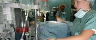
Treatment of pulmonary edema
People with acute respiratory failure are treated in the intensive care unit. First aid consists of measures that reduce venous return to the heart. To do this, the patient must take a semi-sitting position. Applying tourniquets to the limbs and hot foot baths have a positive effect.
It is imperative to monitor blood oxygen saturation levels. Oxygen therapy is used to compensate for its deficiency. If pulmonary edema has developed against the background of mechanical asphyxia, it is necessary to clear the airways of foreign objects. If it is impossible to breathe independently, the patient is connected to a ventilator.
A combination of medications is used to eliminate pulmonary edema. Among them:
- morphine and other narcotic analgesics;
- drugs to stimulate diuresis;
- nitroglycerine;
- drugs to lower blood pressure.
Depending on the underlying disease, patients with pulmonary edema are prescribed antihistamines, antibiotics, hormonal and antiarrhythmic drugs, and cardiac glycosides. The task of the resuscitator is to stop the attack and prevent acute hypoxia, which leads to cell death and death of the patient. After the symptoms of respiratory failure are relieved, treatment of the disease that caused it begins.
It is very important to eliminate the root cause that led to the clinical syndrome. Otherwise, a relapse of the disease is possible.
Pathogenesis
The pathogenesis of pulmonary edema is determined by its type (cardiogenic/non-cardiogenic). Accordingly, a hydrostatic and membranogenic mechanism for the development of edema is distinguished.
Hydrostatic pulmonary edema
The main etiological factor is diseases of the cardiovascular system, in which there is an increase in intracapillary hydrostatic pressure. When the pressure reaches 15 mm Hg. Art. blood plasma is transfused into the interstitium in a volume that exceeds the physiological ability to remove it by lymphatic drainage. When pressure rises to 20 mm Hg. Art. and higher, the liquid enters the surface of the alveoli. The mechanism of development of this type of edema is based on the Frank-Starling law, which describes the patterns of exchange of liquid media in lung tissue. Within the framework of the article, it is not possible to decipher each of the operating factors of the formula. Let us highlight only the main sequentially changing interconnected stages of the pathological process:
- An increase in hydrostatic pressure in the pulmonary circulation, which contributes to an increase in pressure in the pulmonary artery and then inside the capillaries of the lungs.
- Increased filtration of fluid from the pulmonary vessels directly into the tissue.
- The formation of excessive negative pressure in the interstitial space of the lungs and the development of interstitial edema .
- Decompensation of fluid outflow through the lymphatic pathways.
- Development of alveolar edema (a developed clinical picture of pulmonary edema is observed).
- Reduction in the area of gas exchange and disorganization of the gas exchange mechanism between the external air and blood.
- A state of hypoxia , which, in turn, provokes the production of stress hormones, catecholamines , which leads to an even greater increase in blood pressure and a decrease in vascular permeability, further increasing the load on the left ventricle of the heart, thereby forming a vicious circle.
The figure schematically shows the pathogenesis of cardiogenic pulmonary edema
Membranogenic pulmonary edema
Its development is due to a primary increase in permeability and dysfunction of the alveolar-capillary membrane, which is caused by damage to the alveolar epithelium and capillary endothelium. Increased insight occurs under the influence of toxic oxygen radicals, proteinases , prostaglandins , leukotrienes , histamine , formed or released in the tissue under the influence of various types of irritants.
An increase in the permeability of the pulmonary capillaries quickly leads to the appearance of a large amount of edematous fluid, which contains a lot of protein. At the same time, changes in the permeability of the alveolar and capillary parts of the barrier do not occur uniformly. First, the permeability of the endothelial layer increases, which leads to the leakage of vascular fluid into the interstitium, where it temporarily accumulates ( interstitial pulmonary edema ). At the same time, interstitial edema is partially compensated by accelerating lymph drainage. In most cases, this adaptive reaction is insufficient, and gradually the edematous fluid penetrates through the surface of the destructively altered cells of the alveoli into their cavity, filling the entire volume.
Alveolar pulmonary edema develops . The exclusion of part of the alveoli from the gas exchange process is compensated by stretching (emphysema) of the remaining intact alveoli, which leads to compression of the lymphatic vessels and capillaries of the lungs. Alveolar edema develops when the alveoli are filled with fluid and is characterized by the appearance of severe disturbances in the respiratory system, the development of arterial hypoxemia / hypercapnia .
Against this background, microcirculation changes/blood viscosity increases (development of DIC syndrome). Vascular reactivity and bronchial muscle tone are disrupted, surfactant activity decreases, which leads to the development of microatelectasis . Alveolar hypoventilation increases, leading to a mismatch between perfusion and ventilation, changes in carbon dioxide/oxygen diffusion and a decrease in oxygen supply to tissues with the development of hypoxia and metabolic acidosis .
Membranogenic pulmonary edema develops under the influence of various types of damaging factors, one of which is toxicants of various origins, using the example of which we will consider in more detail the pathogenesis of toxic edema.
The figure schematically shows the pathogenesis of toxic pulmonary edema
Toxic pulmonary edema . The pathogenesis of this type of pulmonary edema is due to the effect of toxicants directly on the cells of the alveolar-capillary barrier with disruption of their function. Toxic edema has different mechanisms of damage to lung cells, but the pathological processes that develop behind this are quite similar. When cell damage/death occurs, the permeability of the barrier increases and the production of biologically active substances in the lung tissue sharply increases ( histamine , norepinephrine , angiotensin , acetylcholine , serotonin , prostaglandins ), which contributes to a further increase in permeability/impairment of the function of the alveolar-capillary barrier and the development of hemodynamic disorders in the lungs (blood flow speed decreases, pressure in the pulmonary circulation increases). As the edema progresses, the respiratory and terminal bronchioles fill with fluid, and under the influence of turbulence arising from air movement, foam forms in the airways.
Pulmonary edema of mixed type . The development is based on a combination of the mechanisms of edema of both types.
Predictions and prevention
Pulmonary edema is a deadly condition; Mortality with this clinical syndrome is up to 50%. Patients with anaphylactic shock most often develop fulminant or acute pulmonary edema, which in more than 90% leads to death.
The consequences of acute pulmonary failure are no less serious than the disease itself. Many patients who have suffered pulmonary edema experience ischemic damage to internal organs, the appearance of areas of pneumosclerosis, and congestive pneumonia. This affects the functioning of the respiratory system, in the organs of which irreversible changes occur. The pathology is manifested by shortness of breath, chest pain, and cough. Pneumosclerosis and other consequences of pulmonary edema require long-term treatment, can lead to disability and shorten the patient's life expectancy.
The likelihood of a favorable prognosis and outcome of the disease with pulmonary edema increases if the diagnosis was made in a timely manner and doctors began to stop the attack at the initial stage.
Prognosis for patients
After the onset of cardiogenic pulmonary edema, the prognosis for the patient is extremely rarely favorable. Survival rate, according to various statistics, ranges from 30 to 50%. In this case, edema caused by myocardial infarction leads to death in 90% of cases.
After overcoming a dangerous pathology, many people experience various disorders of the respiratory, cardiac, and central nervous systems. After recovery, a person must regularly visit a doctor for 12 months, monitor his diet, and follow other preventive measures.
A favorable prognosis for the patient is possible only with timely assistance.
An important condition for complete recovery is intensive therapy for the disease that caused the swelling. Only after eliminating the root cause is the risk of a recurrent attack reduced to almost zero.
Important! Only timely diagnosis of cardiogenic pulmonary edema, correctly selected treatment tactics and strict implementation of all doctor’s recommendations provide a favorable prognosis for a person.
List of sources
- Braunwald E. Acute pulmonary edema // Internal diseases / Ed. Braunwald E. Book. 5. Per. from English -Moscow: Medicine, 1995. P. 123-124.
- Inkova A. N. Handbook of emergency and emergency medical care doctors. Rostov n/a: Phoenix; M.: ACT, 2001. - 337 p.
- Stranshnov V.I., Voinov V.A. Respiratory distress syndrome. In: Koryachkin V.A., Strashnov V.I., eds. Intensive therapy of threatening conditions. St. Petersburg Medical Publishing House, St. Petersburg, 2002. 135 p.
- Vlasenko A.V., Ostapchenko D.A., Pavlov D.P. et al. Features of the pathogenesis and effectiveness of therapy for acute respiratory distress syndrome. Abstracts of reports of the XI Congress of the Federation of Anesthesiologists and Resuscitators of Russia 2008; 508-509.
- Acute respiratory distress syndrome in pediatric practice / Aleksandrovich Yu.S., Pshenisnov K.V. // Bulletin of intensive care. – 2014 – No3.