Thrombosis is a pathological process in which a blood clot blocks blood flow in a vessel. In ophthalmology, the phenomenon of retinal vascular thrombosis is rare, and in most cases it requires emergency elimination due to the risk of complete loss of vision. The disease is diagnosed mainly in elderly patients.
Most often, blood clots block the upper branches of the central retinal veins
(abbreviated as CVS). This condition is called peripheral occlusion in ophthalmology. If we are talking specifically about thrombosis of the central retinal vein, we mean central occlusion.
Table of contents:
What is central retinal vein thrombosis Causes of occurrence Main symptoms of the disease Stages of the disease Diagnosis of central retinal vein thrombosis Treatment methods Prevention of the disease
The formation of blood clots prevents blood circulation through the vessels. As a result, they become blocked, leading to the development of the disease. Treatment of central retinal vein thrombosis is possible with medication or surgery. The choice of thrombosis therapy technique depends largely on the degree of damage.
Signs of occlusion in the fundus
“Swollen” convoluted veins of an abnormally dark color, accentuated intersections of veins with arteries - upon visual examination of the fundus structures, these alarming prethrombotic signs of venous stagnation will immediately attract the attention of any ophthalmologist. A special angiographic study confirms the slowdown in venous blood flow.
Ophthalmoscopic symptoms of the first phase of thrombosis are dilation and tension of the veins, swelling of the tissues along them, and the presence of pinpoint hemorrhagic foci.
When complete occlusion of the central vein occurs, subjective complaints about a sudden “failure” of visual functions are added (in the case of arterial occlusion, these symptoms develop even more rapidly and more malignantly). There is swelling of the optic disc (the optic disc extending into the retina) with blurring of its perimeter; veins swollen in caliber can take on the appearance of a dotted line, in some areas as if “drowning” in edematous retinal tissue; spasmodic arteries, on the contrary, are sharply narrowed and seem to be bleeding; there are multiple hemorrhagic foci of various shapes and sizes, to which after some time are added significantly lighter foci of protein degeneration and denaturation.
The clinical picture varies somewhat depending on the nature of the blockage of the ductus venosus - whether it is partial thrombosis or complete occlusion.
Thus, with partial thrombosis, fewer foci of hemorrhage are usually observed; they can resolve during therapy with the restoration (at least incomplete) of blood circulation in the venous basin due to the spontaneous formation of anastomoses (points of communication between vessels); Accordingly, in these cases there may be some restoration of the functions of both central and peripheral vision.
Occlusion is characterized by pronounced neovascularization in the optic disc area and throughout the entire macular region, a sharp increase in the permeability of the vascular walls, multiple and extensive recurrent hemorrhages up to total hemophthalmos.
The consequences of thrombosis and occlusion of the central vein in the long term can be secondary glaucoma, degenerative retinopathy (in particular, maculopathy - degeneration, or macula), various types of growths (proliferation), optic nerve atrophy.
Reasons for appearance
Thrombosis occurs under the influence of other pathologies in the patient that negatively affect the vascular system of the organs of vision:
- atherosclerosis of arteries;
- hypertension;
- infectious viral and bacterial diseases;
- diabetes;
- sepsis;
- swelling of the optic nerve.
The disease can develop due to the patient's sedentary lifestyle, obesity, and endocrine disorders. Improper therapy for the listed diseases also leads to blockage of blood vessels.
Causes
According to statistics, retinal vein thrombosis is initiated by chronic diseases of the cardiovascular or endocrine systems, accompanied by changes in blood viscosity, deterioration of vascular patency, etc. The most common precursors of pathology are:
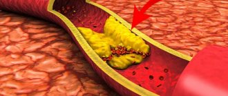
- Vascular atherosclerosis
is a condition in which the level of lipoproteins in the circulatory system increases, depositing on the intima of arteries and veins, forming plaques, and in severe cases completely blocking the lumen of the tubes. Blood clots often form on or near the surface of these plaques, which can break off and float throughout the circulatory system, including the ocular bloodstream. - Diabetes mellitus
is an endocrine disease, the symptom of which is an increase in blood glucose levels. The so-called “sugar” makes blood vessels more fragile and brittle. The body, trying to “patch” the damage, sends blood clotting factors to the damage, resulting in the formation of a blood clot. - Hypertension
is a systemic cardiovascular disease in which the load on small vascular networks, including the eye, increases. As a result, they are damaged, and thrombosis becomes a natural protective reaction (it prevents extensive hemorrhage). - Vasculitis
is an inflammatory disease of the walls of blood vessels of an autoimmune or infectious nature. The formation of blood clots is one of the body's defense reactions. - Tumor processes and metastases occur
in the area where the ocular vessels are located. They create pressure on the vessels and slow down blood flow, resulting in the formation of blood clots. Another scenario is the destruction of the tumor and its fragment entering the bloodstream, followed by blockage of the vessel.
The rarest reason why retinal thrombosis can occur is associated with dysfunction of the thyroid gland - thyrotoxicosis. In the advanced form of this disease, the tissue under the eye grows, causing it to protrude from the eye socket (the so-called thyroid bulging eye). This creates constant tension on the blood vessels, causing them to burst and become clogged with blood clots.
Main symptoms of the disease
Symptoms of thrombosis appear when a patient reaches a certain stage of pathological changes in the retina. The blockage is of a non-ischemic type, in which visual acuity remains at 0.1 diopters. Ischemic thrombosis is characterized by extensive hemorrhages.
The process of development of the disease can last up to several years, while the person’s vision remains normal. The main danger is that the patient does not attach importance to the initial signs of retinal disease. Incomplete thrombosis hardly manifests itself and does not cause concern.
Occasionally a person sees spots before his eyes. Patients experience symptoms in the evenings or mornings when looking at certain objects. Blind spots sometimes occur depending on the extent of the lesion.
Vision deteriorates: patients complain of blurred and blurred lines. Edema gradually develops, which is clearly visible during ophthalmoscopy. The vitreous body becomes covered with hemorrhages. They pass after a few months, but vision is not completely restored. Sometimes the back of the retina is damaged, but the patient does not lose vision.
Treatment of thrombosis of the branch of the central retinal vein
One of the most dangerous and undesirable consequences of central vein thrombosis is neovascularization, i.e. the formation and growth of a new network of small blood vessels, with the help of which the body tries to compensate for chronic ischemia (lack of blood supply) of the affected area of the retina. To stop this process, as well as to reduce swelling and, to a certain extent, reduce the risk of new hemorrhages, they usually try to inject Lucentis, a drug that was specially developed for the needs of ophthalmology, in particular, for the treatment of degenerative processes of the retina; the active substance penetrates quickly and deeply into the retinal tissue and, in general, is considered one of the most effective for such cases.
In the presence of extensive ischemic areas with intense neovascularization and constant hemorrhages, a more powerful remedy is prescribed - excimer laser exposure to the affected areas.
As a rule, treatment takes quite a long time (several months), but in no case should it be stopped: this is the only way to eliminate retinal edema, prevent vascular neoplasms, stop hemorrhages and, by and large, avoid total irreversible blindness.
Stages of the disease
There are several stages of retinal damage:
- Prethrombosis. The veins of the organs of vision are pathologically dilated. Vascular changes and macular edema are noticeable. At this stage, symptoms do not bother the patient. In rare cases, visual acuity decreases or objects appear blurry.
- Thrombosis. The retina is covered with multiple hemorrhages. The pathological focus can affect only one vein. The boundaries of the optic nerve are unclear, and swelling is noticeable around the macula. Possible loss of part of the visual field, decreased acuity.
- Retinopathy. After thrombosis, vision slowly recovers. Blood clots are visible at the bottom of the eye.
In some cases, recurrences of retinal vascular thrombosis are possible.
Forecast
In the absence of a pronounced and prolonged ischemic process against the background of vein thrombosis in the retina, the prognosis is favorable. If it is not possible to avoid ischemia, the likelihood of complications is high:
- neovascular glaucoma;
- recurrence of thrombosis and vitreous hemorrhage;
- traction retinal detachment.
All these pathologies lead to permanent vision loss, which even modern surgical interventions cannot correct. That is why you should not ignore the signs of partial venous obstruction in the retina: the earlier it is diagnosed, the higher the chance of avoiding vision loss.
Treatment methods
Thrombosis therapy begins immediately after diagnosis. The prescribed measures are aimed at resolving the resulting hemorrhages, reducing IOP, improving tissue nutrition and blood circulation.
Sometimes a positive result for the disease can be achieved with the help of drug therapy. The patient is prescribed eye drops that reduce pressure on the retina and accelerate the destruction of thrombotic formations. The use of B vitamins and ascorbic acid is indicated.
If traditional methods of treating the disease do not produce results, the ophthalmologist prescribes laser coagulation. Surgical intervention involves the impact of a laser device on a specific area of the shell.
Laser coagulation is good because it is bloodless, safe and does not require special preparation. The laser has coagulating properties, so it seals blood vessels and accelerates regeneration. After surgery, the rehabilitation period is significantly reduced.
The surgery takes place in a sitting position. The patient is seated on a chair and a drip of anesthesia is injected into the visual organs. With the help of drugs, the pupil is dilated to provide access to hard-to-reach areas of the retina.
A lens is installed on the patient's eye, thanks to which the light radiation will be focused. At this point, the patient may feel tingling or slight discomfort.
The surgeon controls the process of surgical intervention for the disease using a stereomicroscope. The laser beam seals certain areas and glues the membrane to the vascular fundus. Coagulation points keep the retina from further detachment.
Upon completion of the operation, the lens is removed. The patient should sit quietly for some time. At this time, ophthalmologists monitor the patient and monitor his condition. If there are no negative feelings, the patient goes home.
After surgery, you should not overstrain your visual organs. For a while you need to stop watching TV and working on the computer. If the operation was performed on one eye, the patient will not notice any special effects on vision.
Complete rehabilitation takes place in 2 weeks. During this period, it is important to avoid visiting the sauna, swimming pool, and heavy physical activity. It is not advisable to drink alcohol or antidepressants. Do not bend sharply. Since the operated eye is easily susceptible to infectious influences, it is important to promptly treat colds and viral diseases.
Complications after surgical treatment are rare. Sometimes repeated detachment occurs, in which case the operation is repeated. Possible inflammation of the conjunctiva or minor changes in vision.
Treatment of central retinal vein thrombosis
The main and mandatory condition for a favorable prognosis is the immediate initiation of responsive therapeutic measures when thrombosis begins, and even better - in the prethrombotic phase.
In addition to the above-mentioned Lucentis injections and laser coagulation, such measures may include the following prescriptions. Streptokinase and hemase in injections have a partial thrombolytic and good fibrinolytic (resolving hemorrhages) effect. Antihypertensive eye drops are used to stimulate retinal circulation.
Swelling is relieved with injectable dexamethasone and/or diprospan.
However, one of the most important tasks remains the cessation of neovascularization and the hemorrhages caused by it; here the first choice means are laser coagulation of the retina, injections of Lucentis or Ozurdex.
Disease prevention
Compliance with preventive measures will help prevent the development of thrombosis:
- Maintaining and maintaining a healthy lifestyle;
- reduce consumption of foods that increase blood pressure;
- playing sports;
- performing exercises to develop eye muscles;
- high-quality treatment of vascular diseases;
- timely examination by an ophthalmologist.
For the prevention and treatment of pathology, you can make an appointment with a doctor at the Santa Clinic in Chelyabinsk by calling. Operators will select a convenient time for a visit for the patient.
Central retinal artery occlusion
The cause of arterial occlusion of the retina can be hypertension (25%), atherosclerotic changes in the cardiovascular system (35%), rheumatic carditis (7%), and temporal arteritis (3%). In approximately 25-30% of cases, the etiology of the disease cannot be established.
In the mechanism of circulatory disorders in the central retinal artery and its branches, the main role is played by embolism, which can be caused by damage to the heart valve, disintegrating atherosclerotic plaque of the carotid artery or collateral blood flow tracts, as well as blood clots forming in the heart cavity.
Central retinal artery occlusion (CRA) is a predominantly unilateral disease. The age of patients varies from 40 to 70 years. Men are more often affected. The disease begins with a sudden persistent decrease in vision, narrowing or sectoral loss of the visual field, most often in the morning. Sometimes patients note precursors of decreased vision in the form of flickering, the appearance of sparks, short-term and spontaneously disappearing loss of vision.
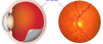
The classic ophthalmoscopic picture of central retinal artery occlusion is widespread ischemic retinal edema with a bright red spot in the macular area (cherry pit sign).
Urgent hospitalization is necessary, since treatment started in the early stages of the disease is most effective. It is usually not possible to determine the exact cause of artery blockage.
Treatment includes massage of the eyeball, administration of active vasodilators (papaverine, compolamine, aminophylline) both systemically and locally in the form of parabulbar injections, dehydration (diacarb, furosemide). Local (parabulbar) corticosteroids are prescribed. To increase perfusion pressure, instillation of beta blockers is indicated. Rheopolyglucin with trental and dexamethasone is administered intravenously. The patient must remember that the effectiveness of treatment is largely determined by the timing of its initiation and is highest in the first minutes and hours of the disease.
The consequences of acute obstruction of the central nervous system are primary atrophy of the optic nerve, central secondary retinal degeneration and a sharp narrowing of retinal vessels. Secondary neovascular glaucoma develops in 1% of patients with central retinal artery occlusion.
What can cause retinal vascular thrombosis
People aged 60 and older are at risk. In addition, such patients often have a history of:
- diabetes;
- arterial hypertension;
- elevated cholesterol levels (dyslipidemia);
- heart rhythm disturbance (atrial fibrillation);
- heart defects;
- cardiac surgery;
- blood coagulation disorder.
Maya Lonskaya:
“Recently, young people aged 30 to 35 years have begun to come to us more often with such thromboses, in whom circulatory disorders in the retinal vessels occurred after severe emotional overstrain or stress. Moreover, there are quite a lot of such patients who come with this problem after suffering a coronavirus infection. Typically, the first complaints appear two to three weeks after COVID-19.
The main symptom is blurred vision, which increases gradually. Often people do not attach any importance to this or do not notice it at all. At the same time, the condition worsens and leads to the development of severe complications.
Diagnosis of pathology
In making a diagnosis, an important role is played by the analysis of medical history: the patient has systemic, inflammatory, cardiovascular diseases, metabolic diseases, eye injuries, other vascular accidents (myocardial infarction, stroke, superficial or deep thrombosis of the leg veins, obliterating endarteritis, etc. ).
The necessary diagnostic examination includes: examination of the fundus, contrast radiography of retinal vessels, laboratory tests. Visometry during occlusion reveals a sharp decrease in visual acuity, which can vary from 0.02-0.1 to 0. The degree of vision loss is directly dependent on the level of occlusion, as well as the size of the ischemic area.
Perimetry reveals defects in peripheral vision - scotomas (sectoral or central, which correspond to the zone of retinal ischemia), as well as concentric narrowing of the visual field.
Biomicroscopy makes it possible to draw a preliminary conclusion about the degree of occlusion. In this case, incomplete occlusion is determined by an afferent pupillary defect (the so-called Marcus-Gunn pupil); total occlusion - a reaction to light that is absent or sharply reduced.
Visual examination of the fundus during ophthalmoscopy reveals swelling, loss of transparency, pallor of the retina and optic disc. Particularly noticeable against this background is the central fovea of the macular region, which has a bright color, since the choroidal blood supply is preserved (cherry pit syndrome). The retinal arterioles are narrowed and have an uneven size. In the first days of development of occlusion, emboli are sometimes visible in them.
Fluorescein angiography clarifies the location of the thrombus or embolus and determines the degree of blockage of the vessel. In this case, radiographic signs of occlusion are a slowdown in blood flow or its segmental nature in the retinal arterioles, as well as the symptom of “vascular breakage” when the branches of the central nervous system are obstructed.
Electroretinography data during occlusion are characterized by a decrease or absence of the amplitude of the recorded wave oscillations, which indicates the death of ganglion cells, with choroidal ischemia.
Specific diagnostics for blockage of the central retinal artery are carried out by performing ultrasound scanning of the vessels of the eye, optical coherence tomography, laser scanning tomography of the retina and tonometry.
Among the necessary laboratory tests, a mandatory coagulogram and lipid profile, as well as blood culture (if there is a suspicion of a bacterial embolism).
Duplex scanning of the carotid arteries and ultrasound of the heart are also necessary. If there are indications, the patient is referred for examination to a cardiologist, vascular surgeon, rheumatologist, endocrinologist, hematologist, or infectious disease specialist.
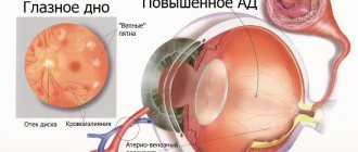
Hypertensive retinopathy
Hypertensive retinopathy reflects changes in the fundus of the eye due to arterial hypertension. Due to the high prevalence of hypertension (23% of the unselected world population), it is expected that the incidence of fundus pathology associated with this disease will also increase.
The main diagnostic method is ophthalmoscopy. Complications of hypertensive retinopathy may include recurrent hemophthalmos and retinal vein thrombosis.
Treatment takes place with the participation of a cardiologist and an ophthalmologist. It is mainly aimed at stabilizing hypertension and its consequences.