Echoencephalography is a diagnostic neurophysiological study of the structure of the brain, aimed at identifying its pathological processes and changes. The method is non-invasive and requires the use of ultrasonic waves. Thanks to it, it is possible to determine intracranial pressure and identify volumetric processes due to which structures are displaced.
At the same time, it does not allow one to accurately determine the type of detected formations, therefore it is accompanied by computed tomography or magnetic resonance imaging. These are more accurate ways to make an accurate diagnosis.
Echo-EG of the brain involves scanning and calculations by a doctor. They are necessary in order to determine the parameters of the internal volume of the skull and the presence of displacements of brain structures, to identify an increase in intracranial pressure after injuries or due to neoplasms.
You can undergo an Echo-ECG in Moscow at the CELT multidisciplinary clinic. We have everything necessary to obtain all the necessary information and correctly interpret research data.
Echoencephalography of the brain (Echo-EG) - 1,200 rubles.
15 minutes
(duration of procedure)
Outpatient
Advantages of echoencephalography of the brain in CELT
We employ specialists with many years of experience, allowing them to carry out diagnostic studies as accurately as possible and obtain all the necessary data. They have modern equipment in their arsenal that allows them to get a complete picture. Thus, Echo-EG of the head in our clinic allows:
- identify neoplasms in the early stages of their development;
- conduct a qualitative study of the midline structures and periosteal space;
- determine ICP indicators and the nature of pulsations;
- minimize the human factor and increase diagnostic accuracy.
You can find out the price of Echo-EG in our clinic in the corresponding section of our website or by calling us!
Consent to the processing of personal data
If, when using the Service, the user provides any information relating to a directly or indirectly identified or identifiable individual (hereinafter referred to as personal data), namely last name, first name, patronymic, telephone number, email address and other information provided on the user’s own initiative, its subsequent processing will be carried out in accordance with the requirements of the Federal Law of June 27, 2006 No. 152-FZ “On Personal Data”.
We receive information directly from you when you make an appointment using the site and when a user requests feedback and evaluation of the company's work. In this case, the user can browse the site and use its other features without disclosing any of his personal information. With respect to all reported personal data, the user gives consent to the Administrator for their processing. The Administrator processes the user’s personal data solely for the purpose of providing the user with the functionality of the Service and the Site, the content posted on it, marketing, advertising, and other information, in order to maintain feedback and process user requests to the Administrator, as well as for the purpose of promoting the user’s goods and services. The Administrator takes all necessary measures to protect the personal data of the Site user, and also provides access to such data only to those employees, contractors and agents of the Administrator who need this information to ensure the purposes of processing personal data by the Site Administrator. Disclosure of personal data provided by the user can only be made in accordance with the legislation of the Russian Federation. With regard to all communications, the User understands and agrees that providing the Administrator with any information about himself that is not contact and not related to the purposes designated by the Site Administrator (not related to the activities of the Administrator, to the goods and/or services promoted by him, to terms of cooperation between the Administrator and the Site user), and exactly the provision of information related to state, banking and/or commercial secrets, information about race and/or nationality, political views, religious or philosophical beliefs, health, intimate life of the Site user or other third party is prohibited.
Photo and video materials on the site Some images on our site were found through search engines, so we cannot determine in advance under what conditions they are distributed and who their author is. If you are the copyright holder of any image used on the santem24.com website and you have complaints about its use by us, write to us and we will immediately remove the copyrighted material or indicate your authorship, if necessary. To do this, contact us and indicate information about the object of copyright, information about the copyright holder and the address of the page on our website that contains data that violates copyright. You can write to us by email
Indications for ultrasound echoencephalography
The study is carried out as part of primary neurological diagnosis when the patient complains of:
- headache;
- constant feeling of nausea;
- frequent loss of consciousness;
- feeling of pressure on the eyes;
- incessant noise in the head;
- disturbances in concentration and memory.
The indication for this is an increase in ICP or the need to measure it, suspicion of the presence of neoplasms, foreign objects, cysts and hematomas in the cranial cavity if computed tomography and magnetic resonance imaging are not available. The study is carried out if necessary:
- confirm the presence of diseases such as epilepsy, brain damage of a degenerative, vascular or inflammatory nature;
- identify the patient’s simulation of hearing and visual impairments;
- assess the brain functioning of comatose patients;
- determine the cause of sleep disturbances and abnormal surges in blood pressure;
- determine the efficiency of functioning of blood vessels and tissues.
More about diagnostics
Many people are interested in what it is – an echoEG of the main organ of the central nervous system. Another question that concerns patients is how it affects their health.
The essence of the method
The examination is completely safe - it is no coincidence that it is performed even on infants.
The method is based on the difference in surfaces and tissue density from which directed waves return. Thus, healthy skin and subcutaneous tissue have one reflected signal, a more liquid cyst or hematoma has another, gray matter has a third, etc. The difference between them gives the specialist a reliable idea of possible disorders in the patient.
Doctor's actions
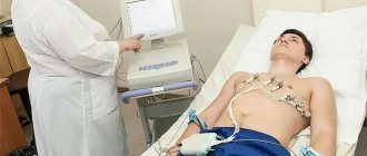
EchoEG is carried out with a sensor equipped with a special plate capable of both sending and receiving data. It is attached to the temple above the ear. First, the sides of the head are lubricated with gel. The patient usually lies or sits during these manipulations
An important condition is that the person should not move.
When the plate is deformed, ultrasound is generated and directed to the tissue elements. Returning, the wave is transformed into an electrical signal and reflected on the monitor. The entire procedure takes a maximum of 15 minutes.
Modes
EchoEG is performed both in one-dimensional M-mode and in two-dimensional (ultrasound). In the first case, a graph with several rises is visible on the screen, showing the relationship between brain structures. Two-dimensional diagnostics gives a flat picture and is used for children.
Types of impulses
The graphic image that appears on the monitor after the examination consists of 3 sets of signals: initial, final and the most important - median, or M-echo
- The initial complex is reflected high-frequency pulses showing the location of bone tissue and external integument, the hard fibrous membrane of the brain, and muscles located in the sensor’s coverage area. Data is captured at the left edge of the monitor.
- The final one is a set of impulses pushed off from another inner wall of the skull and its covers. The data is visible on the right side of the screen.
- The median complex is waves reflected from the 3rd ventricle, the transparent septum, and the epiphysis. The distance correlates with the midline of the skull in the sagittal plane. Deviation in M-echoencephalography from the midline indicates pathological displacements in this organ of the central nervous system.
Data decryption
What the monitor screen shows must be interpreted correctly. This is what a neurologist does. As a rule, deciphering the results is based on the components described above. Occasionally additional bursts (lateral echoes) are taken into account.
Decoding begins with an analysis of the median complex. The permissible displacement of the distance from the selected point to 2 other complexes between which it is located is no more than 1-2 mm in adults, and up to 3 mm in children (maximum 5 mm, and ideally the same).
The ripple limits should not exceed 50%. If this happens, the doctor, if there are relevant patient complaints, diagnoses hypertension.
If the midsellar index is less than certain values (“4”, marks 39), this indicates increased intracranial pressure in adults.
EchoEG is also used for indirect diagnosis of the condition of cerebral vessels. The vector of median deviations gives an idea of the location of the lesion.
The accuracy of interpretation of the obtained EchoEG data depends on the professionalism of the neurologist, the depth of probing and the resolution of the echoencephalograph.
Contraindications to echoencephalography in M-mode
Echoencephalography of the brain in children and adults is a safe procedure. It does not require the use of painkillers (or any other pharmacological drugs) and has no physiological contraindications. However, it should be postponed during acute respiratory diseases accompanied by a runny nose and/or cough. It is not used if the patient has open wounds or suffers from psychological disorders in the areas where the sensors need to be installed. The latter can cause hysterics due to the unfamiliarity of the situation.
How to properly prepare a child for research?
Without preliminary preparation, the research procedure may cause fear in young children due to the specific preparatory manipulations performed. Therefore, it is recommended that parents prepare their baby in advance. Young children can be prepared in a playful way, for example, by telling the child that he will try on the helmet of an astronaut, pilot or superhero. Older children need to be explained in detail how the EEG procedure will be performed, even shown if possible. With such preparation, children come for diagnostics with pleasure and are not afraid of anything, which guarantees a successful recording of the study.
Preparation for the procedure and its implementation
One- and two-dimensional echoencephalography does not require the patient to prepare independently. All he needs to do is visit the diagnostic room at the appointed time. Before starting the examination, the diagnostician will ask him to remove from his head any accessories that may interfere.
During the process, the doctor installs an ultrasound sensor in certain areas of the head. To ensure high-quality contact, a contact gel is first applied to them. This prevents air from entering and causing interference. The sensor is placed:
- above the ear canals;
- near the outer edges of the eyebrow arches (at the temples);
- behind the ears at a distance of four to five centimeters.
By sending ultrasound signals, the sensor catches them in reflected form and transmits them to the device, which displays them on the monitor in the form of a graphic image similar to the teeth of an electrocardiogram.
When carrying out the procedure in a two-dimensional format, the sensor is not rearranged sequentially - it is moved along the surface of the head, providing an image of the slice in a horizontal plane along the line of its movement. An image of the formations located at this level is displayed on the screen. The duration of the procedure is no more than 15 minutes.
ECHO-EG, EEG, REG
ECHO-EG, EEG, REG are studies that are prescribed to patients to study the brain. At first glance, complex abbreviations seem exactly the same or interchangeable. In fact, this is not so, ECHO-EG, EEG and REG are completely different studies, let’s take a closer look.
ECHO-EG - echoencephalogram - is a method of studying the brain, which measures the width of the third ventricle of the brain, the displacement of the midline structures of the brain (M-ECHO).
Indications for this study are: headaches, arterial hypertension, migraine, cerebrovascular accidents, concussion, head injuries, decreased vision.
When performing echoencephalography, no preliminary preparation is required.
EEG - electroencephalography - is a method for studying the functional state of the brain by recording its bioelectrical activity. A special cap with electrodes-antennas, which are connected to the device, is put on the person’s head. Signals coming from the cerebral cortex are transmitted to an electroencephalograph, which converts them into a graphic image (waves).
Conducted to identify brain excitability (epileptiform brain activity).
Indications are: epileptic seizures, fainting, traumatic brain injuries, mental disorders, enuresis, VSD.
REG - rheoencephalography is one of the most effective and simplest ways to diagnose cerebral circulation. REG of cerebral vessels is a safe diagnostic method, has no contraindications and can be prescribed to a patient of any age. The study is carried out using a special recording device - a rheograph. The patient lies down or sits in a comfortable position, closes his eyes, electrodes are applied to certain points on the head and secured with rubber bands. A weak current is passed through the electrodes, and the resistance readings of the brain tissue are recorded and then deciphered.
Rheoencephalography is prescribed for diagnosing cerebral vascular lesions, monitoring blood circulation in the brain after injuries or operations, for assessing hypertension, for assessing the state of the brain (in case of head injury, encephalopathy, pituitary adenoma, after ischemia or stroke) for diagnosing VSD and SVD.
At the Good Doctor clinic, they conduct ECHO-EG and REG brain studies for adults and children over 4 years of age according to indications and referrals from a neurologist, ophthalmologist, otolaryngologist, internist or pediatrician.
Decoding echoencephalography of the brain
The results of the diagnostic study are interpreted by a neurologist. As already mentioned, the norm provides for the same distance from the central signal to both sides. The permissible deviations are no more than two millimeters for adults and three for children.
During volumetric processes in the GM substance, a shift in the central signal occurs, changes in the shape and duration of responses. Thus, the following pathological conditions can be identified:
- intracranial neoplasms of benign and malignant etiology;
- tuberculomas;
- hematomas localized in the intracranial space;
- cerebral infarctions;
- focal accumulations of pus in the GM substance;
- syphilitic granulomas.
Their localization is determined by median deviations. The distance to the central signal on the side of the formation is greater than on the healthy side, although in certain inflammatory diseases and stroke the opposite may be true. A similar phenomenon can be triggered by a decrease in the volume of one hemisphere during recovery processes, when scarring or resorption is observed.
The doctor determines the presence of a problem based on echo-EG readings:
- with tumors of malignant etiology, a serious shift is observed;
- infarction states of the brain are characterized by small transient changes;
- in case of traumatic injuries, displacements of no more than three millimeters are observed due to swelling;
- hydrocephalus is characterized by expansions of the central signal from five minimeters and the appearance of a large number of lateral ultrasound signals;
- suspicion of hemorrhage arises with the most serious asymmetry.
It is important for patients to understand that the diagnostic accuracy of this method directly depends on the qualifications and experience of the specialist, as well as on the characteristics of the device involved in the process.
Make an appointment with our specialists by filling out and sending us an application online or by calling us by phone! Trust the diagnostics to professionals!
Decoding indicators
The results of the examination are interpreted by a neurologist or laboratory specialists.
The main indicator is the same distance to the M-echo on both sides of the head. Normally, deviations should not exceed 1-2 mm (in children - 3 mm). In pathological processes, a shift in the M-echo is observed, and the shape and duration of responses change.
When using echoencephalography, there are 3 types of signals (complexes) that help decipher the results:
- Primary complex. A signal localized directly next to the sensor. It is formed by an ultrasound wave when reflected from muscles, skin, fiber and upper brain structures.
- Median. The signal obtained when ultrasound collides with structures located in the middle of the brain: septum pellucida, cerebral peduncle, third ventricle, pineal gland, falciform process of the cerebrum.
- The final complex. The signal comes from the lateral structures: the bones of the skull, soft tissues and the dura mater of the brain.
Deciphering the diagnostic results by an experienced specialist takes no more than a quarter of an hour.
In what cases is such an examination prescribed?
Symptoms for which a doctor may prescribe an Echo EG:
- periodic headache;
- feeling of dizziness, poor spatial orientation;
- ringing in the ears;
- head injuries accompanied by nausea or vomiting.
In addition, echoencephalography is prescribed to identify the causes of other pathologies:
- pain in the cervical region;
- vegetative-vascular dystonia;
- impaired blood circulation;
- vertebrobasilar arterial system syndrome;
- ischemic disease;
- brain concussion;
- vascular encephalopathy of the head;
- stroke.
An electroencephalogram shows the main parameters of cerebral functions. Using a head echo, a number of diseases can be detected. These include: various tumors, inflammatory processes, cerebral blood flow disorders, intracranial pressure, hydrocephalic syndrome, adenoma of brain areas.
The essence of the study
The human brain plays a major role in the functioning of the entire nervous system. A slight malfunction in the functioning of this organ can lead to disturbances in the coordination of the human body. A thorough examination will help identify the causes of the disease.
Echoencephalography of the brain is a reliable and accessible method of research presented in medicine. The procedure is absolutely painless. It can be done at any age.
Not everyone has an idea about the features of this examination method and where it is carried out. Many people are interested in how this procedure is done and what are the indications for diagnostics.
Echo EG is practically no different from ultrasound (ultrasound). Echoencephalography of the brain or echoencephaloscopy (EchoES) is based on the reflection of ultrasonic waves from the tissues of human organs.
Sensors installed on the patient's head transmit waves to each other, which are reflected from the surface and internal structures of the brain. These waves are converted into an electrical signal, which is recorded by a specialist in the form of an image or diagram on the screen. This method gives an accurate picture of the condition and pathologies of the brain.
Electroencephalography allows you to learn about vascular diseases and the causes of improper cerebral circulation. The method helps to assess the lumen and tone of the veins.
A head echo is a non-invasive type of examination, so it is often used to quickly establish a diagnosis in an emergency. This examination should not be confused with the echocardiography procedure, which is used in heart studies.
results
Decoding Echo ES is the competence of a neurologist, diagnostician or neurophysiologist. Normally, it is believed that the signal from the first sensor should be identical to the signal from the second. A signal that deviates by 1-2 mm from the original value can be called pathological (an error of up to 3 mm in children is acceptable).
If the initial signal changes, it means that within the cranium there is a dislocation of structures due to a volumetric process, which can be:
- tumor;
- intracerebral hemorrhage;
- cyst;
- purulent abscess;
- tuberculoma;
- accumulation of parasites.
- volumetric inflammation.
However, different diseases have specific signs on the monitor:
- Tumors and cysts. They greatly displace brain structures compared to other brain diseases, and therefore increase the difference between the original and received signal.
- Trauma to the skull and brain. They show a difference of within 3mm due to the formation of edema. Later, injuries can provoke the development of cysts, which increases the difference between signals from 3mm.
- Acute cerebrovascular accident. Intracerebral hemorrhage makes a big difference. The value of the lateral echo signal increases due to the presence of a focus of hemorrhage in the tissue. Ischemic stroke, cerebral infarction and softening of brain tissue produce subtle signal differences.
- Hydrocephalus. This pathology produces a signal difference of more than 7 mm.
Decoding the research results
An echoencephalogram is a graphical representation of three signals:
- Initial.
- Of course.
- Middle.
The initial complex is a set of reflected high-frequency impulses that show the location of the external tissues of the head (skin, muscles, bones), as well as the fibrous membrane of the brain. The image is fixed in the left corner of the screen.
The final complex is impulses coming from the other wall of the skull. The data is shown in the right corner of the screen.
The median complex (M-Echo) is waves coming from the third ventricle, the transparent septum, and the epiphysis.
Normal EchoEG indicators in an adult:
- The distance between M-Echo signals on both sides should not exceed 2 ml. Otherwise, there is a suspicion of displacement of brain structures.
- The signal from the third ventricle should be uniform. If it changes, intracranial hypertension should be suspected.
- M-Echo pulsation is not higher than 10–30%. An increase in this indicator indicates an excess of cerebrospinal fluid and cerebral edema.
- The distance between pulses must be the same.
- The average sellar index is at least 4.0. A decrease in the indicator indicates intracranial hypertension.
In various pathological processes, echoencephalography data may change as follows:
- brain tumors cause a significant shift in indicators;
- with head injuries, moderate displacements are detected (up to 3 mm);
- post-traumatic cysts are recorded in the form of lateral echo waves;
- hemorrhage is characterized by significant asymmetry;
- with hydrocephalus, the M-Echo expands up to 0.7 cm, and a large number of lateral waves arise;
- cerebral infarction is recorded in the form of a transient dislocation of the midline structures.