Pulmonologist
Prokhina
Maria Egorovna
Experience 38 years
Pulmonologist
Make an appointment
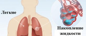
Pulmonary edema is a pathological, very serious condition, which is characterized by the release of transudate into the lung tissue. As a result, gas exchange is disrupted, which leads to serious consequences, including death.
Emergency care for pulmonary edema is the only thing that can increase the patient’s risks of survival and recovery. A person in such a situation requires immediate medical attention.
Pulmonary edema itself is most often a complication that accompanies serious problems of organs and body systems, for example, the cardiovascular system, gastrointestinal tract, etc.
What is pulmonary edema and how does it develop?
Edema occurs due to the accumulation of fluid in the lungs, more specifically in the interstitium and pulmonary alveoli. It causes disruption of gas exchange and the development of hypoxia, as a result of which the blood begins to lack oxygen. Symptoms of pulmonary edema in older people include bluish skin and shortness of breath.
We recommend
“Difficulties in treating congestive pneumonia in the elderly” Read more There
are many reasons that cause this disease. This includes left ventricular failure, pneumonia, foreign body entry into the bronchi, and a sharp decrease in atmospheric pressure. With pulmonary edema, a person is in critical condition requiring emergency care. The process develops so quickly that any delay can cost lives.
A person has two lungs: right and left. Their main purpose is to ensure the exchange of gases between the blood and the alveoli. The walls of the alveoli and the walls of the capillaries take part in this process. Edema develops when fluid from the capillaries moves into the alveoli of the lungs. This may occur due to high blood pressure or low protein levels in the blood. Due to edema, the lungs cease to perform their functions in the human body.
The disease, depending on the causes that provoked it, can be of two types:
- Hydrostatic edema develops as a consequence of diseases during which the fluid pressure inside the vessels increases. Because of this, the liquid part of the blood leaves the vessel into the connective tissue and then enters the alveoli.
- Membranous edema occurs due to internal or external causes related to exposure to toxins. They destroy the walls of the alveoli and/or capillaries, and the fluid begins to freely flow into the space between the vessels.
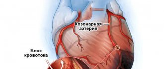
The first type of pulmonary edema is much more common in older people. This happens due to the spread of cardiovascular diseases, the most famous of which is coronary heart disease or myocardial infarction.
Pulmonary edema in the elderly is a difficult diagnosis. Even with a positive result of treatment, the disease can return at any time.
We recommend
“Gymnastics for the elderly: the best exercises for various diseases” Read more
Pulmonary edema is characterized by development in three main directions:
1. Increased hydrostatic pressure or increased blood volume. If the pressure in the capillaries that form the pulmonary circulation unexpectedly increases, the permeability of their walls is disrupted, and the fluid from them leaks into the connective tissue of the lung. The lymphatic system cannot cope with the drainage of such a volume of substance. As a result, the fluid enters the alveoli, which then lose their functionality. They can no longer provide gas exchange in the lungs. This causes a lack of oxygen in the body. Due to the accumulation of carbon dioxide, tissues turn blue and symptoms of severe suffocation appear.
2. Poor blood oncotic pressure associated with low protein levels. In this condition, the blood pressure differs from the pressure of the intercellular fluid. In order to compensate for the difference, fluid from the vessels leaks into the extracellular space, and pulmonary edema begins to develop.
3. Damage to the alveolocapillary membrane itself. Under the influence of various factors, the protein structure of the alveolar capillary membrane is disrupted. Because of this, fluid leaks into the extracellular space, which leads to pulmonary edema.
Recommended articles on this topic:
- Boarding home for the elderly: features and rules of choice
- Low blood pressure in older people: causes, symptoms and treatment
- Health of the elderly: old age is not a problem
Types of pleurisy
Taking into account the clinical manifestations and nature of the effusion in the pleural cavity, the following types of disease are distinguished: fibrinous (dry) pleurisy, exudative, purulent and tuberculous.
Fibrinous pleurisy. The inflammatory process is not characterized by the appearance of exudate in the pleural cavity. The liquid only lubricates the pleural sheets and settles in the form of deposits, which makes it difficult for the sheets to slide. Symptoms of dry pleurisy: fever, painful cough, pain in the affected part.
Exudative pleurisy. The disease is characterized by a gradual increase in symptoms. The patient may complain of headache and low-grade fever, slight weakness and heaviness in the affected part of the chest. Slight shortness of breath bothers the patient even at rest. In the “Treatment” section you will learn about how to treat exudative pleurisy.
Purulent pleurisy is an accumulation of purulent exudate between the layers of the pleura. The disease is a serious pathology that poses a real threat to the patient’s life. Symptoms: high fever, severe pain in the affected part of the chest, shortness of breath, weakness, headache, chills. A doctor will tell you about complications after purulent pleurisy; you can sign up for a consultation with us on our website Dobrobut.com. The specialist will talk about the characteristics of the symptoms of this pathology and methods for diagnosing it.
Tuberculous pleurisy. The disease is characterized by a chronic course with a slow increase in general intoxication of the body and signs of lung damage.
Enclosed pleurisy in oncology is a serious condition characterized by respiratory failure, severe headache, high fever and high blood pressure. Complications are bronchial fistula and empyema.
Causes of pulmonary edema in older people
Due to age-related changes in the body, older people are at risk of pulmonary edema. This disease can develop due to the following reasons:
- Pathological processes in the cardiovascular system that affect the functioning of the left chambers of the heart, which form the pulmonary circulation. With advanced disease and lack of qualified medical care, pressure increases in the pulmonary circulation, which provokes the occurrence of edema.
- A blood clot can block a blood vessel in the pulmonary artery or one of its branches. People with varicose veins and hypertension are predisposed to this process. Blockage can occur due to the formation of a new blood clot or due to the separation of an existing one. The detached thrombus travels through the blood vessels into the pulmonary artery or its branch. Blockage occurs provided that the diameters of the vessel and the thrombus are the same. Because of it, the pressure in the pulmonary artery and capillaries increases.
- Toxins can also cause pulmonary edema in older people. They enter the human body in two ways: external and internal. The first involves the penetration of toxins from the environment. In the second version, they are produced by viruses, fungi, bacteria, parasites and other organisms that are inside a person. This may include an overdose of drugs, for example, Fentanyl or Apressin.
- Other causes of edema include radiation, acute lung disease, and inhalation of drugs such as cocaine. All of these processes disrupt the membrane of capillaries and blood vessels, causing fluid to enter the extravascular space and provoke the occurrence of edema.
- Liver and kidney diseases reduce the protein content in human blood. This results in low oncotic pressure, which leads to the appearance of fluid in the alveoli.
- Conditions such as pneumothorax, pleurisy, or chest injury can also cause pulmonary edema.
- Uncontrolled intravenous infusion of solutions leads to an increase in hydrostatic blood pressure, which makes it possible for fluid to enter the alveoli.
We recommend
“Lonely older people: how to help them” Read more
Diagnostic measures
To identify the syndrome, two types of diagnostics are carried out: simple and differential.
Simple diagnostic methods:
- Collection of information and study of the clinical picture. If the patient is conscious, and pulmonary edema does not occur in an acute form, then the doctor is obliged to collect an anamnesis, measure the patient’s pulse, his pressure, and use chest percussion to confirm the diagnosis.
- BAC tests. They help identify the primary cause of the disease (for example, a heart attack).
- Analysis of gas concentrations in the bloodstream.
- Chest X-ray. This usually helps confirm or deny the presence of fluid in the lung tissue.
- ECG. Helps identify abnormalities of the heart muscle.
- Pulmonary artery catheterization.
The attending physician can make a final diagnosis only after a detailed study of the patient’s clinical picture, carrying out all types of tests and examinations.
First aid for pulmonary edema
At the first stage, before hospitalization of the sick person, it is necessary to stabilize his condition and prevent deterioration of his health. If emergency doctors cannot cope with the development of edema and the person becomes increasingly worse, he must be taken to the hospital, acting very carefully. When transporting in an ambulance, resuscitation actions must be carried out, the purpose of which is to stabilize the pressure and speed of blood movement through the vessels.
In order for the doctor to determine what to do first, how to help the patient, he needs to take into account not only the obvious signs of the disease, but also the type of pulmonary edema. But there is a certain set of actions that are applicable for any type of pathology. It is to them that an ambulance specialist should first resort.
First, the sick person must have access to fresh air. Secondly, the person must be placed in a horizontal position on the floor. Third, clothing that may put pressure on the sternum should be removed.
The most effective way to reduce pressure in the pulmonary circulation is bloodletting. The permissible volume of extracted liquid is three hundred milliliters. Bloodletting can significantly slow down the development of the pathological process in the lungs. But this method has contraindications. These are low blood pressure and bad veins.
Bloodletting can be replaced by using venous tourniquets. They are also able to reduce pressure in the pulmonary circulation. When installing a tourniquet, you need to monitor the pulse below the clamp line so as not to disrupt blood flow. It is allowed to leave the tourniquet for one hour. In this case, limbs should be changed every 20 minutes. You can also use hot foot baths.
First aid for pulmonary edema in the elderly includes the use of the following methods and medications:
- To maintain a satisfactory condition of the patient, oxygen under pressure, insertion of an endotracheal tube, and artificial respiration with a volume of injected air of 800–900 ml and a frequency of 16–18 times per minute can be used. Oxygen, humidified to 100%, should be supplied through nasal tubes. If it is not possible to obtain accurate data in the ambulance, then an indicator of sufficient oxygen saturation in the blood is an arterial blood pressure of 70–80 mm Hg. Art., and venous - at around 35–45 mm Hg. Art.
- To reduce the load on the pulmonary circulation, it is necessary to reduce the volume of fluid circulated and blood pressure. This effect is exerted by diuretic drugs of the saluretic group, for example, Furosemide in a dosage of 40–100 mg. It is strictly not recommended to use osmodiuretics, since they cause tissue fluid to move into the blood and, on the contrary, increase swelling.
Reducing pressure inside blood vessels is also possible with the help of a drug such as Lasix at a dosage of 4–6 ml of solution intravenously.
We recommend
“Interests of older people: how to improve the life of a pensioner” Read more
- Removing fluid from the upper respiratory tract using an aspirator.
- Treatment with antifoam agents. Inhalation with a 30% ethyl alcohol solution or slow intravenous administration of 5 ml of 96% ethyl alcohol along with 15 ml of a 5% glucose solution is used. If the foam comes out too abundantly, then 2 ml of 96% ethyl alcohol is injected endotracheally by puncturing the trachea.
- Heparin therapy has proven itself to normalize blood flow in the lungs. The drug is first administered in a large volume (6000–10,000 units) intravenously, then switched to subcutaneous injections of low molecular weight heparins, for example, Fraxiparine, 0.3 ml twice a day.
- If severe pain is observed during pulmonary edema in elderly people, then Fentanyl (2 ml of 0.005% solution) with Droperidol (4 ml of 0.25% solution) in 10 ml of saline is used.
- In order to reduce excitation of the respiratory center, Morphine is used (1 ml of 1% solution intravenously). When left ventricular failure and accompanying pulmonary edema are observed, Morphine helps to stabilize the functioning of the respiratory system and is therefore prescribed to all patients.
This medicine has a wide range of positive properties. Firstly, it affects the vagus nerve, as a result of which the overexcited respiratory center calms down. Secondly, it increases the lumen of the pulmonary and peripheral veins. Thirdly, it blocks nerve impulses, which reduces vascular tone and, as a result, blood pressure.
Morphine should not be taken for bronchospastic syndrome and cerebral edema. It can be replaced with Droperidol, which is administered intravenously (2 ml of 0.25% solution). A side effect of Morphine is vomiting, so it must be combined with an intramuscular injection of 1 ml of Diphenhydramine or Pipolfen.
- Periodic, uneven Cheyne-Stokes breathing also occurs when the respiratory center is depressed. When it appears, it is recommended to administer Eufillin 10 ml of a 2.4% solution intravenously. The use of this drug reduces blood pressure. This is a mandatory measure for heart failure and hypertension, but it is necessary to take into account the side effects of the drug in the form of tachycardia and the need for additional saturation of the heart muscle with oxygen.
- Injection of corticosteroids, such as Hydrocortisone 125 mg per 150 ml of 5% glucose solution, is used to improve the functioning of the pulmonary membrane.
- Antihistamines: Diphenhydramine 1 ml of 1% solution intramuscularly, Suprastin 1 ml of 2% solution intravenously.
- Emergency actions aimed at localizing pulmonary edema in elderly people caused by heart failure are designed to reduce the overall load on the heart and pulmonary circulation and restore the contractile function of the heart muscle.
Reducing the load depends on reducing the blood flow from the periphery into the pulmonary circulation. In order for this to happen, vasodilators are used, for example, Nitroglycerin. It can be taken in tablet form at intervals of 1 tablet. in 10 minutes and intravenously 0.01% solution at a rate of 1 ml in 4 minutes.
- For pulmonary edema in elderly people, in order to restore the contractile function of the heart muscle, Dopamine is prescribed intravenously at a dosage of 2 mcg/kg/min with Reopoliglucin. Dopamine has side effects. These are increasing tachycardia and shortness of breath, as well as vomiting.
We recommend
“Adaptation of older people: in modern society and boarding homes” Read more
- The volume of 5% glucose solution injected into the bloodstream should be 200–300 ml.
- If the patient has difficulty breathing and can be described as hard, there are spasms in the bronchi and shortness of breath, then it is necessary to administer Prednisolone intravenously at a dosage of 30–60 mg.
- Pulmonary edema in older people can occur due to heart failure and arrhythmias. In this case, electrical stimulation is used, which blocks the arrhythmia.
- In order to reduce the permeability of cell membranes, the drug Contrikal 40–60 thousand units is prescribed intravenously.
- If pulmonary edema is caused by heart failure and is accompanied by high blood pressure, then emergency care should consist of the following: intravenous slow infusion of Nitroglycerin (30 mg per 300 ml of physiological sodium chloride solution) at a rate of 10 drops per minute, with continuous monitoring of blood pressure; intravenous administration of 1 ml of 5% solution of Pentamine, for acute hypertension - 1 ml of 0.01% solution of Clonidine.
ethnoscience
You can treat effusion at home only if there is a small amount of it (as determined by x-ray), mild symptoms, and after consultation with your doctor. You can get rid of effusion in the lungs using folk remedies after eliminating the causative factor - the underlying disease that led to the complication.
To remove excess water, take 1 tbsp. spoons of parsley decoction: 500 g of fresh parsley pour 700 ml of milk, bring to a boil, cool. To speed up the process of removing phlegm, drink honey tincture. Compound:
- 200 g of dried or fresh viburnum berries;
- linden flowers, knotweed, plantain - 2 tbsp. spoons;
- coltsfoot - 1-3 sheets;
- vodka – 500 ml;
- honey – 50 ml.
Herbs and berries are poured with vodka, honey is added, stirred until the honey dissolves. Leave for 2 days in a dark place. Take 1 tbsp. spoons 3 times a day until sputum stops coming out. To get rid of a dry cough, stop an attack of bronchitis, and reduce the severity of inflammatory processes in the lungs, take anise decoction. Compound:
- flax seeds - 1 tbsp. l;
- boiling water – 200 ml;
- cognac – 1 tbsp. l;
- honey - 1 tbsp. l.
Pour boiling water over the seeds, cook over low heat for 10 minutes, cover with a lid to infuse for 15 minutes. Take 4 times a day, 1 tbsp. spoon. To strengthen the immune system and speed up the discharge of sputum, take onion juice once a day, 1 tbsp. l: one finely chopped onion sprinkled with 1-2 tbsp. l. sugar, after 10-15 minutes squeeze the juice through cheesecloth.
Important! Traditional methods of treatment can be combined with drug therapy.
Progress in the treatment of pulmonary edema in the elderly
Treatment of pulmonary edema consists of seven stages:
- use of sedatives;
- elimination of foaming;
- use of vasodilators;
- prescription of diuretics;
- cardiac glycosides for edema caused by heart failure and glucocorticoids for other causes;
- decrease in blood volume;
- after reduction of edema, hospitalization for further treatment of the underlying disease.
To localize the syndrome in 80% of cases, the use of morphine hydrochloride, furosemide and nitroglycerin is sufficient. Then the prognosis for stopping pulmonary edema in older people will be positive.
We recommend
“Centers for pensioners: what they are and how to get there” More
After localizing the edema, treatment of the underlying disease begins:
- for cirrhosis of the liver and increased levels of albumin in the blood, a course of hepatoprotectors is prescribed, such as Heptral, Thioctacid, Berlition;
- when swelling is caused by diseases of the pancreas, drugs that suppress its function, for example, Sandostatin, are prescribed. After this, medications are prescribed that activate the healing of necrosis (Timalin, Immunofan), and potent enzyme preparations, for example, Creon;
- treatment of myocardial infarction consists of the use of beta-blockers Concor or Metoprolol, angiotensin-converting enzyme blocker Enalapril and drugs that prevent thrombus formation, such as Thrombo Ass;
- if the problem is diseases of the bronchi or lungs, then a course of antibiotics is prescribed. Macrolides and fluoroquinolones are most often chosen, since penicillins have lost their effectiveness. Plus they add ambroxol preparations: Lazolvan, Ambrobene. They affect the discharge of sputum and fight inflammation. The patient must be prescribed immunomodulators. After pulmonary edema in older people, the state of the body is unstable. Re-infection can lead to death;
- when swelling occurs due to harmful substances entering the body, detoxification therapy is prescribed. The main thing is to restore the fluid balance that is disrupted by the use of diuretics. Medicines that fight poisoning include Regidron, Enterosgel, Enterodes. In case of severe infection, antiemetics are used;
- if the patient suffers from an asthma attack, then glucocorticosteroids, mucolytics, expectorants and bronchodilators are prescribed;
- for toxic shock, antihistamines such as Cetrin, Claritin, and corticosteroids are prescribed;
- Pulmonary edema in the elderly, caused by any disease, requires the use of potent antibiotics, as well as antiviral and immunomodulatory drugs. Antifungal agents may be needed, as antibiotics promote fungal growth. Terbinafine or Fluconazole will help here;
- To maintain a stable state of the body, enzymes, for example, Wobenzym, and immunomodulators, for example, Polyoxidonium and Cycloferon, are prescribed.
The prognosis after pulmonary edema in older people is most often disappointing. In order for the patient to survive, he must be constantly monitored by doctors for a year. High-quality treatment of the underlying disease that provoked pulmonary edema significantly increases the chances of a further full life.
Thus, to summarize, we can say that treatment of pulmonary edema is aimed at relieving the edema itself, and hospitalization is necessary to eliminate the underlying disease.
We recommend
“Boarding home for the elderly: how to place an elderly relative there” Read more
BIBLIOGRAPHY
- Renkin E: Some consequences of capillary permeability to macro-molecules: tarling's hypothesis reconsidered. Am J Physiol 1986, 250:H706–H710.
- Staub N: The pathogenesis of pulmonary edema. Prog Cardiovasc Dis 1980, 23:53–80.
- Matthay MA: Pathophysiology of pulmonary edema. Clin Chest Med 1985, 6:301–314.
- Lewis FR, Elings VB, Sturm JA: Bedside measurement of lung water. J Surg Res 1979, 27:250–261.
- Sivak ED, Starr NJ, Graves JW, et al.: Extravascular lung water values in patients undergoing coronary artery bypass surgery. Crit Care Med 1982, 10:593–596.
- Sibbald WJ, Warshawski FJ, Short AK, et al.: Clinical studies of mea-suring extravascular lung water by the thermal dye technique in critically ill patients. Chest 1983, 83:725–731.
- Gallagher JD, Moore RA, Kerns D, et al.: Effects of advanced age on extravascular lung water accumulation during coronary artery bypass surgery. Crit Care Med 1985, 13:68–71.
- Bongard FS, Matthay M, Mackersie RC, Lewis FR: Morphologic and physiologic correlates of increased extravascular lung water. Surgery 1984, 96:395–403.
- Staub NC: Clinical use of lung water measurements. Report of a workshop. Chest 1986, 90:588–594.
- Schuster DP: The evaluation of pulmonary endothelial barrier function: quantifying pulmonary edema and lung injury. In: Pulmonary Edema. Edited by Matthay MA, Ingbar DH. New York: Marcel Dekker, Inc., 1998:121–161.
- Snashall PD, Keyes SJ, Morgan BM, et al.: The radiographic detection of acute pulmonary oedema. A comparison of radiographic appearances, densitometry and lung water in dogs. Br J Radiol 1981, 54:277–288.
- Gattinoni L, Presenti A, Torresin A, et al.: Adult respiratory distress syndrome profiles by computed tomography. J Thorac Imaging 1986, 1:25–30.
- Wheeler A, Carroll F, Bernard G: Radiographic issues in adult respi-atory distress syndrome. New Horiz 1993, 1:471–477.
- Halperin B, Feeley T, Mihm F, et al.: Evaluation of the portable chest roentgenogram for quantitating extravascular lung water in critically ill adults. Chest 1985, 88:649–652.
- Eisenberg PR, Hansbrough JR, Anderson D, Schuster DP: A prospecious study of lung water measurements during patient management in an intensive care unit. Am Rev Respir Dis 1987, 136:662–668.
- Hedlund LW, Vock P, Effmann EL, Lischko MM, Putman CE: Hydrostatic pulmonary edema. An analysis of lung density changes by computed tomography. Invest Radiol 1984, 19:254–262.
- Forster BB, Muller NL, Mayo JR, et al.: High-resolution computed tomography of experimental hydrostatic pulmonary edema. Chest 1992, 101:1434–1437.
- 18. Mayo JR: Magnetic resonance imaging of the chest. Where we stand. Radiol Clin North Am 1994, 32:795–809.
- Lancaster L, Bogdan AR, Kundel HL, McAffee B: Sodium MRI with coated magnetite: measurement of extravascular lung water in rats. Magn Reson Med 1991, 19:96–104.
- Cutillo AG, Morris AH, Ailion DC, et al.: Assessment of lung water distribution by nuclear magnetic resonance. A new method for quantifying and monitoring experimental lung injury. Am Rev Respir Dis 1988, 137:1371–1378.
- Morris AH, Blatter DD, Case TA, et al.: A new nuclear magnetic resonance property of the lung. J Appl Physiol 1985, 58:759–762.
- Cutillo AG, Morris AH, Ailion DC, Durney CH, Case TA: Determination of lung water content and distribution by nuclear magnetic resonance imaging. J Thorac Imag 1986, 1:39–51.
- Mayo JR, Muller NL, Forster BB, Okazawa M, Pare PD: Magnetic res-onance imaging of hydrostatic pulmonary edema in isolated dog lungs: comparison with computed tomography. Can Assoc Radiol J 1990, 41:281–286.
- Wexler HR, Nicholson RL, Prato FS, et al.: Quantitation of lung water by nuclear magnetic resonance imaging. A preliminary study. Invest Radiol 1985, 20:583–590.
- Phillips DM, Allen PS, Man SF: Assessment of temporal changes in pulmonary edema with NMR imaging. J Appl Physiol 1989, 66:1197–1208.
- Caruthers SD, Paschal CB, Pou NA, Roselli RJ, Harris TR: Regional measurements of pulmonary edema by using magnetic resonance imaging. J Appl Physiol 1998, 84:2143–2153.
- Rhodes CG: Measurement of lung water using nuclear magnetic resonance imaging [letter]. Br J Radiol 1986, 59:1135–1136.
- Cutillo A, Goodrich K, Krishnamurthy G, et al.: Lung water measure-me by nuclear magnetic resonance: correlation with morphome-try. J Appl Physiol 1995, 79:2163–2168.
- Mayo JR, MacKay AL, Whittall KP, Baile EM, Pare PD: Measurement of lung water content and pleural pressure gradient with magnetic resonance imaging. J Thorac Imag 1995, 10:73–81.
- Berthezene Y, Vexler V, Jerome H, et al.: Differentiation of capillary leak and hydrostatic pulmonary edema with a macromolecular MR imaging contrast agent. Radiology 1991, 181:773–777.
- Cutillo AG, Goodrich KC, Ganesan K, et al.: Alveolar air/tissue inter-face and nuclear magnetic resonance behavior of normal and edematous lungs. Am J Respir Crit Care Med 1995, 151:1018–1026.
- Schuster DP: Positron emission tomography: theory and its application to the study of lung disease. Am Rev Respir Dis 1989, 139:818–840.
- Schuster DP, Marklin GF, Mintun MA, Ter-Pogossian MM: PET measurement of regional lung density: 1. J Comput Assist Tomogr 1986, 10:723–729.
- Rhodes C, Hughes, JMB: Pulmonary studies using positron emission tomography. Eur Respir J 1995, 8:1001–1017.
- Schuster DP, Mintun MA, Green MA, Ter-Pogossian MM: Regional lung water and hematocrit determined by positron emission tomography. J Appl Physiol 1985, 59:860–868.
- Schuster DP, Marklin GF, Mintun MA: Regional changes in extravascular lung water detected by positron emission tomography. J Appl Physiol 1986, 60:1170–1178.
- Velazquez M, Haller J, Amundsen T, Schuster DP: Regional lung water measurements with PET: accuracy, reproducibility, and lin-earity. J Nucl Med 1991, 32:719–725.
- Spinale FG, Reines HD, Cook MC, Crawford FA: Noninvasive estimation of extravascular lung water using bioimpedance. J Surg Res 1989, 47:535–540.
- Zellner JL, Spinale FG, Crawford FA: Bioimpedance: a novel method for the determination of extravascular lung water. J Surg Res 1990, 48:454–459.
- Nierman DM, Eisen DI, Fein ED, et al.: Transthoraic bioimpedance can measure extravascular lung water in acute lung injury. J Surg Res 1996, 65:101–108.
- Adler A, Amyot R, Guardo R, Bates JHT, Berthiaume Y: Monitoring changes in lung air and liquid volumes with electrical impedance tomography. J Appl Physiol 1997, 83:1762–1767.
- Effros RM: Lung water measurements with the mean transit time approach. J Appl Physiol 1985, 59:673–683.
- Sivak ED, Wiedemann HP: Clinical measurement of extravascular lung water. Crit Care Clin 1986, 2:511–526.
- Allison RC, Carlile PV Jr, Gray BA: Thermodilution measurement of lung water. Clin Chest Med 1985, 6:439–457.
- Pfeiffer U, Backus G, Blumel G, et al.: A fiberoptics based system for integrated monitoring of cardiac output, intrathoracic blood volume, extravascular lung water, O 2 saturation, and a–v differ-ences. In: Practical Applications of Fiberoptics in Critical Care Monitoring. Edited by Lewis F, Pfeiffer U. Berlin: Springer-Verlag, 1990: 114–125.
- Schuster DP, Calandrino FS: Single versus double indicator dilution measurements of extravascular lung water. Crit Care Med 1991, 19:84–88.
- Sibbald WJ, Short AK, Warshawski FJ, Cunningham DG, Cheung H: Thermal dye measurements of extravascular lung water in critically ill patients. Intravascular Starling forces and extravascular lung water in the adult respiratory distress syndrome. Chest 1985, 87:585–592.
- Bock JC, Lewis FR: Clinical relevance of lung water measurement with the thermal-dye dilution technique. J Surg Res 1990, 48:254–265.
- Wickerts CJ, Jakobsson J, Frostell C, Hedenstierna G: Measurement of extravascular lung water by thermal-dye dilution technique: mechanisms of cardiac output dependence. Intensive Care Med 1990, 16:115–120.
- Fallon KD, Drake RE, Laine GA, Gabel JC: Effect of cardiac output on extravascular lung water estimates made with the Edwards lung water computer. Anesthesiol 1985, 62:505–508.
- Zeravik J, Borg U, Pfeiffer U: Efficacy of pressure support ventilation dependent on extravascular lung water. Chest 1990, 97:1412–1419.
- Haider M, Schad H: Effect of positive end-expiratory airway pressure (PEEP) on extravascular thermal lung water estimation in the dog. In: Practical Applications of Fiberoptics in Critical Care Monitoring. Edited by Lewis F, Pfeiffer U. Berlin: Springer-Verlag, 1990:96–104.
Consequences of pulmonary edema in the elderly
Pulmonary edema syndrome is dangerous because various complications often develop after it:
- a secondary infection may occur, which can lead to pneumonia, which, in combination with edema, is very difficult to treat;
- The lack of oxygen that accompanies pulmonary edema has a detrimental effect on the functioning of internal organs, especially the brain and heart. The consequences of such exposure can be irreversible and lead to human death;
- development of pneumofibrosis and segmental atelectasis.
The recovery period depends on the underlying disease, the general condition of the body, the patient’s age and other factors. Treatment lasts on average from a week to a month.
If there are no complications and quality medical care is provided, then after ten days the patient will be able to go home.
Toxic pulmonary edema is considered the most complex and prone to recurrence. Patients with this diagnosis spend at least three weeks in the hospital.
But regardless of the causes of pulmonary edema, the disease is considered very serious. In acute alveolar edema, the mortality rate is 20–50%. If swelling accompanies myocardial infarction or anaphylactic shock, the patient dies in 90% of cases.
Even with the timely provision of qualified medical care, there is a high probability of complications such as ischemic damage to the viscera, pneumonia, pulmonary atherosclerosis, etc. If the underlying disease was eliminated in a timely manner, then there is still a risk of resumption of pulmonary edema. In any case, the earlier treatment is started, the greater the chance of survival.
Symptoms of manifestation
The main symptom of fluid stagnation is shortness of breath.
It occurs due to the fact that the blood is not sufficiently saturated with oxygen. With a small amount of fluid, shortness of breath is moderate, but as the lungs fill with fluid, breathing difficulties intensify. The patient's breathing is frequent and difficult when inhaling. Symptoms vary depending on the location of the fluid and its volume. The greater the volume of fluid, the more pronounced the symptoms.
The most common signs:
- Attacks of suffocation;
- Frequent shortness of breath. It appears spontaneously and without any prerequisites, more often in the morning;
- Rapid breathing;
- Lack of air;
- Pain in the chest area, aggravated by coughing;
- Cough with mucus, sometimes blood;
- Numbness of hands and feet;
- Dizziness, tachycardia;
- Bluish color of the skin due to oxygen starvation;
- Sometimes anxiety, nervousness, and nervous disorders appear.
The most dangerous consequences are attacks of acute suffocation, which require immediate medical attention.
Prevention of pulmonary edema in the elderly
Prevention of pulmonary edema in older people includes timely treatment of the underlying disease. You need to be constantly monitored by medical specialists, not to come into contact with harmful substances and toxins, not to abuse alcohol and overeating, follow the instructions for using medications, and lead a healthy lifestyle.
If a person has chronic diseases or suffers from hypertension, then you need to regularly visit the doctor and follow all his prescriptions. Secondary, but no less important, measures to prevent pulmonary edema in older people are: a positive attitude, proper nutrition and an active lifestyle.
It is impossible to predict when an attack will occur, but you can reduce the likelihood of it occurring. You also need to remember that timely medical care is the key to saving lives.
To reduce the risk of pulmonary edema in bedridden elderly people, they need to purchase an orthopedic mattress. Even such, at first glance, a trifle can prevent the development of congestive pneumonia.
The patient will need a varied diet high in vitamins, breathing exercises and therapeutic massage.
The air in the room must be humid. This is important to prevent the occurrence of pneumonia. To increase humidity, you can use a special device or ventilate the room in rainy weather.
You need to constantly listen to the breathing of a person confined to bed. It should be smooth and clean, without gurgling or wheezing. If extraneous sounds appear, you must urgently call an ambulance.
Complications
The main thing that threatens pulmonary edema in an elderly person is the lack of oxygen in the tissues. Even if the disease has been stopped, the brain will experience serious changes, and the tissues of the heart and lungs will suffer.
Also among other serious consequences of suffering from pulmonary edema are:
- formation of congestion in the lungs;
- organ ischemia;
- emphysema.
Due to oxygen starvation, the patient’s memory will deteriorate, during the daytime he will constantly feel sleepy, general lethargy will be felt, and his mood will begin to deteriorate. You will have to carefully monitor your own condition in order to notice serious deterioration in time and consult a doctor.
Pulmonary edema in old age is a serious pathology. Even if the disease manifests itself in a protracted or subacute form, the risk of complications after therapy is high. An immediate or acute syndrome practically does not allow saving the patient. So at the first symptoms indicating the development of the disease, you need to undergo an examination and consult a doctor so that it is not too late.
Thoracentesis
For patients with obvious cardiac failure, thoracentesis should be performed in all patients with pleural effusion of unknown etiology of more than minimal volume (a height of more than 1 cm above bottom with data from visual radiography of the OGK at lateroposition, ultrasound and CT). In case of cardiac failure, diagnostic thoracentesis is indicated only if the following atypical symptoms are present: (1) febrile fever and pleuritic pain in the chest; (2) the sound is either one-sided or two-sided, or of a clearly different size; (3) it is not accompanied by cardiomegaly; (4) is refractory to treatment of heart failure.
If hemothorax or pyothorax is suspected, thoracentesis is performed urgently, since in such a situation drainage of the pleural space is indicated. Since it is important to remove the pleural space, the fragments are small in volume and there are many chambers, performing thoracentesis under ultrasound control minimizes the risk of developing iatrogenic pneumothorax. In most cases, analysis of pleural effusion provides valuable diagnostic information and allows one to clearly establish the etiology of pleural effusion based on the identification of malignant cells and microorganisms. c, lymph and the presence of transudate in heart failure or cirrhosis.
Although there are no absolute contraindications for thoracentesis, the most obvious contraindications include impaired throat blood (especially when the prothrombin hour or the activation of the partial thromboplastin hour doubles the normal values), a small volume of hypostasis, artificial ventilation of the leg and Early illness at the site of a planned puncture. Diagnostic thoracentesis can include the following complications: pain at the puncture site, skin or internal bleeding, pneumothorax, empyema, swelling of the spleen or liver.
Visual consolidation of the pleural area can indicate the etiology of illness (Table 3).
Thus, the diet is affected by the presence of tissue and necrotic tissues (in case of empyema) or high fat content (in case of chylothorax). Ridina, regularly filled with blood (hematocrit >1%), occurs in cases of serious illness, trauma (including recent cardiac surgery), pulmonary artery embolism, and pneumonia. If the pleural hematocrit exceeds half the value of the peripheral blood hematocrit, then the patient has hemothorax. Table 3. Clinical significance of visual characteristics of pleural effusion
| Sign | Clinical significance |
| Bloody | When there is an injury, it is most often caused by a malignant process; may also indicate pulmonary embolism, infection, pancreatitis, tuberculosis, mesothelioma or spontaneous pneumothorax |
| Kalamutniy | Possible mobility of a number of cells or lipids |
| Zhovtiy or bіluvaty, kalamutniy | Presence of lymphoma, cholesterol or empyema |
| Brown (chocolate sauce or anchovy paste) | Perforation of liver abscess into the pleural space (amoebiasis with hepatopleural fistula) |
| Chorny | Pleurisy infection by fungi of the genus Aspergillus |
| Yellow-green color with necrotic fabrics | Rheumatoid pleurisy |
| Very thick | Sustainable pyothorax, called malignant mesothelioma (adjusted hyaluronic acid) |
| Putrid smell | Anaerobic infection of the pleural space |
| Ammonium smell | Urinothorax |
| Putrid | Empyema |
| Yellow and thick, with a metallic (rusty) shine | Violent, high in cholesterol (severe chronic vipitus, for example, with tuberculosis and rheumatoid pleurisy) |
If, after thoracentesis, it is imperative to repeat the visual radiography of the OGK, it is not obligatory, due to the following problems: (1) if the puncture is removed in the back; (2) when a sick person develops a backache, cough or pain in the chest; (3) with a pronounced voice tremors over the upper part of the aspirated hemithorax.