The position of the fetus is the relationship of its axis (which passes through the head and buttocks) to the longitudinal axis of the uterus. The position of the fetus can be longitudinal (when the axes of the fetus and uterus coincide), transverse (when the axis of the fetus is perpendicular to the axis of the uterus), and also oblique (the average between longitudinal and transverse).
The presentation of the fetus is determined depending on the part of it that is located in the area of the internal os of the cervix, that is, at the point of transition of the uterus to the cervix (presenting part). The presenting part can be the head or pelvic end of the fetus; in a transverse position, the presenting part is not determined.
Up to 34 weeks, the position of the fetus may change, after this period it usually becomes stable.
What options for placing an EOS can a healthy person have?
In children, the position of the EOS changes as they grow and develop
In infants under 12 months of age, the electrocardiogram shows the direction of the axis to the right. During the course of a year, children's EOS changes and becomes vertically positioned.
By the age of 2-3 years, the axis in 60% of children is vertical, in the rest it changes to normal. This occurs due to growth, enlargement of the left ventricle and reversal of the heart. In preschoolers and older children, the normal position of the EOS dominates.
Bacterial endocarditis: all about the disease and how to deal with it
Determination of the EOS (electrical axis of the heart) by ECG in normal conditions and with abnormalities – meduniver.com
The correct location of the axis in children is considered:
- Babies up to 12 months - EOS is from 90 - 170 degrees
- Children 1-3 years old – vertical direction
- Schoolchildren and teenagers note normal EOS in 60% of children
Most often, the identified deviations in EOS are a variant of the norm and arise due to the individual characteristics of human anatomy. But there are cases when the displacement is too large - this may indicate diseases, including:
- pulmonary hypertension;
- pulmonary stenosis;
- pathology of the atrial septum;
- cardiac ischemia.
Stenosis is determined on the electrocardiogram due to myocardial hypertrophy. Both congenital and acquired forms are detected. In the first case, the diagnosis can be established in early childhood when performing the first ECG.
Atrial septal defects cause a vertical position of the EOS. This happens when the hole size is sufficiently large.
With ischemia of the disease, the lumen of the coronary arteries narrows, which causes insufficient blood supply to the myocardium. In severe form, there is a risk of pathology developing into a heart attack.
During pregnancy, the EOS becomes upright quite rarely. This is due to the physiological characteristics of the body of the woman carrying the baby.
The uterus is constantly enlarging, thereby beginning to affect other internal organs. Because of this, the EOS shifts in most cases in the horizontal direction.
If the ECG showed a vertical position of the axis, the patient will require additional examination. The cause may be heart disease.
- ECG for determining EOS, interpretation of indicators, norms and deviations
In children, such placement is usually attributed to age-related characteristics. As the body matures, it acquires the proper structure, and after complete formation, the electrical axis of the heart returns to its normal location.
EOS position options
When fixing the electrical axis of the heart, the above-described positions are rarely observed. Semi-horizontal and semi-vertical axis positions are recorded in the majority of cases.
The results of the electrocardiogram may record rotations of the EOS around the coordinate axis, which may be normal. Such cases are considered individually, depending on the patient’s symptoms, condition, complaints and the results of other examinations.
Violations of the norm indicators are deviations to the left or right.
For infants, a clear axis shift is noted on the ECG; during growth, it normalizes. For the period of one year from birth, the indicator is usually located vertically.
Standards for children:
- Infants - from ninety to one hundred and seventy degrees;
- Children from one to three years old - vertical position of the axis;
- Adolescent children – normal axis position.
ECG of a newborn
ECG signs in the analysis of EOS are determined by the rightogram and the leftogram.
A rightogram is finding a vector between indicators 70-900. On electrocardiography it is demonstrated by long R waves in the QRS group. The vector of the third lead is larger than the wave of the second.
Phramogram
The levogram on an ECG is the alpha angle passing between 0-500. Electrocardiography helps to determine that the usual lead of the first QRS group is characterized by an R-type expression, but already in the third lead it has an S-type shape.
Levogram
What is the electrical axis of the heart?
EOS is the total (prevailing) vector of all electrical impulses that are observed in the cardiac conduction system during one contraction cycle.
Often this indicator is identical to the electrical position of the heart (EPS) - the orientation of the resulting vector of impulses from the ventricles relative to the axis of lead I on the ECG. In the myocardium, like other muscles of the body, bioelectric currents (action potentials) arise during contraction. It is their electrocardiograph that registers and records them on specialized film in the form of an ECG.
The impulse is generated by the pacemaker (sinus node), from where the excitation reaches the atrium along the nerve pathways of the heart, and then the atrioventricular node (AV). This connection inhibits transmission so that contraction follows relaxation of the atria, which ensures one-way and continuous flow of blood through the cardiac chambers.
On an ECG, electrical impulses are displayed in the form of multidirectional teeth:
- positive – P, R, T – directed upward relative to the isoline;
- negative – Q, S.
An electrocardiographic recording is a recording of changes in potential differences during the process of excitation and relaxation of the atria and ventricles, caused by the electromotive force of the heart (EMF) from the surface of the human body.
EMF is an unstable value; its direction changes throughout the entire cardiac cycle. When summing up all the instantaneous orientations of the impulses (according to the rules of addition), a vector is obtained corresponding to the average EMF during the full period of depolarization - EOS (direction of the electric motor force during the registration of QRS on the ECG).
When recording an ECG, the electrodes are located in three leads that record the potential difference:
- I – left-right hand;
- II – left leg – right hand;
- III – left leg – left arm.
This placement forms a three-dimensional arrangement of EMF vectors on the torso, which forms the “Einthoven triangle”. If you put the EDS in this form, then the angle α (alpha) between the electromotive force and the horizontal of the first lead will express the deviation of the EOS.
Also, the angle α is approximately determined using the six-axis Bailey coordinate system or using special tables. In the absence of the above devices at hand, the orientation of the EOS is established by measuring the height of the R and S waves in standard leads I and III:
- RII=RI+RIII – normal axis location;
- RI>RII>RIII, SIII>RIII – left-sided deviation of the EOS;
- RIII>RI, SI>SIII – the EOS deviates to the right.
ECG formulation
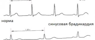
The conclusion of the ECG may include the following wording: “Vertical EOS, sinus rhythm, heart rate per minute. – 77” – this is normal. It should be noted that the concept of “rotation of the EOS around an axis”, the mark of which may be in the electrocardiogram, does not indicate any violations. Such a deviation in itself is not regarded as a diagnosis.
There is a group of ailments that differ precisely in the vertical sinus EOS: various types of cardiomyopathy, especially in the dilated form; ischemia; congenital abnormalities; chronic heart failure.
With these pathologies, a violation of the sinus rhythm of the heart occurs.
The concept of EOS norm
The position of the EOS depends on:
- The speed and correctness of impulse movement through the cardiac systems.
- Quality of myocardial contractions.
- Conditions and pathologies of organs that affect the functionality of the heart.
- Heart condition.
For a person who does not suffer from serious diseases, the axis is characteristic:
- Vertical.
- Horizontal.
- Intermediate
- Normal.
The normal position of the EOS is located according to Died at coordinates 0 – 90º. For most people, the vector passes the limit of 30 - 70º and is directed to the left and down.
- What problems will the electrical axis of the heart tell you about?
In an intermediate position, the vector ranges from 15 to 60 degrees.
According to the ECG, the specialist sees that the positive waves are longer in the second, aVF and aVL leads.
Alpha Angle Definition
You can also determine the position of the electrical axis using a pencil. This method is not accurate enough, and is used in many cases by students.
After this, the sharp side of the pencil is directed to the R-wave, to the lead where it is largest.
| Normal position | Determined if the sharp end of the pencil is in the lower left corner, and its reverse side is in the upper right corner |
| Horizontal | With the pencil positioned almost horizontally |
| Shift left | Determined when the pencil is practically positioned horizontally, it is impossible to accurately determine |
| Offset right | The location of the pencil is determined closer to the vertical position |
The disease is not diagnosed based on the displacement of the electrical axis of the heart alone. This factor is one of the parameters on the basis of which abnormalities in the body can be diagnosed.
These include:
- Insufficient blood supply to the heart;
- Primary damage to the heart muscle, not associated with inflammatory, tumor, ischemic lesions;
- Heart failure;
- Heart defects.
Normal position of the EOS
The EOS angle is considered one of the main parameters that is studied when deciphering ECG indicators. For a cardiologist, this parameter is an important diagnostic indicator, the abnormal value of which clearly signals various disorders and pathologies.
By studying the patient's ECG, the diagnostician can determine the position of the EOS by examining the waves of the QRS complex, which show the work of the ventricles on the graph.
An increased amplitude of the R wave in the I or III chest leads of the graph indicates that the electrical axis of the heart is deviated to the left or right, respectively.
In the normal position of the EOS, the greatest amplitude of the R wave will be observed in the II chest lead.
- Features of conducting and interpreting ECG during pregnancy
Head presentation
Head presentation is determined in approximately 95-97% of cases. The most optimal is the occipital presentation, when the fetal head is bent (the chin is pressed to the chest), and when the baby is born, the back of the head moves forward. The leading point (the one that goes first through the birth canal) is the small fontanelle, located at the junction of the parietal and occipital bones. If the back of the fetal head is facing anteriorly and the face is posterior, this is an anterior view of the occipital preposition (more than 90% of births occur in this position), if it is the other way around, then it is a posterior view. In the posterior form of occipital presentation, childbirth is more difficult; during the birth process, the baby can turn around, but labor is usually longer.
With a cephalic presentation, the pelvic end of the fetus may deviate to the right or left, it depends on which direction the back of the fetus is facing.
There are also extension types of cephalic presentation, when the head is extended to one degree or another. With slight extension, when the leading point is the large fontanelle (it is located at the junction of the frontal and parietal bones), they speak of an anterior cephalic presentation. Childbirth through the natural birth canal is possible, but it takes longer and is more difficult than with an occipital presentation, since the head is inserted into the small pelvis with a larger size.
Therefore, anterior cephalic presentation is a relative indication for cesarean section. The next degree of extension is frontal presentation (it is rare, in 0.04-0.05% of cases). If the fetus is of normal size, delivery through the birth canal is impossible; surgical delivery is required. And finally, the maximum extension of the head is a facial presentation, when the fetal face is born first (it occurs in 0.25% of births). Childbirth through the natural birth canal is possible (in this case, the birth tumor is located in the lower half of the face, in the area of the lips and chin), but it is quite traumatic for the mother and fetus, so the issue is often decided in favor of a cesarean section.
Diagnosis of extensor presentations is carried out during vaginal examination during childbirth.
Concept and specifics
There are several options for the location of the electrical axis of the heart; it changes its position under certain conditions.
This does not always indicate disorders and diseases. In a healthy organism, depending on the anatomy and body composition, EOS deviates from 0 to +90 degrees (+30...+90 is considered the norm, with normal sinus rhythm).
The vertical position of the EOS is observed when it is within the range from +70 to +90 degrees. This is typical for people of thin build and tall stature (asthenics).
Intermediate types of body composition are often observed. Accordingly, the position of the electrical axis of the heart changes, for example, it becomes semi-vertical. Such displacements are not a pathology; they are inherent in people with normal body functions.
An example of wording in the conclusion of an ECG may sound like this: “EOS is vertical, sinus rhythm, heart rate - 77 per minute.” - this is considered normal. It should be noted that the term “rotation of the EOS around its axis,” which may be noted in the electrocardiogram, does not indicate any pathologies. In itself, such a deviation is not regarded as a diagnosis.
The vertical position of the EOS is observed when it is within the range from +70 to +90 degrees.
There is a group of ailments that are characterized by vertical EOS:
- ischemia;
- cardiomyopathy of various nature, especially in the dilated form;
- chronic heart failure;
- congenital anomalies.
Sinus rhythm is disturbed in these pathologies.
Diseases associated with displacement of the OES
Deviation of the electrical position is not an independent pathology. If such a violation is observed, but there are no other pathological symptoms, this phenomenon is not perceived as a pathology. In the presence of other symptoms of cardiovascular diseases, in particular lesions of the conduction system, a shift in the EOS may indicate a disease.
Possible diseases:
- Ventricular hypertrophy.
Marked on the left side. There is an increase in the size of the heart, which is associated with increased blood flow. It usually develops against a background of prolonged hypertension, which simultaneously increases vascular resistance. Hypertrophy can also be triggered by ischemic processes or heart failure. Ventricular hypertrophy - Valve lesions. If damage to the valve apparatus develops in the area of the ventricle on the left side, axis displacement may also occur. This usually occurs due to a blockage in the blood vessels that prevents the release of blood. Such a disorder may be congenital or acquired.
- Heart block. A pathology associated with a disturbance in the rhythm of the heartbeat, which is caused by an increase in the interval between the conduction of nerve impulses. The disorder can also occur against the background of asystole - a long pause, during which the heart does not contract with further ejection of blood.
- Pulmonary hypertension. It is noted when the EOS deviates to the right side. Usually occurs against the background of chronic diseases of the respiratory system, including asthma, COPD. The long-term effect of these diseases on the lungs causes hypertrophy, which in turn provokes a change in the position of the heart.
- Hormonal disorders. Against the background of hormonal imbalance, an increase in the heart chambers may occur. This leads to disruption of nerve patency and deterioration of blood emission.
Vascular crisis: symptoms and causes of a dangerous pathology
In addition to these reasons, deviations may indicate congenital heart defects, atrial fibrillation. A shift in EOS is often observed in people who engage in intense sports or subject the body to other types of physical activity.