Read information about other diseases starting with the letter “L”: Lactostasis, Laryngitis, Laryngotracheitis, Cor pulmonale, Pulmonary fibrosis, Leukemia, Leukemia in children, Leukoplakia, Leucinosis, Leprosy, Dental treatment, Lymphadenitis, Lymphangitis, Lymphogranulomatosis, Lymphoma, Lymphostasis, Lipoma, Listeriosis, Lichen, Pediculosis pubis.
Lymphangitis is a type of inflammation that affects the capillaries and trunks of the lymphatic system. The disease occurs in the presence of purulent-inflammatory processes. It is characterized by visual swelling and is accompanied by painful sensations along the location of the inflamed lymph vessels. I receive complaints from a patient about a feeling of weakness in the body and elevated temperature.
The disease can affect blood vessels of different sizes. Phlebologists and lymphologists note frequently diagnosed lymphangitis of the legs. Less commonly, the pathology affects the pulmonary lobes, mammary glands and other parts of the body. Some patients experience the development of specific venereal lymphangitis and non-venereal lymphangitis, spreading throughout the penis.
General information
Lymphangitis (lat. lymphangoitis) is an acute inflammatory process of lymphatic vessels of various locations/calibers (lymphatic capillaries, vessels, trunks), developing as a complication of local purulent-inflammatory foci or infected skin lesions. That is, lymphangitis is a secondary disease, most often developing against the background of an infected wound , boil / carbuncle , panaritium , phlegmon , abscess , trophic ulcers , erysipelas, etc. or skin damage (microtraumas, abrasions, scratching, bites). As a rule, lymphangitis is accompanied by lymphadenitis (enlargement of the submandibular, cervical, supra/subclavian, axillary, inguinal, popliteal and other lymph nodes). The place of primary localization of the purulent-inflammatory process and lymphangitis is the upper/lower extremities (photos of lymphangitis are given below), but since the human lymphatic system is extremely extensive, the pathological process can develop on any part of the body.
Lymphangitis is accompanied by painful swelling / hyperemia of inflamed lymphatic vessels along their course, regional lymphadenitis , edema , weakness , chills , and increased body temperature.
In andrology/urology, a condition such as “non-venereal lymphangitis of the penis” is often diagnosed. Non-venereal lymphangitis develops when the tissues of the penis are injured (during prolonged sexual intercourse/frequent masturbation). Its manifestation is the appearance of a painless, compacted (on palpation) lymphatic vessel localized along the shaft of the penis or parallel to the coronary groove. As a rule, such manifestations of lymphangitis do not require treatment, and they disappear without a trace/spontaneously within 1-2 days.
Treatment of lymphangitis
Any therapeutic measures for the disease in question begin with eliminating the main source of infection. In case of suppuration, treatment is carried out in a hospital setting; the suppurating lymph node is opened. If lymphangitis of the extremities is diagnosed, then it is necessary to ensure their immobility - the arm or leg must be fixed in an elevated position, which allows for complete drainage of lymph. The patient is recommended to be sedentary and drug therapy is prescribed:
- antibiotics;
- cephalosporins;
- aminoglycosides;
- antihistamines.
To alleviate the patient's condition against the background of general intoxication of the body, infusion therapy is carried out. If the pathology occurs in a chronic form with a vague clinical picture (this is different, for example, from pulmonary lymphangitis), then compresses with dimethyl sulfoxide, physiotherapeutic procedures - mud therapy, irradiation with ultraviolet rays will be effective.
Damage to the penis requires special attention. In case of non-venereal origin, no specific treatment is required, but for 2-3 days it is recommended not to have sexual relations and avoid injury. If lymphangitis of the penis is diagnosed against the background of a progressive sexually transmitted infection, then it is treated.
More information about the pathology and what epizootic lymphangitis is can be found on the pages of our website https://www.dobrobut.com/.
Pathogenesis
In the development of lymphangitis, a significant role is played by the anatomical and physiological specificity of lymph circulation in one or another part of the body, the localization/depth of the wound or primary inflammatory focus, the virulence of pathogenic microflora and the state of general immunity . Pathogens/their toxins initially enter the interstitial spaces, and then move into the lymphatic vessels from the primary site of infection with the lymph flow into large-caliber lymphatic vessels or adjacent lymph nodes.
Their action is manifested by a reactive inflammatory process of the walls of lymphatic vessels with swelling of the vascular endothelium, increasing its permeability, release of exudate and loss of fibrin with the formation of clots (thrombi), which contributes to the development of local lymph circulation disorders (lymphostasis). The pathological process develops as endolymphangitis / panlymphangitis with its spread towards the lymph nodes.
As the inflammatory process progresses, purulent lymphangitis , and in the absence of treatment, purulent fusion of blood clots. perilymphangitis develops with damage to muscles, blood vessels, joints, etc.
Why is lymphangitis dangerous?
Long-term lymphangitis can lead to:
- lymph circulation disorder (the qualitative composition of lymph changes, its quantity increases significantly);
- obliteration of lymphatic vessels (the lumen of the vessel becomes overgrown);
- development of elephantiasis (skin and subcutaneous tissue thicken due to chronic inflammation of the lymphatic vessels), lymphostasis (tissue swelling resulting from impaired lymph outflow);
- abscess (purulent inflammation of tissues with their melting, which leads to the formation of a purulent cavity);
- sepsis (a systemic inflammatory reaction diagnosed as a result of infectious agents entering the blood);
- cellulite (changes in the structure of the subcutaneous fat layer, which lead to disturbances in lymphatic outflow and lymph circulation).
Classification
The classification is based on several factors, according to which they distinguish:
- Depending on the etiological factor, nonspecific and specific lymphangitis .
- Depending on the course – acute and chronic.
- According to the depth of the affected vessels - superficial and deep.
- The nature of the inflammation is serous (simple) and purulent.
- According to the caliber of the affected vessels - capillary (reticular/mesh), in which an extensive network of small capillaries is involved in the pathological process, and truncular (stem), which is based on inflammation of one/several large vessels.
Diagnostics
To diagnose acute reticular lymphangitis, as a rule, a simple examination is sufficient, but it should be separated from erysipelas and superficial phlebitis. It is very important to correctly identify the primary source of inflammation.
Acute truncular lymphangitis is differentiated from superficial thrombophlebitis.
Deep lymphangitis causes difficulties in diagnosing the disease. To determine it, a comprehensive examination is carried out.
Causes
The etiological factor of nonspecific lymphangitis is pathogenic pyogenic microflora. The most common causative agents of the disease include coccal flora (Staphylococcus aureus/beta-hemolytic streptococcus), Pseudomonas aeruginosa , Escherichia coli , Proteus , both in the form of a monoculture and their combinations, and in patients with immunodeficiency - anaerobes and gram-negative bacteria .
Specific lymphangitis in a patient is associated primarily with the presence of tuberculosis infection (Koch bacillus), syphilis (treponema pallidum), plague (plague bacillus), actinomycosis (actinomycete bacteria), genital herpes (herpes simplex virus type 1/2), etc.
Predisposing factors are the presence of local purulent-inflammatory foci/infected skin lesions and decreased immunity .
How does lymphoma develop?
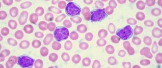
Oncology begins with the appearance in the body of just one altered cell of the immune system. In total, there are 2 main types:
- B lymphocytes
: produce antibodies - proteins that protect the body from bacteria and viruses. This is where most lymphomas form. - T lymphocytes
, one part of which destroys germs and abnormal cells, and the second helps to increase or slow down the activity of the immune system.
Almost all abnormal cells are identified and destroyed by our immune system, but some of them manage to survive. They gradually multiply, spread throughout the body, create tumors, accumulate in internal organs and disrupt their functioning.
The disease can occur in any area where lymphatic tissue is located, the main areas of which are:
- Lymph nodes
are small, pea-sized organs that are collections of cells of the immune system, including lymphocytes. There are more than 500 of them in the human body. - The spleen
, located under the lower ribs on the left side of the body. It produces lymphocytes, stores healthy blood cells and filters out damaged ones, and destroys microbes and foreign substances. - Bone marrow is
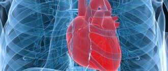
the spongy tissue inside certain bones. Here new blood cells are formed, including some lymphocytes. - The thymus, or thymus gland, is
a small organ located behind the upper part of the sternum in front of the heart. It is where some lymphocytes mature and develop. - Tonsils, or tonsils, are
collections of lymphatic tissue in the back of the throat. These organs help produce antibodies, proteins that prevent inhaled or ingested microorganisms from multiplying. - Digestive Tract:
The stomach, intestines and many other organs also contain lymphatic tissue.
Symptoms
Acute lymphangitis is characterized by severe general intoxication accompanying the purulent-inflammatory process, manifested by weakness , chills , high temperature (39-40°C), sweating and headache . With reticular lymphangitis, superficial pronounced hyperemia first appears in the area of the source of infection ( abscess , boil , etc.) with a pronounced mesh pattern against the background of increasing erythema . At the same time, reticular lymphangitis in clinical symptoms resembles erysipelas , but hyperemia has unclear boundaries uncharacteristic of erysipelas.
A local manifestation of stem lymphangitis is the presence on the skin of red stripes of narrow inflamed lymphatic vessels towards the regional lymph nodes. There is induration, swelling and pain in the inflamed cord-type cords, tension/swelling of the surrounding tissues, and often the development of regional lymphadenitis.
When deep vessels are damaged, there is no local hyperemia, but pain and swelling in the affected area quickly increases; on palpation - severe pain, lymphedema .
In the chronic course of lymphangitis, the clinical symptoms are erased and manifest mainly by persistent edema and lymphostasis caused by blockage of the deep lymphatic trunks with minimal appearance of a general nature.
Treatment of axillary lymphadenitis
Treatment tactics for axillary lymphadenitis depend on the form and cause of the disease. In most cases, lymphadenitis does not require special therapy and goes away on its own after the underlying disease is eliminated. If the process is complicated by suppuration, then the treatment tactics completely change.
General recommendations: maintain rest, immobilize the affected area (do not rub or injure the affected lymph nodes), proper nutrition, take painkillers, anti-inflammatory medications. Therapy for acute axillary lymphadenitis depends entirely on the stage of the disease. Conservative treatment is used for the initial stages: UHF therapy, sanitation of the source of infection (opening of abscesses, leaks, cellulitis, drainage of the abscess), antibiotic therapy taking into account the sensitivity of the microbial flora in the main focus.
Surgical intervention is necessary for purulent axillary lymphadenitis: adenophlegmon, abscesses are opened, pus is removed, and the wound is drained. It happens that a biopsy confirms the presence of a tumor process - benign or malignant. Treatment may include radiation and chemotherapy. In the case of lymphadenitis, as in the presence of any other diseases, it is extremely dangerous to self-medicate.
Back to articles
Tests and diagnostics
Diagnosis of superficial lymphangitis is not difficult and is based on clinical symptoms and the presence of a focus of primary infection. To diagnose lymphangitis of deep lymph nodes, additional laboratory and instrumental studies are carried out:
- Complete blood count (increased ESR/moderate leukocytosis).
- Blood test (bacteria culture) for sterility.
- Bacterial culture of discharge from the primary source of infection in order to identify the pathogen and determine its sensitivity to antibiotics.
- Doppler ultrasound/duplex scanning/CT. Allows you to visualize the narrowing of the lumen of the lymphatic vessels/hardening of soft tissues, the extent of the pathological process.
Differential diagnosis with deep vein thrombophlebitis , soft tissue phlegmon , osteomyelitis , erysipelas .
How is it diagnosed?
The development of regional lymphangitis is diagnosed during the examination. In order not to confuse it with superficial phlebitis, it is necessary to do laboratory tests and conduct instrumental studies.
Diagnostic signs are considered:
- the number of leukocytes in the peripheral blood is increased;
- duplex scanning and Doppler ultrasound visualize the heterogeneity of lymph vessels, a change in the lumen towards narrowing;
- CT scan determines how deep the lymphangitis has spread;
- analysis of pus allows you to determine the pathogenic microorganism.
A complicated form of the disease requires a blood test for sterility.
Prevention
Prevention consists of preventing purulent infection. A healthy lifestyle and strong immunity play a big role in this. If you have cuts or other wounds, it is important to promptly treat them with antiseptics at home. If ulcers appear ( furuncle , carbuncle , abscesses after insect bites, etc.), you need to contact a surgeon in time to sanitize the lesions and follow his instructions regarding taking antibiotics (if necessary).
Treatment options
The main therapy is designed to eliminate the primary focus. The patient's wounds are treated, opened, the contents of the inflammation are removed and sanitized.
It is forbidden to heat or massage inflamed areas. The patient must remain in bed.
Drug treatment involves taking:
- antibacterial agents;
- antiallergic drugs;
- anti-inflammatory complexes.
Physiotherapy methods include irradiation of the patient's blood with laser and ultraviolet light.
Surgical methods are used when it is necessary to drain abscesses.
List of sources
- Ermolaev V. L., Makarova N. P. Diseases of lymphatic vessels and nodes. Ekaterinburg: GBOU VPO USMU Ministry of Health of Russia, 2015. – 264 p.
- Shlyapnikov S.A. Surgical infections of soft tissues - an old problem in a new light // Infections in surgery, 2003, No. 1(1). – P. 14-21.
- Belousova T.A., Kayumova L.N., Goryachkina M.V. Systemic antibiotics in the treatment of bacterial infections of the skin and soft tissues: focus on macrolides / breast cancer. - 2011. - No. 5.
- Guchev I.A., Sidorenko S.V., Frantsuzov V.N. Rational antimicrobial chemotherapy for skin and soft tissue infections. Antibiotics and chemotherapy. 2003, vol. 48, 10, pp. 25–31.
- Surgical infection: A guide for general practitioners / Stolyarov E.A., Grachev B.D., Kolsanov A.V., Batakov E.A., Sonis A.G. - 2004.
Symptoms and signs of lymphoma
As a rule, in the early stages this type of oncology does not manifest itself in any way, and its owner feels well and is not aware of the disease - almost all of its symptoms appear later, in advanced stages.
One of the most common signs is the appearance of swelling in the neck, armpits, groin, or above the collarbone.
, which is an enlarged lymph node. Typically, such a tumor does not hurt, but over time it often increases, and new lumps appear next to it or in other areas of the body.
Lymphomas that begin to develop or grow in the abdominal cavity can cause swelling
or
abdominal pain, nausea,
and
vomiting
. Such sensations occur due to enlargement of the lymph nodes or internal organs, such as the spleen or liver, or the accumulation of large amounts of fluid.
An enlarged spleen can put pressure on the stomach, causing loss of appetite.
and
a feeling of fullness after a small amount of food
.
Enlarged thymus Thymus, or thymus gland -
a small organ located behind the upper part of the sternum in front of the heart.
It is where some lymphocytes mature and develop. or lymph nodes in the chest may put pressure on the trachea, which carries air to the lungs. This leads to coughing
,
difficulty breathing, pain
or
heaviness in the chest
.
Brain lesions can cause headaches, weakness, personality changes, and problems with thinking
and
seizures
.
Other types of the disease can spread to the tissues surrounding the brain and spinal cord, causing the patient to see double and have a numb face
and
speech deteriorates
.
Lymphomas of the skin often appear as itchy red bumps
or
cones
.
In addition, symptoms may include:
- weight loss;
- chills;
- night sweats;
- elevated temperature;
- severe fatigue;
- bloating;
- frequent or severe infections;
- Easy bruising or bleeding.
Lymphoma stages
Immediately after detecting the disease, doctors determine its stage - find out how far it has spread and what tissues it has damaged. This information is extremely important for specialists, since it allows not only to understand the patient’s prognosis, but also to select the most appropriate treatment for him.
Stages of Hodgkin lymphoma:
I
: Changed cells are found in only one group of lymph nodes or one lymphoid organ, such as the tonsils.
II
: they are present in 2 or more groups of lymph nodes located on one side of the diaphragm, or have spread from one damaged lymph node to an adjacent organ.
III
: lymphoma cells are present in the lymph nodes on both sides of the diaphragm;
or not only in the lymph nodes above the diaphragm, but also in the spleen. IV
: The disease has spread to at least one organ outside the lymphatic system, such as the liver, bone marrow, or lungs.
Stages of non-Hodgkin's lymphomas:
I
: altered cells are found only in 1 group of lymph nodes or one lymphoid organ, for example, tonsils;
or in 1 area of one organ outside the lymphatic system. II
: they are present in 2 or more groups of lymph nodes on 1 side of the diaphragm;
either in the lymph nodes and 1 area of an adjacent organ, or in another group of lymph nodes on the same side of the diaphragm. III
: lymphoma cells are present in the lymph nodes on both sides of the diaphragm;
or they are present in both the lymph nodes above the diaphragm and the spleen. IV
: The disease has spread to at least one organ outside the lymphatic system, such as the liver, bone marrow, or lungs.
Lymphoma treatment
Treating lymphoma is not an easy task.
To solve it, you need not just one doctor, but a whole team of professionals - a chemotherapist, radiologist, surgeon, oncologist, hematologist and others. The oncology department has all the necessary specialists - world-class doctors who conduct a full diagnosis of the disease and any necessary therapy. With us, you don’t have to retake tests, redo studies and ask the question “what to do next?”
We fully guide the patient and give him a clear action plan, following which he gets the best possible result. Several methods are used to combat this type of cancer:
The main one is chemotherapy
– drugs that destroy altered cells. They are taken in pill form or injected into a vein, enter the bloodstream and spread throughout the body. Treatment is carried out in cycles, each lasting several weeks, followed by a period of rest during which the body recovers.
Bone marrow transplantation
or stem cells from which blood cells are formed. The procedure allows higher doses of chemotherapy, sometimes along with radiation, to be given, which helps kill the lymphoma more effectively. Transplantation is possible not only with donor material, but also with your own material, collected several weeks before the intervention.
Radiation therapy
– destruction of altered cells using radiation. This method is suitable for most patients, and works especially well if the disease has affected a small amount of tissue. It is used both independently and in combination with chemotherapy.
Immunotherapy
– drugs that help a person’s own immune system better recognize and destroy abnormal cells. There are several types used for lymphomas. These include:
- monoclonal antibodies are proteins designed to attack a specific substance on the surface of lymphocytes;
- Immune checkpoint inhibitors - drugs that prevent altered cells from masquerading as healthy ones;
- T-cell therapy: removal of immune cells from the patient’s blood and modification in the laboratory, their reproduction and return to the body, where they find and destroy foci of the disease.
Surgery
: Often used to obtain samples of suspicious tissue and determine its type, but rarely for therapy itself. In rare cases, operations are prescribed for lesions of the spleen or other organs that are not part of the lymphatic system - for example, the thyroid gland or stomach.