The medical term retinal angiodystonia describes a pathological process accompanied by a deterioration in the blood supply to the fundus of the eye, which is responsible for the perception of visual information. Despite the fact that this anomaly is accompanied by certain symptoms and can lead to irreversible deterioration of vision, angiodystonia is not an independent disease. According to the ICD classification, it refers to a kind of “side effect” of systemic diseases.
Classification
In ophthalmology, it is customary to classify angiodystonia of retinal vessels only according to the origin of vascular insufficiency. Based on this, several groups of the disease are distinguished:
- Diabetic angiodystonia
- a complication of diabetes mellitus. It develops against the background of thinning of the vascular walls, loss of elasticity and their destruction. Diagnosed in patients who do not receive quality therapy or violate their diet. It is considered the least common form of pathology.
- Traumatic angiodystonia
- develops as a result of abnormal pressure on the vessels in the neck or impaired blood supply to the head, accompanied by increased intracranial pressure. The only form that is accompanied by visually noticeable symptoms is hemorrhage in the eyes.
- Hypertensive angiodystonia
- a condition that occurs in patients with chronic high blood pressure. Deterioration of vision provokes a prolonged spasm of the retinal arteries, as a result of which the general blood circulation in the vessels of the eye is disrupted. More often than other forms it leads to irreversible loss of vision.
- Hypotonic angiodystonia
- a pathology that occurs against the background of a slowdown in venous blood flow and a decrease in blood pressure. Accompanied by the formation of blood clots and hemorrhages in the eyes. It is also accompanied by neurological symptoms, including dizziness and fainting.
Angiodystonia, which develops during pregnancy, is classified into a separate class. Unlike previous options, it is transient in nature, that is, it does not require special treatment measures and elimination of the disease. Due to several temporary factors:
- increasing the volume of circulating blood;
- changes in vascular tone under the influence of hormones;
- increasing the overall load on the body.
Quite often, this disorder goes away on its own after childbirth, but in some cases the birth process provokes a sharp increase in the pressure of the blood vessels in the eyes and their rupture. As a result of this, a woman in labor may lose her vision within 3-4 months, but only if the problem is not detected in time.
Good to know! If in a previous birth a woman encountered a dangerous form of retinal angiodystonia, the doctor may insist on terminating the subsequent pregnancy due to the risk of complete loss of vision.
Rheoencephalography for angiodystonia of cerebral vessels
If there is a suspicion of angiodystonia of the cerebral vessels, rheoencephalography is performed. This method is also used when examining children. Rheoencephalography can detect circulatory disorders in the brain. This diagnostic method is safe, but it is prohibited to use it if there are wounds on the skin in the place where the electrodes need to be applied. People suffering from fungal diseases of the scalp should avoid rheoencephalography. It is not recommended to drink coffee or strong tea before the study, otherwise the accuracy of the study may decrease.
During the process of rheoencephalography, a person must be in a relaxed state: he sits comfortably on a special couch. The specialist places electrodes on the patient’s head, which are pre-treated with gel. The electrodes are fixed with a tape made of elastic material. During the procedure, special signals are sent to the brain: the monitor displays certain indicators characterizing the condition of the blood vessels. In modern devices, data is transferred directly to paper tape.
Causes
Retinal angiodystonia can develop against the background of diseases and conditions of a general (systemic) nature, in which significant fluctuations in vascular tone occur. Normally, these processes are strictly controlled by the nervous and hormonal systems: if it is necessary for certain tissues to actively work, more blood is sent to them by relaxing the walls of the arteries and increasing their diameter, and less “important” organs are switched to saving mode by increasing vascular tone and reducing their diameter
Failures of the regulatory mechanism lead to uncontrolled spasm of arteries and veins, which causes oxygen deficiency in the tissues. This is exactly what happens with retinal angiodystonia: the tissue does not receive enough nutrients and oxygen and begins to atrophy.
There are several reasons for the development of vascular insufficiency in the eyes:
- mechanical head injuries, resulting in increased intracranial pressure, including birth injuries in children;
- hypertension or hypotension;
- frequent stress that provokes hypertensive crises;
- chronic alcoholism and drug addiction, as well as long-term smoking;
- intoxication with chemicals and heavy metals;
- intoxication of the body due to infectious diseases;
- menopausal changes in the body of women;
- endocrine diseases - thyrotoxicosis, diabetes mellitus, tumor diseases of the adrenal glands.
Age-related weakening of blood vessels plays a major role in the development of vascular insufficiency. Due to a lack of collagen, their walls lose elasticity and firmness, which is why retinal vascular angiodystonia is often diagnosed in people suffering from varicose veins.
Preventive techniques
To reduce the likelihood of developing pathology, you need to adhere to the rules of prevention. When angiodystonia develops in people, such actions can help get rid of the subsequent development of the disease or slow it down.
Adviсe:
- Exercise more.
- You need to devote enough time to rest.
- Walk outdoors more often.
- Properly selected diet.
- Lose or gain weight if necessary.
- You cannot smoke or drink alcohol.
- Constant use of medications, visiting a specialist for examination.
Many people do not understand what cerebral angiodystonia is, they think that they are just tired and feel slightly unwell. The disease develops calmly, complications and serious consequences are not observed, the quality of life of patients worsens many times . Therefore, you should immediately contact a medical specialist if you experience regular headaches.
Diseases affecting the brain significantly worsen the well-being of patients, and therefore require regular treatment. If therapy is not used in a timely manner, diseases can significantly disrupt the processes in such an organ. This negatively affects all body systems. Vascular diseases disrupt the blood supply to the head and other parts. Due to this, the patient suffers from pain and associated symptoms.
Symptoms
Angiodystonia of the visual organs rarely manifests itself as a disturbance in the perception of visual images.
, especially at the initial stage of development. This is why the disease is almost never detected in the early stages. As it progresses, the patient may notice:
- sensation of painless pulsation in one eye;
- the appearance of sudden flashes of light in the field of vision;
- gradual deterioration of vision - narrowing of its fields (darkening or clouding of the image at the edges), general blurriness of the picture;
- development of myopia.
Important! One of the visually recognizable signs of retinal angiodystonia is a change in the color of the sclera: yellowish spots form on it, pinpoint hemorrhages or a clear pattern of capillaries periodically appear.
The disease may also be accompanied by additional symptoms indicating the root cause of the pathology:
- in the traumatic form - headaches, paresthesia, dizziness and fainting, deterioration of cognitive functions and memory, chronic fatigue;
- with hypertension - acute headaches, sudden jumps in blood pressure to a maximum, accompanied by nausea;
- with hypotension - apathy, weakness, dizziness and nausea.
The higher the degree of pathological changes in the retina, the more intense the symptoms of the disease.
Diagnostics
Retinal angiodystonia is diagnosed using a standard set of procedures
designed to detect vascular anomalies:
- rheoencephalography - study of cerebral vessels to determine the intensity of blood supply;
- Dopplerography of blood vessels - ultrasound examination of individual basins of the bloodstream, which reveals arterial and venous insufficiency;
- electrocardiography is a classic method for recognizing signs of systemic cardiovascular disorders;
- retinal angiography.
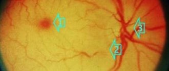
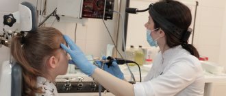
The most informative research method is ophthalmoscopy - examination of the fundus of the eye using a special lens. This method allows the doctor to visualize vascular changes in the retina, establish their location and determine the degree of complexity. To find out the causes of the disease, laboratory blood tests are carried out (for sugar, hormones, etc.).
How can you help yourself without pills?
There are two simple ways. The first can be used by each person independently without preparation. The second one is a little more difficult, but regular viewers and readers of Dr. Shishonin will master it very quickly.
It is better to use both methods: working on the points on the sides of the front of the neck, and working under the back of the head. This way, you will not only get rid of headaches and other symptoms of impaired venous outflow, but you will also feel more energetic, your mood and memory will improve. The main thing is to regularly devote time to yourself and help your body recover. The capabilities of the human body are almost limitless, but you need to develop them by doing gymnastics and self-massage.
Treatment methods
Retinal angiodystonia is always treated comprehensively. The main direction of therapy is to eliminate the cause of vascular insufficiency. Treatment also includes correction of some habits:
- quitting smoking and alcohol or minimizing them;
- stabilization of physical activity - increasing activity with obvious physical inactivity and restrictions with increased loads;
- changing the daily routine, alternating activity and rest;
- following a diet appropriate to the underlying disease.
Treatment also includes taking medications that help restore blood supply to the retina. Local remedies in the form of drops that improve eye nutrition are often used - Mildronate, Trental and their analogues. More focused therapy is prescribed depending on the type of disease.
Treatment of hypertensive angiodystonia
Since hypertensive angiodystonia of the retina can result in a catastrophe for vision with a sharp increase in blood pressure, patients are recommended to take medications on an ongoing basis that regulate this indicator - Captopril, Tenoric and their analogues. Additionally, a complex of drugs is prescribed to help stabilize vascular tone and eliminate factors contributing to their spasm:
- sedatives - motherwort and valerian, Corvalol;
- antiarrhythmic drugs - "Verapamil";
- diuretics (if necessary, quickly reduce blood pressure) - Enalapril or Hypothiazide.
To reduce current symptoms, analgesics and antispasmodics can be prescribed.
Treatment of hypotonic angiodystonia
In the hypotonic form of the disease, drugs are used to increase and stabilize blood pressure. The drugs chosen are predominantly of natural origin containing extracts of lemongrass, ginseng and echinacea. These plants are powerful adaptogens, strengthen the immune system and help improve vascular tone.
Additionally, the following may be prescribed:
- means for improving cerebral blood circulation - “Piracetam”, “Pantocalcin”;
- local drugs to enhance microcirculation - “Taufon” and “Emoxipin”;
- vitamins with components beneficial for vision - “Lutein Complex” and “Aevit”.
For any type of disease, the effectiveness of conservative therapy increases with the use of physiotherapeutic methods: magnetic therapy, acupuncture, electrophoresis. Any medications and procedures can only be used after consulting a doctor!
