Infographics for the “bio/mol/text” competition: A clinical blood test is the most common laboratory test prescribed by a doctor when we come to an appointment and complain of feeling unwell. “Blood from a finger, tomorrow from 8:00 to 9:30, on an empty stomach, Nth office,” several generations have invariably heard this phrase. However, blood testing technology has undergone great changes in recent decades and has moved from manual methods to automatic ones. Let's figure out how your grandmother's blood was analyzed and why things are done differently now.
Competition "bio/mol/text"-2019
This work was published in the category “Visually about the beloved” of the “bio/mol/text” competition-2019.
The general sponsor of the competition and partner of the Skoltech nomination is the Skoltech Center for Life Sciences.
Competition sponsor: the largest supplier of equipment, reagents and consumables for biological research and production.
The audience award was sponsored by BioVitrum.
"Book" sponsor of the competition - "Alpina Non-Fiction"
Microchips in vaccines? Blood test gives surprising results
Discussions about vaccines and vaccination against COVID-19 are not dying down, but, on the contrary, are becoming increasingly heated. Even serious doctors doubt that they have a complete understanding of the composition of the drugs used for vaccinations. What is really there?
What are people dying from?
The skepticism of Russian doctors only intensified after the recent statement by Academician A. Ginzburg (Gamaleya Institute, developer of the Sputnik line). He mentioned some “markers” in the Sputnik V drug that make it possible to determine who was vaccinated and who just bought a vaccination certificate. The official information about Sputnik V says nothing about these “markers”.
A conference of pathologists, which took place on September 20 this year in Germany at the Institute of Pathology in Reutlingen (Pathologischen Institut in Reutlingen), added fuel to the fire of controversy and doubts regarding the composition of vaccination preparations. According to media reports, the event was attended by 30 to 40 specialists, including from Austria. The key figures were:
- Prof. Dr. Arne Burkhardt. He headed the Institute of Pathology in Reutlingen for 18 years, after which he worked as a practicing pathologist. The professor has published more than 150 articles in specialized journals and medical reference books. He also certified pathological institutes.
- Prof. Dr. Walter Lang. He worked as a pathologist at Hannover Medical School from 1968 to 1985. For 25 years he headed the private institute of pathology in Hannover.
- Prof. Dr. Werner Bergholz. Unlike the first two professors, he is not a physician, but a specialist in the field of microelectronics: he worked for 17 years at Siemens Corporation. Recently he has also acted as an expert on medical statistics.
Screenshot of the page pathologie-konferenz.de/en/
The conference focused on the results of autopsies on eight people who died after vaccination against COVID-19, which were carried out this year under the direction of Professor Arne Burckhardt. The results of these autopsies surprisingly confirm the conclusions of Arne Burckhardt's colleague, Professor Dr. Peter Schirmacher. The latter performed autopsies on more than 40 deceased people who were infected with the covid virus. Peter Schirmacher confidently stated that about a third of them died not from Covid, but from vaccination against Covid.
These statements were made in the summer, the authorities and the media controlled by them tried to silence or refute the professor’s conclusions. And then a conference of pathologists in Reutlingen arrived, which again revealed the mortal danger of vaccinations against Covid.
They are already in us
The conference was broadcast via video link. It presented numerous photographs and drawings that clearly complemented the picture described by the speaking pathologists.
The analysis of thin tissues of the deceased was carried out using a special, so-called “dark-field” microscope. It made it possible to identify the content of foreign microparticles in the tissues, which in shape are clearly non-living structures of a fairly regular geometric shape. Outwardly they look... like microcircuits!
Screenshot of video frame Cause of death after COVID-19 vaccination & Undeclared components of the COVID-19 vaccines / odysee.com
There are two versions of the appearance of such foreign objects. Either they were introduced into the bloodstream ready-made, or they were formed in the human body from nanoparticles contained in the vaccine. Accidental entry of foreign particles into the human body is excluded, since the same foreign objects were detected in all those who died after vaccination.
The above-mentioned professor, Dr. Werner Bergholz, as a microchip specialist, expressed his opinion on the “discovery” of pathologists. He does not rule out the possibility of using particles identified in the tissues of deceased people as those very “markers” and “identifiers”, the presence of which in vaccines was suspected by supporters of the so-called “conspiracy theory”.
Pfizer with extras
This reflection of the professor fully corresponds with the opinion of those specialists who have tried and are trying to identify the “markers” of vaccines without opening them, through an in-depth chemical and physical study of the drugs themselves. There are a number of studies that report the discovery of graphene (also graphene oxide) in the composition of at least two drugs - Pfizer and Moderna (mRNA vaccines), which does not have any medical role, but is quite suitable for the role of a “marker”, “ identifier". Adding fuel to the fire was a statement from Karen Kingston, a former Pfizer employee. Kingston says that although graphene oxide is not mentioned in the patents for the Pfizer vaccine, it appears in a number of accompanying documents.
Screenshot of a video frame from Stew Peters show “Former Pfizer Employee Confirms Poison in COVID 'Vaccine'”/ redvoicemedia.com
Another area of study by “inquisitive skeptics” of components and properties of drugs unannounced by vaccine manufacturers is attempts to identify people who have received vaccines using special technical means. The furious energy with which the Silicon Mafia (the leading IT corporations that control the Internet and social networks) deletes publications of this kind also suggests that there is no smoke without fire.
It is hard to believe that what was said at the Reutlingen conference regarding foreign particles in vaccination preparations is just “smoke” that will quickly dissipate. There is no smoke without fire. It’s just that this fire is carefully hidden. Until the moment when a universal fire begins, which can no longer be stopped.
The conference participants adopted a resolution calling on the authorities of Germany, Austria and other countries to begin conducting mass post-mortem studies of those who died after vaccinations against Covid, make appropriate requests to drug manufacturers and, of course, immediately stop the further process of vaccinations against COVID-19 until the issue is fully clarified .
It would seem, what does Gates have to do with it?
The idea of implanting a microchip into the human body through a vaccination injection has been nurtured by the world elite for a long time. In “Prevent Disease.Com” (a US electronic publication specializing in exposing the plans of the American and international “medical mafia”) back in 2009, an article “Are Populations Being Primed For Nano-Microchips Inside Vaccines?” appeared. The title of the article in Russian: “Is the population being pushed to accept nanochips hidden in vaccines?” As noted in this article, in the last years of the twentieth century it was possible to develop a new generation of microchips based on the use of nanotechnology. Ultra-compact (no larger than a speck of dust, radius about 5 micromillimeter, which is about 10 times smaller than the radius of a hair) and inexpensive. Here is what, in particular, was said in the above article: “The swine flu pandemic scenario launched by the World Health Organization is perfectly suited for propaganda and forcing the population to voluntarily agree to the introduction of microchips through nanovaccines. All this will be done under the slogan of the “highest good” for humanity.”
Five years ago, a public-private initiative codenamed ID2020 was launched. Its initiator was Bill Gates, founder and head of the IT corporation Microsoft, at the same time the founder and head of the largest charitable foundation in the United States. The initiative was supported by the UN. Its essence is simple - to carry out global digital identification of the population so that the world elite can keep it under their control. In the first speeches of Bill Gates as the main enthusiast of total digital identification, he did not hide the fact that identification through chipization is the simplest and most reliable way to solve the problem.
But having encountered misunderstanding and even angry protests from a number of politicians and public figures, Gates no longer voiced this idea. And, according to some experts, he continued to push it forward, giving money for the development of nanochips, which will become a “free additive” to vaccination drugs. The solution to the problem of “a nanochip and a vaccine in one bottle” was carried out jointly, in close cooperation, by two structures under the control of Bill Gates: the above-mentioned public-private partnership “ID2020” and the GAVI Vaccination Alliance (also a public-private partnership). Already in 2018, all references to nanochips in vaccines were removed from the ID2020 and GAVI websites.
So what?
Although almost two months have passed since the conference in Reutlingen, you probably haven’t heard anything about it - and this is a clear example of the control established by the Silicon Mafia over the channels of information dissemination.
Videos and other materials of the conference are blocked in every possible way, and where it is impossible to block, they come up with poster “exposures” of the “fakes” voiced there.
What can you do to instill in people confidence in “saving” vaccines!
What will we explore?
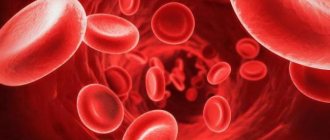
Blood is a liquid connective tissue of the body, consisting of plasma and three types of formed elements: red blood cells, platelets and leukocytes. Leukocytes, in turn, come with granules in the cytoplasm - these are neutrophils, eosinophils and basophils - and without granules - lymphocytes and monocytes. In order to distinguish pathology from the norm, you need to know what the concentration of blood cells is, what they look like and what function they perform. It's time to remember who we are dealing with.
Figure 1. Formed elements of blood [1–3]
Figure 2. The figure shows: the concentrations of blood cells are normal; leukocyte formula - the percentage of different types of leukocytes in the blood; erythrocyte sedimentation rate; hemoglobin concentration; hematocrit is normal [4].
So, experts analyze the relative and absolute content of cells, their morphological characteristics, distribution over blood volume and many other parameters. These indicators can tell whether cells are able to fully perform their functions, and if not, then indicate the reason for their “inoperability” and serve as the basis for making a diagnosis.
Figure 3. Blood testing: then and now
What does blood look like under a microscope?
There are several types of blood cells that have different tasks. Some of them function only within the circulatory system, others go beyond its boundaries. The only thing they have in common is that they are all formed in the bone marrow from stem cells. The process of their formation is continuous, and their lifespan is limited. Leukocytes, platelets and erythrocytes are 3 formed elements contained in biological fluid. Their number depends on the age of the person and the state of the body at the moment.
Red blood cells
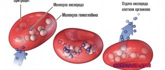
Red blood cells “take” oxygen from the lungs and deliver it to all organs and tissues. On the way back, carbon dioxide is transported to the air breathing organ. These cells contain a unique protein - hemoglobin, consisting of a divalent iron atom. This is what causes the red pigment of red blood cells.
About 2-3 million of these bodies die every second, but the same number are produced every second. They live for approximately 4 months. In 1 cubic ml of blood there are about 25 million red blood cells. Their total number is 25 billion. In just one drop of blood there are about 250 million red blood cells.
An erythrocyte under a microscope has a discoid-concave shape, the average diameter of which is about 7-7.5 microns, and the thickness at the edges is 2.5 microns. This shape facilitates the “smooth” passage of red blood cells through small vessels. Over time, red blood cells lose this property, which is why they linger in the small vessels of the spleen and liver. They are destroyed in these organs.
Up to 80% of red blood cells have a spherical biconcave shape. The remaining 20% can be oval, crescent-shaped, spherical (simple), cup-shaped and other shapes. Changes in appearance are often caused by pathology (vitaminosis, anemia, etc.).
Leukocytes
These are white blood cells that have a protective function. They are usually divided into granulocytes and agranulocytes. The first group includes granular cells:
- Neutrophils. The most numerous group of leukocytes - up to 70% of the total number of white cells. The main task is to capture pathogenic microbes. The nuclear cell has a round shape, the diameter of which can reach 10 microns. The core may be rod-shaped or consist of several segments connected to each other. If the number of segments is more than 8, then this indicates the development of the disease.
- Basophils. A small group, which is represented by no more than 1% of the total number of leukocytes. The main task is to maintain the functioning of the immune system. They have a round shape and a segmented or rod-shaped nucleus. Their diameter reaches 7-11 microns.
- Eosinophils. The total number of white blood cells accounts for 2-5% of eosinophils. Their granules are stained with an acidic dye, eosin. They have a round shape and a slightly colored core, consisting of 2-3 segments of the same size. Eosinophils reach 10-11 microns in diameter. Their cytoplasm is pale blue.
In 1 cubic ml of blood there are 5-10 thousand leukocytes. The average size of white blood cells is 15 microns. Their total number in the human body reaches 35 billion.
The second group consists of cells without granules in the cytoplasm:
- Lymphocytes. Leading component of the immune system. Knowing what lymphocytes are in a blood test, doctors determine the patient’s immune status and receive information about the presence of infection and other diseases in the body. These are round cells with a large nucleus occupying most of the cytoplasm. Their diameter ranges from 7 to 10 microns. The kernel can be round, oval or bean-shaped and has a rough structure. Consists of lumps of oxychromatin and basiromatin, resembling blocks.
- Monocytes. The largest of leukocytes, responsible for nonspecific defense of the body. Their number ranges from 2 to 9% of all white blood cells. The diameter reaches 20 microns. The nucleus is large, occupies almost the entire cytoplasm, can be round, bean-shaped, mushroom-shaped, or butterfly-shaped.
Interesting! If you arrange the leukocytes in one row, you can get a distance of 525 km.
Leukogram, or leukocyte formula, is the percentage ratio of different types of leukocytes, determined by counting them in a stained blood smear under a microscope. Deciphering the leukocyte formula provides undoubted benefit in a diagnostic search, but it cannot always fully satisfy the doctor’s interest in relation to some well-masked disease. For example, the more pronounced the inflammatory process of bacterial etiology, the more neutrophils in the leukocyte formula. The presence of neutrophils of varying degrees of maturity indicates the severity of the bacterial infection. The more acute the process, the more band neutrophils in the blood.
Platelets
Blood platelets are “hidden” under the platelets, which regulate the blood clotting process. They secrete the enzyme thromboplastin. When a cut or injection occurs, the platelet membrane is destroyed, thromboplastin is released and enters the plasma, and the blood clots. A blood clot forms, which protects the body from blood loss.
In 1 cubic There should be about 200-300 thousand ml of blood. The size of each platelet reaches about 3 microns. Therefore, they can only be examined in detail using a professional microscope. It turns out that the total number of these red platelets in human blood is approximately 1250 billion.
Forward to the past!
1965, 8 am, there is a line for tests at the local clinic. Your grandmother donated blood, and the laboratory assistant takes a number of test tubes for testing. Let's follow him to the laboratory and see what's there and how. In the laboratory we see doctors bending over microscopes or working with test tubes. It is no exaggeration to say that in your grandmother’s youth, a specialist’s eye and a microscope were the main tools for blood analysis. The following basic characteristics of blood are determined: the concentration of each type of formed element, the number of different types of leukocytes, the erythrocyte sedimentation rate and the concentration of hemoglobin. In addition, the specialist calculates the hematocrit - the ratio of the volume of red blood cells to the total volume of blood [5].
Calculate in order!
The first step is to count the cells and determine their concentration in the blood. The counting of red blood cells, leukocytes and platelets is carried out in the Goryaev chamber, named after its inventor. Goryaev’s chamber is a glass with a recess and a mesh applied, into which a drop of blood diluted in saline solution is placed. To determine the number of formed elements, the camera is placed under a microscope and the cells located in large and small grid squares are counted. Each type of cell has its own counting rules and formula by which their initial concentration is calculated, taking into account blood dilution and the number of grid squares [6]. Changes in the number of formed elements serve as an important criterion for the diagnosis of anemia, inflammatory and viral diseases, disorders of the blood coagulation system and other pathological conditions [7].
Who are you?
Another stage of blood testing is the differentiation of leukocytes into populations. Special attention is paid to it: a change in the concentration of a certain type of cell indicates a specific pathology. Bacterial infection, viruses or allergies? Leukocytes will tell you what diagnosis to make and what treatment to prescribe. The distinction of leukocytes is trusted only to a highly qualified specialist. To begin with, a blood smear is fixed in alcohol and stained using the Romanovsky-Giemsa method. The composition of the dye is selected in such a way that different cell structures are painted in different colors. Coloring depends on the ability of the components of the color mixture to bind to structures containing acids or bases. For example, hemoglobin and eosinophil granules acquire a red-pink color due to eosin, and the nuclei of formed elements and basophil granules (having an affinity for bases) are stained with methylene blue and blue [1]. When the smear is ready, a specialist examines it under a microscope and determines by its appearance what type of different cells they belong to [8]. The presence of colored granules, features of the shape of the nucleus, cell size - all parameters must be kept in mind for error-free classification. Typically, one hundred leukocytes were counted, followed by calculation of the percentage, and in order to avoid confusion, an 11-key counter was used [9]. If you see a cell under a microscope, press the key indicating the cell of this type, and at the end of the count, the number of leukocytes of each type will be displayed on the counter screen [10].
Precipitated
Another characteristic of clinical significance is the erythrocyte sedimentation rate (ESR). This is an indicator that evaluates the rate of separation of blood into plasma and formed elements. What is the reason for this division? Macromolecules found in blood plasma can simultaneously bind two red blood cells to each other, resulting in the formation of “coin columns” [11]. Such complexes, under the influence of gravity, settle to the bottom of the test tube, leaving a layer of transparent plasma above them - this is called erythrocyte sedimentation. An increase in erythrocyte sedimentation rate indicates pathological processes occurring in the body, such as inflammatory, infectious or oncological diseases [12].
To determine ESR, the Westergren method received worldwide recognition, but the Panchenkov method was also widespread in Russia. The operating principle of the methods is the same, only the types of tubes used differ. The blood is mixed with an anticoagulant - sodium citrate - and placed in a capillary - a thin glass tube. Red blood cells settle to the bottom of the tube within an hour, and then the height of the plasma column formed on top is measured [13]. In this way, the erythrocyte sedimentation rate is obtained, expressed in mm/h.
Taste and color
Hemoglobin is the red pigment of red blood cells that binds and transports oxygen and carbon dioxide. A decrease in hemoglobin content in red blood cells is the cause of anemia that accompanies a number of diseases. Hemoglobin concentration is determined visually using a Sali hemometer. The device looks like this: in the center is a test tube for the blood being analyzed, and on the sides are colored reference tubes. The laboratory technician adds hydrochloric acid to the blood being studied - hemoglobin turns into brown hemin. Then the blood is diluted with distilled water until its color (in the subjective opinion of the laboratory assistant!) matches the color of the standard. The liquid level obtained in the central tube corresponds to the hemoglobin concentration [14].
As you may have guessed, 50 years ago it was very easy to make a mistake when testing blood. Incorrect identification of the type of leukocyte or failure to count the formed elements - all this led to inaccurate analysis results. What has been done to prevent errors? Let's go back to our time and find out how blood is studied today.
Objectively about pseudoscientific. Dark Blood Fields: Diagnosis
4619 April 24
IMPORTANT!
The information in this section cannot be used for self-diagnosis and self-treatment.
In case of pain or other exacerbation of the disease, diagnostic tests should be prescribed only by the attending physician. To make a diagnosis and properly prescribe treatment, you should contact your doctor. Dark fields of blood: Diagnostics / Author: Alexey Vodovozov Source: / January 2010
Blood is an amazing creation of nature. It can be said without exaggeration that it is the source of life. After all, it is through the blood that we receive oxygen and nutrients, and it is through the blood that “production waste” is removed from the cells. Any illness is necessarily reflected in the blood. A number of diagnostic techniques are based on this. And charlatan ones too.
Blood was one of the first liquids that inquisitive doctors placed under the newly invented microscope. More than 300 years have passed since then, microscopes have become much more advanced, but doctors’ eyes still look at blood through eyepieces, looking for signs of pathology.
On glass
Antonie van Leeuwenhoek would definitely have received several Nobel Prizes had he lived today. But at the end of the 17th century this award did not exist, so Leeuwenhoek is content with the worldwide fame of the designer of microscopes and the fame of the founder of scientific microscopy. Having achieved 300-fold magnification in his instruments, he made many discoveries, including the first to describe red blood cells.
Leeuwenhoek's followers brought his brainchild to perfection. Modern optical microscopes are capable of magnification up to 2000 times and allow us to examine transparent biological objects, including the cells of our body.
Another Dutchman, physicist Fritz Zernike, noticed in the 1930s that the acceleration of light in a straight line makes the image of the model being studied more detailed, highlighting individual elements against a light background. To create interference in the sample, Zernike came up with a system of rings that were located both in the objective and in the condenser of the microscope. If you correctly configure (adjust) the microscope, then the waves that come from the light source will enter the eye with a certain phase shift. And this allows you to significantly improve the image of the object being studied.
The method was called phase-contrast microscopy and turned out to be so progressive and promising for science that in 1953 Zernike was awarded the Nobel Prize in Physics with the wording “For the justification of the phase-contrast method, especially for the invention of the phase-contrast microscope.” Why was this discovery so highly regarded? Previously, in order to examine tissues and microorganisms under a microscope, they had to be treated with various reagents—fixatives and dyes. It was impossible to see living cells in this situation; the chemicals simply killed them. Zernike's invention opened a new direction in science - intravital microscopy.
In the 21st century, biological and medical microscopes have become digital, capable of operating in different modes - both in phase contrast and in dark field (the image is formed by light diffracted by the object, and as a result the object appears very light against a dark background), as well as in polarized light, which often makes it possible to reveal the structure of objects that lies beyond the usual optical resolution.
It would seem that doctors should rejoice: a powerful tool for studying the secrets and mysteries of the human body has fallen into their hands. But this high-tech method was of great interest not only to serious scientists, but also to charlatans and scammers from medicine, who considered phase-contrast and dark-field microscopy a very successful way to extract certain amounts of money from gullible citizens.
She's alive and moving
From a patient who decides to undergo examination using the “Living Drop of Blood Diagnostics” method (variants of the name are “Testing on a dark-field microscope” or “Hemoscanning”), a drop of blood is taken, not stained, not fixed, applied to a glass slide and studied by viewing the sample on the monitor screen. Based on the results of the study, diagnoses are made and treatment is prescribed.
Hemoscanning can be considered the crowning achievement of fraudulent thought, a masterpiece and the highest aerobatics of near-medical quackery. Firstly, a real existing physical phenomenon is used (remember about the Nobel Prize?) and real complex medical equipment. And really expensive. The cost of a diagnostic complex costs at least 3–4 thousand dollars, and it is sold by reputable suppliers of serious medical equipment. The equipment has all the necessary - genuine and completely deserved - certificates and certificates. Secondly, no problems with licensing. Laboratory diagnostics is a completely legitimate type of medical activity, and a microscope that allows phase-contrast or dark-field microscopy is a completely legitimate medical diagnostic equipment. Moreover, it is widely used in medicine, that is, there are certified and certified specialists. Thirdly, under a microscope you can indeed detect a lot of signs of certain diseases. For example, changes in the shape of red blood cells in sickle cell anemia. You can also see intracellular parasites, all in the same red blood cells, called Bartonella. And even helminth eggs can theoretically be detected in the blood.
I see arba - I sing arba
So what's the catch? In interpretation. In how the “Dark Polish people” explain certain changes in the blood, what the discovered artifacts are called, what diagnoses are made and how they are treated. It is difficult even for a doctor to figure out that this is a hoax. You need special training, experience working with blood samples, and hundreds of examined “slides” – both stained and “live.” As in a regular field, so in a dark one. Fortunately, the author of the article has such experience, as does the experts with whom the results of the investigation were verified.
It’s rightly said – it’s better to see once. And a person will believe his own eyes much faster than all verbal exhortations. This is what the “laboratory assistants” are counting on. A monitor is connected to the microscope, which displays everything that is visible in the smear. When was the last time you saw your own red blood cells? That's it. It's interesting. And while the fascinated visitor admires the cells of his own beloved blood, the “laboratory assistant” begins to interpret what he sees. Moreover, he does this according to the principle of akyn: “I see an arba, I sing an arba.” Read in detail in the sidebar about what kind of “arba” charlatans can sing about.
After the patient is frightened and confused by incomprehensible, and sometimes downright scary pictures, he is given “diagnoses”. Most often there are many, and one is more terrible than the other. For example, they will tell you that the blood plasma is infected with fungi or bacteria. It doesn’t matter that seeing them even at such magnification is quite problematic, much less distinguishing them from each other. Microbiologists have to sow pathogens of various diseases on special nutrient media, so that later they can say exactly who has grown, what antibiotics they are sensitive to, etc. Microscopy is used in laboratory research, but either with specific dyes or even with fluorescent antibodies that attach to bacteria and thus make them visible.
But even if, purely theoretically, such a giant of the bacterial world as Escherichia coli (1–3 microns long and 0.5–0.8 microns wide) is found in the blood under a microscope, this will mean only one thing: the patient has sepsis, infection blood. And he should lie horizontally with a temperature of under 40 and other signs of a serious condition. Because normally blood is sterile. This is one of the main biological constants, which is checked quite simply by inoculating blood on various nutrient media.
They may also tell you that the blood is “acidified.” A shift in the pH (acidity) of the blood, called acidosis, does occur in many diseases. But no one has yet learned how to measure acidity by eye; you need contact between the sensor and the liquid being tested. They can detect “toxins” and tell you about the degree of slagging in the body according to WHO (World Health Organization). But if you look through the documents on the official website of this organization, there is not a word about either slags or the degree of slagging. Diagnoses may include dehydration syndrome, intoxication syndrome, signs of fermentopathy, signs of dysbacteriosis and a host of others that are not related either to medicine or to this particular patient.
The apotheosis of diagnosis, of course, is the prescription of treatment. By a strange coincidence, it will be carried out with biologically active food additives. Which, in essence and by law, are not medicines and cannot be treated in principle. Especially such terrible diseases as fungal sepsis. But this does not bother hemoscanners. After all, they will not treat the person, but the very diagnoses that were given to him out of thin air. And with repeated diagnostics, rest assured, the indicators will improve.
What you can't see with a microscope
No matter what the “experts” tell you, using a microscope you cannot see the pH of the blood in a drop of blood taken from your finger; deficiency of enzymes for protein breakdown; level of water-salt metabolism; food mutagenic/teratogenic toxins; damage to red blood cells by kidney toxins/free radicals; parasites, fungi, bacteria, worm eggs, cysts; activity, quantity and quality of immune cells.
Live blood testing originated in the United States in the 1970s. Gradually, the true essence and value of the technique became clear to the medical community and regulatory authorities. Since 2005, a campaign has begun to ban this diagnosis as fraudulent and unrelated to medicine. “The patient is deceived three times. The first time is when a disease that does not exist is diagnosed. The second time is when a long and expensive treatment is prescribed. And the third time is when a repeat study is falsified, which will necessarily indicate either an improvement or a return to normal” (Dr. Stephen Barrett, vice president of the American National Council Against Medical Fraud, scientific consultant to the American Council on Science and Health).
Are bribes smooth?
Proving that you were deceived is almost impossible. Firstly, as already mentioned, not every doctor will be able to suspect forgery in the technique. Secondly, even if the patient goes to a regular diagnostic center and they don’t find anything there, you can, as a last resort, blame everything on the operator who performed the diagnostics. Indeed, the visual assessment of complex images depends entirely on the qualifications and even the physical condition of the person doing the assessment. That is, the method is not reliable, since it directly depends on the human factor. Thirdly, you can always refer to some subtle matters that the patient cannot understand. This is the last frontier where all medical scammers usually face their death.
What do we have in the bottom line? Unprofessional laboratory technicians who pass off random artifacts (maybe even orchestrated) in a drop of blood as terrible diseases. Then they suggest treating them with nutritional supplements. Naturally, all this for money, and quite a lot.
Does this technique have diagnostic value? It has. Undoubtedly. The same as traditional smear microscopy. You can see, for example, sickle cell anemia. Or pernicytosis anemia. Or other really serious illnesses. But, to the great regret of scammers, they are rare. And you can’t sell crushed chalk with ascorbic acid to such patients. They need real treatment.
And so - everything is very simple. We discover a non-existent disease and then successfully cure it. Everyone is happy, especially the citizen over there, who had a fragment of the bell-mosquito’s space communications antenna expelled from his blood... And no one feels sorry for the money that was wasted, or rather, to enrich the swindlers.
However, not everyone. Some defend their rights in all possible instances. The author has at his disposal a copy of a letter from the Office of Roszdravnadzor in the Krasnodar Territory, where victims of hemoscanning “doctors” turned. The patient was diagnosed with a bunch of diseases that were proposed to be treated with no less than a bunch of biologically active food supplements. Based on the results of the inspection, it turned out that the medical institution that carried out the diagnostics violated licensing requirements, did not enter into an agreement for the provision of paid services (the doctor takes money in cash), and the rules for maintaining medical records were violated. Other violations were also identified.
I would like to end the article with a quote from a letter from the Central Office of Roszdravnadzor: “The 'Hemoscanning' technique was not submitted to Roszdravnadzor for consideration and permission to use as a new medical technology and is not permitted for use in medical practice.” Couldn't say it any clearer.
IMPORTANT!
The information in this section cannot be used for self-diagnosis and self-treatment. In case of pain or other exacerbation of the disease, diagnostic tests should be prescribed only by the attending physician. To make a diagnosis and properly prescribe treatment, you should contact your doctor.
Times change
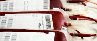
Changes are visible already at the stage of blood sampling: if previously the doctor collected blood in several test tubes with reagents, a glass capillary and made a smear on the glass, now very small volumes are used - from 12 to 150 μl [15] of blood is enough to examine it for all parameters.
Let's take a look into a modern hematology laboratory. Wow! Everything is cluttered with equipment, and the laboratory assistant is nowhere to be seen... Maybe he went off to make himself some coffee? He won't make it in time! The blood test will be ready in a minute, and the device will give the result in the form of a paper tape with numbers and abbreviations, behind which all sorts of parameters are hidden.
Modern hemoanalyzers are divided into several classes, depending on what they can do. Each subsequent class is a new stage of evolution - faster, more accurate, more perfect. The use of a combination of technologies works wonders: if the first analyzers could determine eight blood parameters and did not distinguish between types of leukocytes [16], then the latest devices are able to differentiate up to seven populations of leukocytes [17] and in total examine more than 40 blood characteristics.
As Arthur C. Clarke said, “Any sufficiently advanced technology is indistinguishable from magic.” Indeed, the most detailed results in such a short period of time cannot but surprise. But all magic is based on physical laws. And although names like electrical impedance, light scattering and photometry are a little intimidating at first glance, now we will understand what principles underlie each analysis technology.
Population census
In the middle of the last century, Wallace Coulter made a revolution by patenting the technology of automatic cell counting. One of the leaders in the production of hematology analyzers, the Beckman Coulter company, is named after him [18]. The aperture-impedance method (or Coulter method) is based on the registration and analysis of impulses that occur when a cell passes through an aperture from one container to another, each of which contains an electrode. When there is no cell in the hole, current flows freely through the electrolyte between the electrodes under the influence of an electric field. To direct the cells to the aperture, a pump is used that pumps out liquid from one container, and the formed elements rush into it. Passing through the aperture, the cell displaces a volume of electrolyte equal to its volume from one container to another. In this case, a pulsed change in resistance (impedance) occurs - the cell membrane creates an obstacle to the free flow of current. At the same time, the current strength recorded by the meter also changes. The number of generated impulses corresponds to the number of formed elements, and the height of the impulse is proportional to the volume of the cell [19]. Using information about the number and volume of formed elements, the device can calculate hematocrit, the average concentration of hemoglobin in an erythrocyte, the width of the distribution of cells by volume and many other parameters [15].
Divide and rule
Differentiation of leukocytes into populations can be carried out using a Coulter counter, but a problem arises - different types of leukocytes are similar in volume and similar pulse amplitude does not always allow one to accurately determine the cell type. What should I do? To solve this problem, combinations of reagents are selected that change the size of cells to varying degrees so that it becomes possible to separate them [15].
But the most common method of differentiation is flow cytometry [20]. The method works as follows: cells in the flow are alternately irradiated with a laser, and the resulting light scattering and fluorescence signals are recorded by detectors and analyzed. In order to correctly determine membership in a population, several parameters are examined at once. Thus, light scattering at a small angle provides information about the relative size of cells, and light scattering at a right angle allows you to “look” inside the cell and study its internal structure - the presence of granules and the shape of the nucleus. Another parameter - fluorescence - can tell about the number of antigens and their appearance on the surface of cells - this cannot be accurately determined by eye. Unlike manual differentiation methods, not 100–200 cells are analyzed, but tens of thousands per second! And each leukocyte receives an individual approach: hydrodynamic focusing ensures that the cells line up in a row and are irradiated one by one in the flow cell. The result of the count appears on the screen in the form of scatter diagrams, where cells with similar properties form clusters.
Precipitated: 2.0
Modern instruments can measure ESR in two fundamentally different ways. The first is the modified Westergren method. The principle of operation has not changed since your grandmother's time, but due to automation it has become faster and more accurate. The second is measuring the kinetics of erythrocyte aggregation using an optical method [21]. It happens like this: an anticoagulant is added to the blood, the tubes with blood are placed in a rotor, where automatic mixing occurs. After this, the analyzer takes part of the blood into a microcapillary, where it accelerates and abruptly stops (the so-called “stopped stream” method). The stop causes aggregation of red blood cells, and at this moment the optical density of the blood is determined using a photometer - the denser the red blood cells are located, the less light will pass through the sample. The device uses the data obtained and builds a sedimentation curve - its analysis will allow you to present the result in the usual units of ESR measurement [22], [23].
Photo for memory
To determine hemoglobin concentration, the International Committee for Standardization in Hematology recommends the methemoglobin-cyanide method. However, a different test that does not use toxic cyanide is now widely used. Meet the SLS method. It is named after the main reagent - sodium laurithyl sulfate. SLS destroys red blood cell membranes, after which it binds to heme groups and forms stable complex compounds. They are analyzed photometrically - laser light is passed through the blood sample. Complex compounds absorb part of the light, as a result of which the intensity of the output light flux weakens. Attenuation is measured using a photosensor and the resulting data is converted into hemoglobin concentration units [24].
What can and cannot be learned from a drop of blood
Just type “diagnosis by a drop of blood” into a search engine and a lot of contradictory information will pop up. It turns out that with a drop of blood you can know yourself from head to toe: find out your genetic profile, predisposition to certain diseases, get your psychological portrait, find out about compatibility with your partner and get a life expectancy forecast. How to distinguish bogus studies from real ones? What can you tell from a drop of blood?
General blood analysis
Once blood was first put under a microscope and found to be made up of cells, scientists realized it could be used for diagnosis. A general (aka hematological) blood test is based on cellular analysis.
A general blood test is:
- cell count (how many leukocytes, red blood cells, neutrophils, platelets, etc.);
- cell differentiation (percentage ratio of cells of one type to cells of another type). This allows us to make assumptions about the processes occurring in the body: for example, viral infection, the effect of toxic substances;
- assessment of cell quality (cell and nucleus size, maturity, saturation of red blood cells with hemoglobin, etc.).
The paradox is that there is not a single diagnosis that could be based solely on a general blood test. But at the same time, a hematological analysis gives a picture of the blood, without which it is impossible to carry out diagnostics.
Everything except cells
Blood is made up of cells and a liquid part (called plasma or serum, depending on whether it contains fibrin, which is responsible for blood clotting, or not).
Diagnostics using plasma or serum is the area that is currently developing the fastest in the world: there are about 1000 types of tests: biochemical, immunochemical, diagnostics for infections. Plasma is a molecular mirror of all our organs and tissues, a marker of all diseases and infectious processes.
Wait for an immune response. Many types of serum diagnostics are based on the principle of an immune response: any bacteria, any virus that invades our body leaves a trace in the blood in the form of a response from the immune system, regardless of whether we are sick or not. By doing a blood test, we can find out whether our immune system is familiar with this pathological agent (accordingly, whether immunity has already been developed or not).
Using the principle of the immune response, you can study a huge number of parameters: find out what causes your allergies, identify oncology, hormonal disorders, cardiovascular diseases, brain tissue disorders and much more.
It's all biochemistry. Cholesterol, bilirubin, blood enzymes, albumin and globulins are all the subject of a biochemical blood test. Using biochemical indicators, you can evaluate the functioning of the main organs and systems (liver, kidneys, protein metabolism), obtain information about metabolism (lipid and carbohydrate metabolism), and determine the need for minerals.
Deep into the core
Relatively recently emerging diagnostic methods involve the study of cell nuclei and DNA fragments.
PCR diagnostics . This is a method that allows you to detect even small concentrations of certain DNA fragments, achieving a significant increase in their concentration in the sample. Using the PCR method and other DNA methods, it is possible, for example, to find the immunodeficiency virus and hepatitis in the blood and confirm the fact of infection.
Bacteriological research . Allows you to isolate pathogens from biological materials. “Knowing the enemy by sight” is necessary in order to test him for sensitivity to antibiotics and select therapy that will be effective for a particular patient.
When tests are unnecessary
Tests may be unnecessary for two reasons: due to the patient’s excessive curiosity and due to the doctor’s insufficient qualifications.
With a curious patient, everything is clear: in principle, an analysis for something new and interesting can be done at least every day - fortunately, now there is a great variety of markers, and new ones appear almost every day.
The situation is different with the doctor’s qualifications. Indeed, an indifferent doctor may say: “Get tested for all infections.” If we take his words literally, we will have to take tests for more than 200 infections, including such exotic ones as borelliosis and malaria. Hence the conclusion: a good specialist will not direct the patient to donate blood for “all infections” or “all allergens.” But a competent patient will not go, if only because this frontal approach will cost him several salaries.
Blood diagnostics is an area in which the desire to find out “everything at once” can turn out to be either ruinous or too labor-intensive. Therefore, there is a rule: it is necessary to do screenings that can tell us something specific, and then, if necessary, expand the boundaries of the study. In other words, there is no point in making insulin antibodies without measuring glucose levels.
Diagnostic skill
Any ultra-modern equipment, any supernova markers are just tools that are just an addition to the clinician’s head. To the very head that makes decisions: what studies to order and how to interpret the information received. This will require experience, flair, and talent as a diagnostician.
The doctor finds himself caught between two fires: on the one hand, there is nothing sadder than missing a disease and not prescribing additional screening. On the other hand, it is no less sad for a doctor to rely on his experience and not double-check his assumption. For example, after a single determination of antibodies to infectious agents, a diagnosis should not be made: this is always the subject of repeated research, comparison of the results obtained over time and the intellectual work of the clinician.
The doctor’s experience is also useful in less critical cases - for example, when the patient is not worried about anything and he wants to “just get checked”: a preliminary consultation with a specialist will be useful. Word from Lyubov Stankevich:
“For example, in front of me is a 16-year-old boy. Most likely, he will not need a tumor marker if nothing bothers him. It is also not worth asking for an extended lipid profile unless the young person is obese and unless I suspect he has a metabolic disorder. Hematological blood test, standard biochemistry, urinalysis - that's what I can prescribe for him. This is the minimum that will never really hurt anyone.”
Not everything can be learned from a drop of blood.
But this is all medicine. People want to use the capabilities of blood testing for simpler, everyday tasks, if possible not related to diseases. The advertisement meets these popular aspirations and offers: diets based on blood type, find out your life expectancy based on a blood test, get to know yourself and find out what the blood type of your other half should be so that you live happily ever after.
How can we understand when the technique works and when our brother is being fooled - especially if something new is constantly appearing in blood diagnostics?
Lyubov Stankevich, medical director of the Ditrix Medical laboratory, says:
“First of all, any theory must be supported by practical developments. For example, there is a theory that each blood group has its own diet; they even try to base this on an evolutionary genetic theory: the first group are hunters, the second are gatherers, and so on. But where is the scientific experimental confirmation of these considerations in the conditions of the modern world?
We may notice some fact. For example, in Japan, when applying for a job, you must indicate your blood type: on this basis, the employer makes conclusions about the employee’s character. Suppose there is such a connection: but have we interpreted the observed fact correctly? This can only be understood after many years of observation. Therefore, any technique has an important criterion: data obtained in one laboratory must be reproducible in another with exactly the same results. If a technique is not reproducible, such a technique is worthless.”
Thank you for your consultation
Medical Director of the Ditrix Medical laboratory Lyubov Stankevich