| Ultrasound examination | Price | Last minute | Record |
| Ultrasound of the abdominal aorta | 2200 rub. | 1900 rub. |
* Check the details of the promotion and price with the operator by calling 8(812)317-70-73.
Record
Ultrasound of the abdominal aorta and its branches is a painless examination that does not imply any adverse consequences
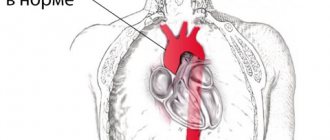
The abdominal aorta, part of the largest vessel in the human body, is subject to a number of changes that do not always manifest symptoms. The main clinical sign, abdominal pain, is not specific and can be present in many diseases. If the cause of the disease is unclear, an ultrasound of the abdominal aorta in St. Petersburg can be done in diagnostic centers where there is the appropriate equipment, if the results of other studies are ambiguous. The price for Doppler testing is set by the clinic, so the cost varies depending on the institution. Doppler ultrasound of the abdominal aorta is a non-invasive, highly informative study that uses the principle of echolocation and the Doppler effect to construct images. Over several decades of practice, no adverse effects on the body have been identified, so ultrasound of the abdominal aorta and its branches can be performed on any person, regardless of gender and age, as many times as the clinical situation requires. Before choosing a medical center, you should study the price lists that are available on specialized websites and patient reviews. There you can also get information about discounts and promotions; in this case, the price for ultrasound examination of abdominal vessels will be lower.
What it is
We should start by defining the aorta. The aorta is the largest vessel in our body, and accordingly its importance is high. An aneurysm is an enlargement of a blood vessel. An aortic aneurysm can form in the thoracic or abdominal regions. Moreover, aneurysm of the abdominal aorta is much more common. This condition of the vessel may not cause any harm for a long time, however, an aneurysm is very dangerous due to the unpredictable course of expansion and the threat of rupture of the vessel walls when they become thinner - and this condition already poses a threat to life due to severe internal bleeding. In addition, blood clots can form at the site of vessel expansion due to changes in blood flow - the danger of this condition is the risk of the blood clot breaking off and clogging a smaller vessel.
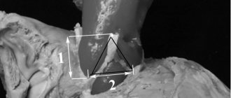
Structure of the aortic bifurcation
The aorta is one of the most important vessels in the human body. The structure of its walls does not differ from other arteries, but it exceeds them in diameter. In addition, in the aorta, blood pressure exceeds the average values in other vessels and reaches 100 mm Hg. Art. Its walls are strong and elastic, pulsating simultaneously with the release of blood from the left ventricle of the heart and its passage through the bloodstream. They have a complex structure and consist of several layers:
- endothelium (intima) - the inner layer, which is formed by one row of connective tissue cells;
- muscle layer - consists of long and strong smooth muscle cells, which provide the ability of the walls to change diameter and pulsate as blood moves;
- adventitia is an outer protective layer of connective tissue cells.
The aorta and other large vessels are of the elastic type. This means that their walls contain virtually no muscle fibers and are dominated by connective tissue cells. Arteries are also very strong, can withstand high blood pressure, are easily stretched during fluid movement and quickly return to their original shape.
Causes of aortic aneurysm in the abdominal region
Experts identify several reasons for the formation of an abdominal aortic aneurysm:
- heredity (it is worth undergoing additional examinations if one of your close relatives had such a diagnosis);
- trauma or surgery on the aorta;
- atherosclerosis (approx. link to article);
There are also risk factors for aneurysm formation:
- smoking,
- excess weight,
- high blood pressure,
- The disease is most often encountered by men over 50 years of age.
Quitting smoking and maintaining a normal weight remain factors that you can get rid of, protecting yourself from the risk of not only an aneurysm, but also other serious diseases.
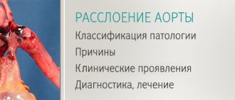
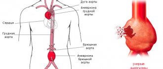
Aortic aneurysm
The aorta is the largest, most powerful blood vessel in the human body. Powerful, therefore, it seemed that nothing “took” him. However, aortic aneurysm is the scourge of modern cardiovascular surgery. In normal conditions, in adult women and men, the diameter of the lumen of the ascending aorta is about 3 cm, the descending part is 2.5 cm, the abdominal segment of this large vessel is even smaller - 2 cm. The diagnosis of an aneurysm is announced only if the diameter of the affected aorta increases by 2 or more times compared to the norm.
An aneurysm is an abnormal bulge that occurs in the walls of an artery. The walls of the arteries are quite thick and strong, the muscle fibers of which they are composed allow them to withstand intense blood pressure. However, if there is a weak area in the artery wall, the pressure causes the area to bulge, thereby forming an aneurysm.
An aortic aneurysm can develop in two parts of this artery:
- the abdominal part passing through the lower part of the abdominal cavity is an abdominal aortic aneurysm;
- A thoracic aortic aneurysm developing in the chest cavity. This type of aneurysm is less common, but both types are equally dangerous to human health and life.
Depending on the appearance, the aneurysm can be: 1. fusiform 2. saccular.
Small aneurysms usually pose no threat. However, they can increase the risk of: the formation of atherosclerotic plaques at the site of the aneurysm, which cause further weakening of the artery walls; formation and separation of a blood clot, therefore increasing the risk of stroke; an increase in the size of the aneurysm, which means compression of nearby organs, which causes pain; aneurysm rupture. The main complication of aneurysms of any location is their dissection followed by possible rupture (mortality rate - 90%).
Causes and risk factors
The main causes of aneurysm are diseases and conditions that reduce the strength and elasticity of the vascular wall:
- atherosclerosis of the aortic wall (according to various sources, from 70 to 90%); inflammation of the aorta (aortitis) of a syphilitic, giant cell, mycotic nature;
- traumatic injury;
- congenital systemic connective tissue diseases (for example, Marfan or Ehlers-Danlos syndrome);
- autoimmune diseases (nonspecific aortoarteritis);
- iatrogenic causes caused by medical manipulations (reconstructive operations on the aorta and its branches, cardiac catheterization, aortography).
Risk factors for the development of atherosclerosis and aneurysm formation:
- male gender (the incidence of aneurysms in men is 2–14 times higher than in women);
- smoking (during screening diagnostics of 455 people aged 50 to 89 years in the Department of Vascular Surgery of the Moscow Regional Research Clinical Institute, it was revealed that 100% of patients with abdominal aortic aneurysms had a smoking history of more than 25 years, and as a result of the Whitehall study it was proven that life-threatening complications of aneurysms occur 4 times more often in smokers than in non-smokers);
- age over 55 years;
- family history;
- long-term arterial hypertension (blood pressure above 140/90 mm Hg);
- physical inactivity;
- excess body weight;
- increased blood cholesterol levels.
They also talk about a dissecting aneurysm, which is formed as a result of rupture of the inner membrane with its subsequent dissection and the formation of a second false channel for blood flow.
Depending on the location and extent of the dissection, 3 types of pathology are distinguished: 1. Dissection begins in the ascending aorta and moves along the arch (50%). 2. Dissection occurs only in the ascending aorta (35%). 3. Dissection begins in the descending aorta and moves down (more often) or up (less often) along the arch (15%). Depending on the duration of the process, a dissecting aneurysm can be: acute (1–2 days from the appearance of the endothelial defect); subacute (2–4 weeks); chronic (4–8 weeks or more, up to several years).
SYMPTOMS OF AN AORTIC ANEURYSM
Aortic aneurysm manifests itself in different ways - it mainly depends on the size of the aneurysmal sac and its location (below is a visual clinical picture using the example of an aneurysm of the sinus of Valsalva). In some cases, no symptoms are observed at all (in particular, before the aneurysm ruptures, but this will be a different diagnosis), which makes early diagnosis difficult. The most common complaints from patients with an aneurysm of the ascending aorta: pain in the chest (in the area of the heart or behind the sternum) - due to the fact that the aneurysmal protrusion presses on nearby organs and tissues, as well as due to the pressure of the blood flow on thinned and weak wall; shortness of breath, increasing over time; feeling of palpitations (“As if something is pounding in the chest” - a comment from patients); dizziness; with large aneurysms, attacks of headaches, swelling of the soft tissues of the face and upper half of the body are disturbing - due to the development of the so-called superior vena cava syndrome (because the aneurysm presses on the superior vena cava).
An aortic arch aneurysm is characterized by:
- difficulty swallowing (due to pressure on the esophagus);
- hoarseness of the voice, sometimes coughing - if the aneurysm puts pressure on the recurrent nerve, which is “responsible” for the voice;
- suddenly increased salivation and slow pulse - if pressure spreads to the vagus nerve, which controls salivation and pulse rate;
- strained breathing, and later shortness of breath if the trachea and bronchi are compressed by a huge aneurysm;
- unilateral pneumonia - if an aneurysm, pressing on the root of the lung, interferes with its normal ventilation, then, as a result, congestion occurs in the lungs, which, when an infection occurs, develops into pneumonia.
With an aneurysm of the descending aorta, the following appear:
- pain in the left hand (sometimes up to the fingers) and shoulder blade;
- with pressure on the intercostal arteries, a lack of oxygen supply to the spinal cord may develop, which is why paresis and paralysis are inevitable;
- in case of constant long-term pressure of a large aneurysm on the vertebrae, they may even be displaced;
- in milder cases, due to pressure on the intercostal nerves and arteries - pain, as with radiculitis or neuralgia.
The most common complaints with an aneurysm of the abdominal aorta:
- a feeling of fullness in the stomach and heaviness in the epigastrium (upper floor of the abdomen), which the patient initially tries to explain by overeating or stomach pathology;
- belching;
- in some cases, reflex vomiting (appears as a reaction to the pressure of the aortic aneurysm on nearby organs and tissues);
- When palpated, a tense, tumor-like pulsating formation is felt. Sometimes patients can independently detect this pulsation.
DIAGNOSIS OF AORTIC ANEURYSMS AND ITS COMPLICATIONS
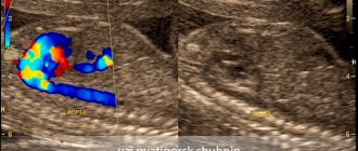
An aortic aneurysm in the period before rupture has rather meager clinical manifestations: murmurs that are heard on auscultation; the doctor listens not only to the chest, but also to the abdominal cavity; a tumor-like pulsating formation, which is found with deep but careful palpation (sometimes it is actually regarded as a tumor, since it is quite dense to the touch); incomprehensible discomfort at the site of aneurysmal protrusion formation. Therefore, to clarify the pathology before it “gives birth” to dangerous complications, instrumental diagnostic methods are used: fluoroscopy and radiography of the chest and abdominal cavity - they visualize a tumor-like formation (its pulsation is visible during fluoroscopy); echocardiography - if an aneurysm of the ascending aorta is suspected; Doppler ultrasound (USDG) - for signs of aneurysm in other parts of the aorta; CT and MRI.
TREATMENT AND SURGERY FOR AORTIC ANEURYSM
If an aneurysm is diagnosed, but its progression is not observed, doctors adopt conservative tactics: further careful observation by a vascular surgeon and cardiologist - monitoring the general condition, blood pressure, pulse, repeated electrocardiography and other more informative methods to monitor the possible progression of the aneurysm and notice in time the prerequisites for complications of an aneurysm; antihypertensive therapy - in order to reduce blood pressure on the thinned wall of the aneurysm; anticoagulant treatment - to prevent the formation of blood clots and possible subsequent thromboembolism of medium and small vessels; reducing the amount of cholesterol in the blood (using both drug therapy and diet). Surgical intervention is resorted to in the following cases: large aneurysms (at least 4 cm in diameter) or with a rapid increase in size (by half a centimeter in six months); complications that threaten the patient’s life - aneurysm rupture and others; complications that, although not critical from the point of view of death, sharply reduce the patient’s quality of life - for example, pressure on nearby organs and tissues, which causes pain, shortness of breath, vomiting, belching and similar symptoms.
PROGNOSIS FOR AORTIC ANEURYSM
Aortic aneurysm is a nosology that should be constantly under close monitoring by doctors. The reason is possible complications, which in most cases threaten human life. Over time, the aneurysm progresses morphologically (the altered wall becomes thinner and thinner, the protrusion increases). The life and health of a patient can be saved only through careful monitoring of the course of the disease and, if necessary, immediate surgical intervention.
PREVENTIVE MEASURES
Prevention, thanks to which it is possible to prevent the occurrence of aortic aneurysm in healthy people, is nonspecific (that is, effective not only in the case of this pathology) and includes: complete cessation of smoking; reducing alcohol standards to the level of “only for holidays”, or better yet, a complete refusal; physical education and sports; elimination of factors that cause a rise in blood pressure (stress, kidney disease); treatment and prevention of pathology that contributes to the formation of aortic aneurysm (atherosclerosis); immediate alertness in the event of a sudden, at first glance inexplicable appearance of interruptions in the functioning of the heart, gastrointestinal tract and respiratory system and immediate examination by specialized specialists to exclude an aortic aneurysm; regular, high-quality, not just for show, medical examinations with a vascular surgeon and cardiologist. If an aortic aneurysm is already present, preventive measures are indicated in order to prevent complications of this disease: well-chosen anticoagulant therapy to prevent the formation of blood clots in the lumen of the aneurysm; a significant reduction in physical activity - otherwise they can cause overstrain of the thinned wall of the aneurysm, which will result in its rupture; sometimes a complete refusal of physical activity is necessary until the doctor clarifies the diagnosis and assesses the risk; antihypertensive treatment - thanks to it, it is possible to avoid an increase in the pressure of the blood flow on the thinned wall of the aneurysm, which can rupture at any moment; careful psychological control - in some patients, even minor stressful situations pushed the aortic aneurysm to rupture.
Symptoms of an abdominal aortic aneurysm
Most often, an aneurysm is accidentally diagnosed during an examination for other reasons of concern. The dilation of the vessel usually does not manifest itself in any way, but an aneurysm should be excluded during an examination by a doctor if you experience a dull, aching pain in the chest, under the ribs, or feel pulsation in the abdomen.
If the aneurysm is not detected in a timely manner, there is a risk of its rupture. This condition manifests itself in sharp, severe pain in the abdominal area, a dramatic drop in blood pressure, and fainting. In such a situation, it is necessary to immediately call an ambulance: aortic rupture is life-threatening.
Diagnostics
Due to the absence of clear symptoms and gradual expansion of the aorta, most often the suspicion of an aneurysm appears to the doctor during an examination for another reason. The diagnosis is confirmed using ultrasound or CT scan of the abdominal cavity, angiography - which examination is suitable for a particular patient is decided by the attending physician. Examinations will provide information about the size of the aneurysm, the presence of a blood clot in this location, and the condition of the vessel walls.
In many patients, the identified dilatation of the aorta does not require surgical intervention by a specialist for a long time - the main task will be observation. In this case, the patient will be given a list of recommendations so as not to worsen the condition of the blood vessels (first of all, quitting smoking, if such an addiction is present), and will also be offered to undergo periodic examinations to monitor the condition of the aorta. But in any case, the doctor must exclude the risk of aortic rupture at the site of expansion. For this purpose, examination data is used: the width of the aorta and the rate of its expansion over time. If there is a threat of rupture, the doctor may decide to perform an operation, and this will require additional tests of blood, blood vessels, and urine to prepare for the operation.
Preparing for the study
To successfully perform an ultrasound of the abdominal cavity, preparation is necessary aimed at reducing the amount of gases in the intestines that interfere with seeing the retroperitoneal organs, including the aorta. The preparation rules for examining the abdominal aorta and its branches using ultrasound are simple:
- 2 days before the examination, exclude legumes, potatoes, cabbage, melon, dairy products, soda and all foods high in carbohydrates from the diet. These products may cause increased gas formation.
- On the eve of the ultrasound, start taking medications to improve bowel function. For these purposes, taking espumisan or regular activated carbon is suitable.
- It is advisable to skip dinner the evening before the procedure, and breakfast in the morning.
Treatment
As already noted, the most dangerous complication of an aortic aneurysm is the threat of its rupture and internal bleeding. If the patient did not consult a doctor and the aneurysm was not detected in a timely manner, then in the event of a rupture, the only option would be open surgery: the surgeon will remove the affected area of the aorta and install a prosthesis in its place. This method of emergency treatment of an aneurysm has contraindications; in addition, the operation is a serious intervention with associated risks and a long rehabilitation period (up to three months). Therefore, it is so important to contact a specialist if you are at risk and feel periodic pain and/or throbbing in the abdominal area.
Once an aneurysm is diagnosed, its further expansion is difficult to predict; regular monitoring by an experienced specialist, lifestyle adjustments, and control of risk factors are required. When the attending physician realizes that there is a risk of rupture, an alternative to emergency open surgery will be the planned installation of a stent graft. The installation is minimally invasive, i.e. the stent is inserted through the vessel, advanced to the site of the aneurysm and secured there. The structure of the stent resembles a vessel; it is fixed with one edge above the expansion of the aorta, and with the other below. After the operation, blood will flow through the stent, and the resulting cavity between it and the aortic wall will decrease over time. The stent graft is inserted under local anesthesia and requires only a couple of days of recovery.
Of course, any surgical intervention carries its own risks, which is why it is so important to choose a good medical center with experienced cardiovascular surgeons. Understanding the danger of aortic aneurysm as a disease, not only doctors in the cardiology department, but also in other departments of our center, when conducting routine examinations or examinations due to other diseases, monitor the patient’s condition and analyze the entire volume of data received: if a problem with the aorta is suspected, the patient within one center will be transferred for consultation with a cardiologist. For our patients, we try to organize the most convenient logic for staying in our center and consulting with specialists in order to avoid double examinations and unnecessary manipulations. Comprehensive patient management is one of the main principles of our work. And the quality of surgical and conservative care is maintained thanks to a team of experienced specialists, provided with the necessary diagnostic and treatment facilities.
You can sign up for a consultation using a special form on the website or by phone.
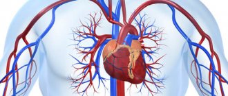
Sections and topography of the aorta
The aorta is a vital vessel that arises from the left ventricle of the heart. It begins at the level of the third rib, behind the left corner of the sternum. It contains blood enriched with oxygen, which is sent to organs and tissues. The vessel is anatomically divided into 3 main sections:
- ascending;
- aortic arch;
- descending (includes thoracic and abdominal aorta).
REFERENCE! The bifurcation of the aorta is located in the descending section, at the level of the fourth lumbar vertebra. Here the main vessel is divided into two large symmetrical branches - the iliac arteries.
Ascending section and aortic arch
The length of the ascending section is only 6 centimeters. It originates in the left ventricle of the heart and begins with an extension - the bulb. Despite its small size, this section gives off two vital branches - the coronary arteries. They are involved in the blood supply to the heart.
In the area of the manubrium of the sternum, the aorta forms a bend in the postero-left direction. This section is called its arc. Here the vessel bends around the left bronchus. The aortic arch also gives off several branches:
- brachiocephalic trunk;
- left common carotid artery;
- left subclavian artery.
IMPORTANT! If a person has a pyramidal part of the thyroid gland, the aortic arch gives off an additional branch - the inferior thyroid artery. It is found in no more than 30% of the population, but with surgery there is a risk of damage and death due to blood loss.
Descending department
This section continues from the 4th thoracic to the 4th lumbar vertebrae. This is the longest section, which gives off a large number of branches along its length. It is usually divided into two sections: the thoracic and abdominal aorta. The first passes into the second in the region of the diaphragm when it passes through the aortic opening.
The thoracic aorta begins in the chest cavity. Initially, it is located in front of the esophagus, but then forms a bend and moves to its posterior part (in the area of \u200b\u200bthe 8th and 9th thoracic vertebrae). It gives off various branches that take part in the blood supply to the organs of the thoracic cavity: intercostal, esophageal, pericardial and others.
The abdominal aorta is the terminal section of the vessel that ends at a bifurcation. It is located on the anterior surface of the lumbar vertebrae and continues to the 4th of them. Important branches necessary for blood circulation of the abdominal organs depart from this area:
- the celiac trunk is a large vessel through which blood flows to the stomach, spleen and liver;
- two mesenteric arteries (superior and inferior), which go to the intestines;
- two renal arteries (right and left) and other vessels.
Aortic bifurcation is a section where a vessel divides into two equal vessels. The right and left common iliac arteries form a large number of branches that supply blood to the pelvic organs, muscles and skin, as well as the membranes of the spinal cord.