The right aortic arch in the fetus is a congenital heart defect, which can occur alone or be combined with other, sometimes severe, defects. In any case, during the formation of the right arch, disturbances in the normal development of the fetal heart occur.
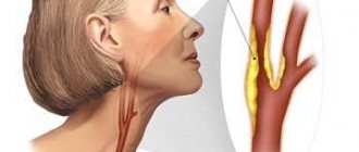
The aorta is the largest vessel in the human body, the function of which is to move blood from the heart to other arterial trunks, up to the arteries and capillaries of the whole body.
Phylogenetically, the development of the aorta undergoes complex changes during evolution. Thus, the formation of the aorta as an integral vessel occurs only in vertebrates, in particular in fish (two-chamber heart), amphibians (two-chamber heart with an incomplete septum), reptiles (three-chamber heart), birds and mammals (four-chamber heart). However, all vertebrates have an aorta, into which arterial blood mixed with venous, or entirely arterial, flows.
During the process of individual development of the embryo (ontogenesis), the formation of the aorta undergoes changes as complex as the heart itself. Starting from the first two weeks of embryo development, there is an increased convergence of the arterial trunk and the venous sinus, located in the cervical part of the embryo, which subsequently migrated more medially, towards the future thoracic cavity. The arterial trunk gives rise not only to two ventricles subsequently, but also to six branchial (arterial) arches (six on each side), which, as they develop, within 3-4 weeks, are formed as follows:
- the first and second aortic arches are reduced,
- the third arch gives rise to the internal carotid arteries that supply the brain,
- the fourth arch gives rise to the aortic arch and the so-called “right” part,
- the fifth arc is reduced,
- the sixth arch gives rise to the pulmonary trunk and the arterial (Botallov) duct.
The heart becomes completely four-chambered, with a clear division of the cardiac vessels into the aorta and pulmonary trunk, by the sixth week of development. A 6-week embryo has a fully formed, beating heart with large vessels.
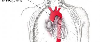
After the formation of the aorta and other internal organs, the topography of the vessel looks like this. Normally, the left aortic arch begins from the aortic bulb in its ascending part, which, in turn, originates from the left ventricle. That is, the ascending part of the aorta passes into the arch approximately at the level of the second rib on the left, and the arch bends around the left main bronchus, heading posteriorly and to the left. The uppermost part of the aortic arch projects onto the jugular notch just above the upper part of the sternum. The aortic arch goes down to the fourth rib, located to the left of the spine, and then passes into the descending part of the aorta.
In the case when the aortic arch “turns” not to the left, but to the right, due to a failure in the formation of human vessels from the branchial arches of the embryo, they speak of a right-sided aortic arch. In this case, the aortic arch extends through the right main bronchus, and not through the left, as it should normally be.
Why does vice occur?
Any malformation is formed in the fetus if a woman is influenced by negative environmental factors during pregnancy - smoking, alcoholism, drug addiction, ecology and unfavorable background radiation. However, genetic (hereditary) factors play an important role in the development of the child’s heart, as well as existing chronic diseases in the mother or past infectious diseases, especially in the early stages of pregnancy (influenza, herpes infection, chickenpox, rubella, measles, toxoplasmosis and many others) .
But, in any case, when any of these factors influence a woman in the early stages of pregnancy, the normal processes of ontogenesis (individual development) of the heart and aorta, formed during evolution, are disrupted.
So, in particular, the period of pregnancy of approximately 2-6 weeks is especially vulnerable to the fetal heart, since it is at this time that the formation of the aorta occurs.
Classification of the right-sided aortic arch
variant of the right aortic arch with the formation of a vascular ring
Depending on the anatomy of the duct anomaly, there are:
- The right aortic arch without the formation of a vascular ring, when the arterial ligament (overgrown arterial, or Botallov, duct, as it should be normally after childbirth) is located behind the esophagus and trachea,
- The right arch of the aorta with the formation of a vascular ring, code arterial ligament, or patent ductus arteriosus, is located on the left of the trachea and esophagus, as if surrounding them.
- As well as a separate similar form, the double arch of the aorta is distinguished - in this case, the vascular ring is formed not by the connective ligament, but by the inflow of the vessel.
Figure: a variety of options for the atypical structure of the aortic arch
Depending on whether any other structures of the heart were damaged during its formation, the following types of defect are distinguished:
- An isolated type of defect, without other developmental anomalies (in this case, if the right-sided aorta is not combined with the DiGeorge syndrome characteristic of it in some cases, the prognosis is as favorable as possible);
- In combination with dextraposition (mirror, right position of the heart and great vessels, including the aorta), (which is also usually not dangerous),
- In combination with a more serious heart defect - in particular tetralogy of Fallot (dextraposition of the aorta, ventricular septal defect, pulmonary stenosis, right ventricular hypertrophy).
Tetralogy of Fallot combined with the right arch is an unfavorable development option
Symptoms of aortic aneurysm
Aortic sinus aneurysm may be accompanied by aortic valve insufficiency or narrowing of the lumen of the coronary arteries of the heart. Reaching a large size, such an aneurysm can compress the pulmonary trunk, right ventricle and right atrium, which leads to the formation of subacute right ventricular heart failure, characterized by liver enlargement, swelling of the jugular veins and the appearance of edema. Rapid compression of the pulmonary trunk by an aneurysm can lead to sudden death of the patient.
An aneurysm of the ascending aorta is usually manifested by dull retrosternal pain, which in some patients is accompanied by reflex attacks of shortness of breath. If the aneurysm reaches a large size, it can cause atrophy of the adjacent areas of the sternum and ribs, with pathological vascular pulsation appearing in the second or third intercostal space to the right of the sternum. Compression of the superior vena cava by an aneurysm or a breakthrough of the aneurysm into it leads to the development of superior vena cava syndrome, which in turn causes swelling of the neck, face, arms, and swelling of the jugular veins.
Aneurysm of the aortic arch most often manifests itself as shortness of breath (and, as a rule, it is more difficult to take a breath), caused by compression of the trachea and bronchi. Compression of the left main bronchus by an aneurysm can lead to atelectasis (collapse) of the left lung. Sometimes hemoptysis appears, which may precede the rupture of an aneurysm. Compression of the left inferior laryngeal nerve by an aneurysm is manifested by a dry cough, attacks of suffocation, and a change in voice timbre (hoarseness). The development of superior vena cava syndrome is possible. When an aneurysm compresses the brachiocephalic trunk, left subclavian and left common carotid arteries, symptoms of a gradually worsening disturbance of the blood supply to the upper extremities and head appear. A rupture of an aortic aneurysm into the esophagus or trachea is possible, which, as a rule, develops gradually, which is initially manifested by the appearance of scanty bloody vomiting or hemoptysis, but then massive bleeding develops.
An aneurysm of the descending aorta leads to compression of the nerve roots, vertebral bodies, esophagus and left lung. Compression of the nerve roots leads to intense pain that is resistant to the administration of the most powerful painkillers. Pressure on the vertebral bodies and posterior parts of the ribs leads to their deformation, to the point that the aneurysmal sac can protrude between the inner edge of the left scapula and the spinal column. These patients may develop lower paraplegia (complete loss of voluntary movements of both lower extremities). Compression of the left lung by an aneurysm leads to its atelectasis and creates favorable conditions for the occurrence of pneumonia. Compression of the esophagus in some cases can lead to difficulty passing food through it (dysphagia). When the wall of the esophagus is destroyed due to prolonged pressure on it from the aneurysm, small bleeding from the esophagus occurs, after which, as a rule, the aneurysm ruptures into its lumen with the development of massive bleeding. When a descending aortic aneurysm ruptures into the pleural cavity, rapidly increasing anemia (anemia) and large hemothorax (accumulation of blood in the pleural cavity) develop.
Aneurysm of the thoracoabdominal (thoracoabdominal aorta) aorta is rare and is usually caused by syphilis. The aneurysm compresses the esophagus and upper stomach, which leads to pressing pain in the epigastric region, which can be associated with food intake, sometimes belching, vomiting, and disruption of the passage of food through the esophagus. An aneurysm of the thoracoabdominal aorta can cause narrowing or complete occlusion of the lumen of the superior mesenteric artery and the celiac trunk, which supply blood to the abdominal organs, which is manifested by attacks of painful abdominal pain (the so-called abdominal pain). Due to the reasons described above, an aneurysm of this location leads to weight loss for the patient.
An abdominal aortic aneurysm over time manifests itself as pain caused by the pressure of the aneurysm on the nerve plexuses and nerve roots located directly next to it. Pain may be in the lumbar or epigastric region. A large aneurysm located below the origin of the renal arteries can compress the ureters, causing the development of hydronephrosis and anuria. If compression of the renal arteries occurs, symptomatic hypertension occurs. When the duodenum is compressed by an aneurysm, the passage of food through it is disrupted, which is manifested by vomiting and weight loss. Most often, an abdominal aortic aneurysm is manifested by the presence of a pulsating tumor-like formation in the abdominal cavity at the level of the navel or just below and slightly to the left of it. A thrombosed aneurysm does not pulsate, and therefore can be mistaken for a tumor. Sometimes there is a rise in body temperature. An aneurysm ruptures into the abdominal cavity quickly and, as a rule, painlessly, and into the retroperitoneal tissue - with severe pain in the abdomen and lower back, with the development of shock. After some time, the patient may die due to increasing blood loss.
Dissecting aortic aneurysm is manifested by sudden onset of acute chest pain that is not relieved by painkillers and collapse. Sometimes a complete loss of the ability to voluntarily move both lower extremities develops, which can be temporary or permanent. Due to the localization and nature of the pain that occurs, the clinical manifestations of dissecting aortic aneurysm can be mistaken for acute myocardial infarction.
WHEN TO CONTACT THE DOCTOR
When the above symptoms appear.
You can make an appointment with a cardiologist at Medical by calling 8 (49244) 9-32-49.
How to recognize a vice?
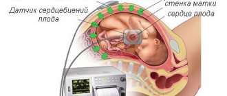
Diagnosis of the defect is not difficult even during pregnancy. This is especially true in cases where the right aortic arch is combined with other, more severe anomalies of heart development. However, to confirm the diagnosis, a pregnant woman is repeatedly examined, including with expert-class ultrasound machines, and a council of geneticists, cardiologists and cardiac surgeons is assembled to make a decision on the prognosis and the possibility of delivery in a specialized perinatal center. This is due to the fact that with some types of defects combined with the right aortic arch, the newborn baby may require heart surgery immediately after delivery.
Regarding the clinical manifestations of the right aortic arch, it should be mentioned that an isolated defect may not manifest itself at all, only sometimes accompanied by frequent obsessive hiccups in a child. In the case of a combination with tetralogy of Fallot, which accompanies the defect in some cases, the clinical manifestations are pronounced and appear in the first days after birth, such as increasing pulmonary heart failure with severe cyanosis (blue discoloration of the skin) in the baby. That is why tetralogy of Fallot is classified as a “blue” heart defect.
What screening shows a defect in pregnant women?
The diagnosis of a right-sided aortic arch can be made already at the first screening, that is, at 12-13 weeks of pregnancy. More accurate information about the condition of the fetal heart can be obtained at the second and third screening ultrasound examinations (20 and 30 weeks of pregnancy).
An analysis of fetal DNA can further clarify the absence of a connection between the formation of a right-sided aorta and severe genetic mutations. In this case, chorionic villus material or amniotic fluid is usually collected through a puncture. First of all, DiGeorge syndrome is excluded.
Complications of aortic aneurysm
1. Aortic valve defects and heart failure . With an aneurysm of the ascending aorta of syphilitic origin, cardiac decompensation may develop due to a defect of the aortic valve or blockage of the mouth of the coronary arteries.
2. Rupture of an aneurysm with bleeding . Bleeding can occur into the respiratory organs (bronchi, trachea), pleural cavity, cardiac sac, into the esophagus, large blood vessels located in the chest cavity, and sometimes even out through the skin when the sternum is destroyed. In case of bleeding into the pericardial cavity, cardiac tamponade occurs. Bleeding results in rapidly increasing blood loss.
3. Acute and subacute thrombosis of the aortic aneurysm. Most often it develops in the abdominal aorta and leads to the closure of its branches located here.
The listed complications quickly lead to the death of the patient if appropriate measures are not taken in time.
Treatment
In the event that the right aortic arch is isolated and is not accompanied by any clinical manifestations after the birth of the child, the defect does not require surgical treatment. All you need is a monthly examination by a pediatric cardiologist with regular (every six months - once a year) ultrasound of the heart.
When combined with other heart defects, the type of surgical intervention is selected based on the type of defect. Thus, with tetralogy of Fallot, surgery is indicated in the first year of a child’s life, carried out in stages. At the first stage, palliative (auxiliary) shunts are applied between the aorta and the pulmonary trunk to improve blood flow into the pulmonary circulation. At the second stage, open heart surgery is performed using a cardiopulmonary bypass machine (ACB) to eliminate pulmonary stenosis.
In addition to surgery, cardiotropic drugs that can slow the progression of chronic heart failure (ACE inhibitors, diuretics, etc.) are prescribed for auxiliary purposes.
Causes of aortic aneurysm
Most often, an aortic aneurysm develops as a result of an atherosclerotic process or is of syphilitic origin. Recently, atherosclerosis has taken first place among the causes of aortic aneurysm development, which is due to advances in the treatment of syphilis and an increase in average life expectancy. In addition, syphilis is more often the cause of the development of a thoracic aortic aneurysm, while atherosclerosis more often leads to the formation of an abdominal aneurysm.
Other causes of aortic aneurysm development are medianecrosis and nonspecific aortoarteritis. Traumatic aneurysms are also possible (for example, after a closed abdominal injury) and false anastomotic aneurysms after operations on the aorta. Aneurysms of mycotic (fungal) origin are also described in the scientific medical literature.
The most common cause of the development of dissecting aortic aneurysm is long-standing arterial hypertension against the background of atherosclerosis. In this case, on the inner lining (intima) of the aortic wall, as a rule, there are already various pre-existing small defects. Less commonly, the causes of dissecting aortic aneurysm can be hypertension due to coarctation of the aorta (a congenital defect manifested by segmental narrowing of the aortic lumen); arterial hypertension caused by other factors; Marfan syndrome (hereditary pathology of connective tissue), which is accompanied by severe weakness of the aortic wall. It is possible to form an acute dissecting aneurysm of the ascending aorta due to its rupture caused by a closed injury (for example, a car injury). Sometimes dissecting aortic aneurysm can occur as a result of iatrogenicity: as a complication of cannulation of the arteries and aorta for the purpose of perfusion during cardiopulmonary bypass.
Forecast
The prognosis for an isolated right-sided aortic arch is favorable, since in most cases surgical intervention is not even required. So, in general, we can say that an isolated right aortic arch is not life-threatening for the child.
With combined types, the situation is much more complicated, since the prognosis is determined by the type of concomitant heart defect. For example, with tetralogy of Fallot, the prognosis without treatment is extremely unfavorable; unoperated children with this disease usually die in the first year of life. After surgery, the duration and quality of life increase, and the prognosis becomes more favorable.
Prognosis for aortic aneurysm
In the absence of timely treatment and the occurrence of severe complications of aortic aneurysm, the prognosis is unfavorable. Death can occur as a result of decompensation of cardiac activity caused by the development of aortic valve defects with an aneurysm of the ascending aorta, cardiac tamponade due to the breakthrough of an aneurysm into the pericardial cavity, massive blood loss as a result of a breakthrough of the aneurysm into the hollow organs and the pleural or abdominal cavity.
However, the successes currently achieved in the surgical treatment of aortic aneurysms make it possible, in the case of timely and adequate surgical intervention, to save the lives of the majority of patients. With a planned operation, the mortality rate is 0-5%, and in the case of aneurysm rupture, even with emergency surgery, it is 50-80%. The five-year survival rate among operated patients is 80%, and among non-operated patients it is 5-10%.