Abnormally low blood oxygen levels
| Hypoxemia | |
| Other names | Hypoxemia |
| Speciality | Pulmonology |
Hypoxemia
is an abnormally low level of oxygen in the blood. [1] [2] More specifically, it is a lack of oxygen in the arterial blood. [3] Hypoxemia has many causes and often causes hypoxia because the blood does not supply enough oxygen to the body tissues.
Definition[edit]
Hypoxemia
refers to low levels of oxygen in the blood, and the more general term
hypoxia
is an abnormally low level of oxygen in any tissue or organ or the body as a whole. [2] Hypoxemia can cause hypoxia (hypoxemic hypoxia), but hypoxia can also occur through other mechanisms such as anemia. [4]
Hypoxemia is usually defined as a decreased partial pressure of oxygen (mmHg) in arterial blood, but also as a decreased oxygen content (mL of oxygen per dL of blood) or percentage saturation of hemoglobin (the oxygen-binding protein in red blood). cells ) with oxygen, which is found either individually or in combination. [2] [5]
Although there is general agreement that an arterial blood gas measurement that shows that the partial pressure of oxygen is below normal constitutes hypoxemia,[5][6][7] there is less agreement as to whether blood oxygen content is relevant in determining hypoxemia. This definition includes oxygen carried by hemoglobin. Thus, blood oxygen content is sometimes considered an indicator of tissue delivery rather than an indicator of hypoxemia. [7]
Just as extreme hypoxia can be called anoxia, extreme hypoxemia can be called hypoxemia.
How to recognize the problem?
Hypoxemia is represented by a number of symptoms that you should definitely pay attention to in order to consult a doctor in time. After all, a lack of oxygen can be an extremely dangerous condition that leads to an imbalance in the body as a whole.
So, hypoxemia will be indicated:
- increased heart rate and breathing;
- drowsiness;
- change in skin color towards pallor;
- weakness in general;
- dizziness.
If you ignore such signals from the body, later manifestations may develop, fraught with serious complications. For example, disruptions in the functioning of the heart, the development of severe swelling, problems with the functioning of the brain, as it does not receive enough nutrition. The skin may also take on a bluish tint. A person may experience anxiety, forgetfulness, and sleep problems. Potential complications include decreased blood pressure, stroke conditions, risks of developing pulmonary edema, convulsive effects, and heart rhythm disturbances.
Signs and symptoms[edit]
Main article: Hypoxia (medical) § Signs and symptoms
In an acute context, hypoxemia can cause symptoms such as respiratory distress. These include shortness of breath, rapid breathing, using the chest and abdominal muscles to breathe, and pursing the lips. [8] : 642
Chronic hypoxemia can be compensated or uncompensated. Compensation may cause symptoms to be ignored initially, but further illness or stress, such as any increase in oxygen demand, may ultimately reveal existing hypoxemia. In the compensated state, blood vessels supplying less ventilated areas of the lung may selectively contract, redirecting blood to areas of the lung that are better ventilated. However, in a chronic context and if the lungs are usually poorly ventilated, this mechanism can lead to pulmonary hypertension, overloading the right ventricle of the heart and causing cor pulmonale and right-sided heart failure. Polycythemia may also occur.[8] In children, chronic hypoxemia may result in delayed growth, neurological and motor development, and decreased sleep quality with frequent awakenings during sleep. [9]
Other symptoms of hypoxemia may include cyanosis, digital discomfort, and symptoms that may be related to the cause of the hypoxemia, including cough and hemoptysis. [8] : 642
Severe hypoxemia usually occurs when the partial pressure of oxygen in the blood is less than 60 mm Hg. Art. (8.0 kPa), this is the beginning of the steep portion of the oxygen-hemoglobin dissociation curve, where a small decrease in the partial pressure of oxygen leads to a large decrease in the oxygen content of the blood. [6] [10] Severe hypoxia can lead to respiratory failure [8]
What could be the consequences?
Mild and moderate pathologies are relatively easy to treat. If treatment is not started on time, complications such as:
- encephalopathy;
- hypotension;
- Stroke;
- pulmonary edema;
- arrhythmia;
- convulsions.
If the fetus experiences hypoxemia, the following may occur:
- intrauterine growth retardation;
- death of a child in the womb during or shortly after birth;
- early or difficult labor;
- Delays in mental and physical development in later life.
Acute, fulminant hypoxemia can cause the most unfavorable complications, as it contributes to a hypoxic coma of the body.
Reasons [edit]
Hypoxemia means a lack of oxygen in the blood. Thus, any cause that affects the rate or volume of air entering the lungs ( ventilation
), or any cause that affects the transport of air from the lungs to the blood can cause hypoxemia. In addition to these respiratory causes, cardiovascular causes such as shunts can also lead to hypoxemia.
Hypoxemia is caused by five categories of etiology: hypoventilation, ventilation-perfusion mismatch, right-to-left shunt, impaired diffusion, and low PO 2 . Low PO 2 and hypoventilation are associated with a normal alveolar-arterial gradient (Aa gradient), whereas the other categories are associated with an increased Aa gradient. [11] : 229
Ventilation[edit]
If alveolar ventilation is low, the alveoli do not receive enough oxygen for the body to use. This can cause hypoxemia even if the lungs are normal because the cause lies in the control of ventilation by the brainstem or the body's inability to breathe effectively.
Respiratory engine[edit]
Breathing is controlled by centers in the medulla that influence the rate of breathing and the depth of each breath. This is influenced by the level of carbon dioxide in the blood, which is determined by central and peripheral chemoreceptors located in the central nervous system and the carotid and aortic bodies, respectively. Hypoxia occurs when the respiratory center does not work properly or when the signal is not appropriate:
- Stroke, epilepsy, and cervical fractures can damage the brain's breathing centers, which generate rhythmic impulses and transmit them along the phrenic nerve to the diaphragm, the muscle responsible for breathing.
- Decreased respiratory activity can also result from metabolic alkalosis, a state of low carbon dioxide in the blood.
- Central sleep apnea. During sleep, the brain's respiratory centers may shut down, leading to long periods of apnea with potentially serious consequences.
- Hyperventilation followed by prolonged breath holding. This hyperventilation, which some swimmers try, reduces the amount of carbon dioxide in the lungs. This reduces the desire to breathe. However, this also means that the drop in blood oxygen levels is not felt, which can lead to hypoxemia. [12]
Physical conditions[edit]
Various conditions that physically restrict airflow can lead to hypoxemia.
- Choking, including temporary interruption or cessation of breathing, as in obstructive sleep apnea, or bedding can interfere with breathing in infants, a suspected cause of SIDS.
- Structural deformities of the chest, such as scoliosis and kyphosis, which can limit breathing and lead to hypoxia.
- Muscle weakness, which may limit the function of the diaphragm, the main muscle that draws new air into the lungs. This may be the result of a congenital condition, such as motor neurone disease, or an acquired condition, such as fatigue in severe cases of COPD.
Environmental oxygen[edit]
Oxygen-hemoglobin dissociation curve.
Under conditions where the proportion of oxygen in the air is low or when the partial pressure of oxygen decreases, less oxygen is present in the alveoli of the lungs. Alveolar oxygen is transferred to hemoglobin, the carrier protein inside red blood cells, with an efficiency that decreases as the partial pressure of oxygen in the air increases.
- Height . The external partial pressure of oxygen decreases with altitude, such as at high altitude or during flight. This decrease leads to a decrease in the transport of oxygen by hemoglobin. [13] It is especially thought to be a cause of cerebral hypoxia and altitude sickness in climbers of Everest and other very high altitude peaks. [14] [15] For example, at the peak of Everest, the partial pressure of oxygen is only 43 mmHg. Art., While at sea level the partial pressure is 150 mm Hg. [16] For this reason, the aircraft cabin pressure is maintained at 5,000 to 6,000 ft (1,500 to 1,800 m).[17]
- Diving. Hypoxia during diving can occur as a result of a sudden ascent. Partial pressures of gases increase during diving by one atm every ten meters. This means that sufficient oxygen partial pressure to maintain good hemoglobin transport is possible at depth, even if it is insufficient at the surface. A diver who remains underwater will slowly consume his oxygen, and upon ascent the partial pressure of oxygen may be insufficient (shallow water blackout). At depth this may appear as darkening in deep water.
- Choking. A decrease in the concentration of oxygen in the inhaled air, caused by a decrease in the replacement of oxygen in the respiratory mixture.
- Anesthetics. Low partial pressure of oxygen in the lungs during the transition from inhalation anesthesia to atmospheric air due to the Fink effect or diffuse hypoxia.
- The lack of oxygen in the air also proved fatal. In the past, anesthesia machines malfunctioned, delivering low-oxygen gases to patients. Additionally, oxygen in a confined space can be consumed if carbon dioxide scrubbers are used without sufficient attention to replenishing the oxygen consumed.
Perfusion [edit]
Ventilation-perfusion mismatch[edit]
This refers to a ventilation/perfusion imbalance. Oxygen entering the lungs usually diffuses through the alveolar capillary membrane into the blood. However, this balancing does not occur when the alveolus is insufficiently ventilated and, as a consequence, the blood leaving the alveolus is relatively hypoxic. When such blood is added to blood from well-ventilated alveoli, the mixture has a lower partial pressure of oxygen than the alveolar air and therefore an Aa difference occurs. Examples of conditions that can cause ventilation-perfusion mismatch include:
- Exercise. Although moderate activity and exercise improve ventilation-perfusion, [18] hypoxemia can develop during intense exercise as a result of pre-existing lung disease. [19] During exercise, nearly half of hypoxemia is due to diffusion restrictions (again, on average). [20]
- Aging. With age, there is a deterioration in the correspondence between ventilation and perfusion, as well as a decrease in the ability to compensate for hypoxic conditions. [8] : 646
- Diseases affecting the pulmonary interstitium can also lead to hypoxia, affecting the ability of oxygen to diffuse into the arteries. An example of these diseases is pulmonary fibrosis, in which, even at rest, a fifth of hypoxemia is due to diffusion restrictions (on average). [20]
- Diseases leading to acute or chronic respiratory failure can lead to hypoxia. These diseases may be acute in onset (such as inhalation obstruction or pulmonary embolus) or chronic (such as chronic obstructive pulmonary disease).
- Cirrhosis may be complicated by refractory hypoxemia due to high blood flow rates through the lungs, resulting in ventilation-perfusion mismatches. [21]
Shunting [edit]
Shunting refers to blood that bypasses the pulmonary circulation, meaning the blood does not receive oxygen from the alveoli. Typically, the shunt may be in the heart or lungs and cannot be corrected by administering oxygen alone. Shunting can occur in normal conditions:
- An anatomical bypass that occurs through the bronchial circulation, which supplies blood to the lung tissues. Shunting also occurs through the smallest cardiac veins, which drain directly into the left ventricle.
- Physiological shunts caused by gravity. The highest concentration of blood in the pulmonary circulation occurs at the base of the pulmonary tree compared to the highest gas pressure at the apex of the lungs. The alveoli cannot be ventilated during shallow breathing.
Shunting can also occur for painful conditions:
- Acute respiratory distress syndrome (ARDS). Alveolar collapse, which can increase the volume of physiologic shunting, can be controlled using a combination of increased oxygenation as well as positive pressure to attract collapsed alveoli. [22] [23]
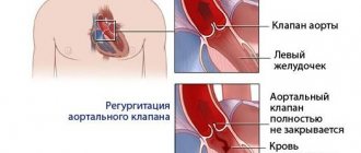
- Pathological shunts such as patent ductus arteriosus, patent foramen ovale and atrial septal defects or ventricular septal defects. These are conditions where blood from the right side of the heart moves directly to the left without first passing through the lungs. This is called a right-to-left shunt, which is often congenital.
Exercise [edit]
Exercise-induced arterial hypoxemia occurs during exercise when a trained person's arterial oxygen saturation is below 93%. It occurs in healthy people of different ages and genders in good physical shape. [24] Exercise-induced adaptations include increased cardiac output due to cardiac hypertrophy, improved venous return and metabolic muscle vasodilation, and increased VO 2 Max. There must be a corresponding increase in VCO 2, hence the need to scavenge carbon dioxide to prevent metabolic acidosis. Hypoxemia occurs in these people due to increased pulmonary blood flow causing:
- Decreased capillary transit time
due to increased blood flow in the pulmonary capillaries. The capillary transit time (tc) at rest is about 0.8 s, which allows sufficient time for oxygen to diffuse into the bloodstream and CO 2 to diffuse out of the bloodstream. After training, capillary volume remains the same, but cardiac output increases, which leads to a decrease in capillary transit time, which in trained people at maximum speed is reduced to approximately 0.16 s. This does not allow enough time for gas to diffuse and results in hypoxemia. - Intrapulmonary arteriovenous shunts
are dormant capillaries in the lungs that
are activated
when venous pressure becomes too high. They are usually located in a dead zone where gas diffusion does not occur, so the blood passing through them is not saturated with oxygen, which leads to hypoxemia.
Preventive measures
To prevent the development of a pathology such as hypoxemia, it is necessary to adhere to simple recommendations, which are as follows:
- daily walks in the fresh air;
- moderate physical activity;
- performing breathing exercises;
- Taking a vitamin-mineral complex, especially in the autumn-winter period;
- Eating fruits and vegetables;
- Early diagnosis of pathologies of the cardiovascular and respiratory systems.
Hypoxia can be prevented. The main thing is to follow the rules for preventing oxygen deficiency, and if symptoms of the disease are detected, you should immediately seek medical help. If the pathology is not treated in time, the development of irreversible effects that can occur in the lungs, brain and the body as a whole is possible.
Physiology [edit]
Main article: Hypoxia (medical) § Physiological compensation
The key to understanding whether the lungs are involved in a particular case of hypoxemia is the difference between alveolar and arterial oxygen levels; this difference Aa is often called the gradient
Aa, and it's usually small. The partial pressure of oxygen in arterial blood is obtained directly from the determination of arterial blood gases. The oxygen contained in the alveolar air can be calculated because it is directly proportional to its fractional composition in the air. As the respiratory tract moistens the (already dilute) inhaled air, the barometric pressure in the atmosphere decreases the water vapor pressure.
Links[edit]
- Pollack, Charles P.; Torpey, Michael J.; Yager, Jan (2010). Encyclopedia of Sleep and Sleep Disorders
(3rd ed.). New York, New York. paragraph 104. ISBN 9780816068333. - ^ a b c
Martin, Lawrence (1999).
Everything You Really Need to Know to Interpret Arterial Blood Gases
(2nd ed.). Philadelphia: Lippincott Williams and Wilkins. paragraph xxvi. ISBN 978-0683306040. - Ekman, Margaret (2010). Professional Guide to Pathophysiology
(3rd ed.). Philadelphia: Wolters Kluwer/Lippincott Williams & Wilkins. p. 208. ISBN 978-1605477664. - Robert J. Mason, W. Courtney Broaddus, Thomas R. Martin, Talmadge E. King, Dean E. Schraufnagel, John F. Murray and Jay A. Nadel (eds.) (2010) Murray and Nadel's Textbook of Respiratory Medicine, 5- e ed. Philadelphia: Saunders Elsevier. ISBN 1-4160-4710-7.
- ^ a b
Morris, Alan;
Kanner, Richard; Crapo, Robert; Gardner, Reed. (1984) Clinical study of pulmonary function.
Manual of Common Laboratory Practices , 2nd ed. - ^ a b
Lorenzo Del Sorbo, Erica L. Martin, V. Marco Ranieri (2010) "Hypoxemic respiratory failure" In: Murray and Nadel Textbook of Respiratory Medicine, Robert J. Mason, W. Courtney Broaddus, Thomas R. Martin, Talmadge E King, Dean E. Schraufnagel, John F. Murray, and Jay A. Nadel (eds.) 5th ed. Philadelphia: Saunders Elsevier. ISBN 1-4160-4710-7. - ^ a b
Wilson, William S.;
Grande, Christopher M.; Hoyt, David B., ed. (2007). Critical Help
. New York: Informa Healthcare. ISBN 978-0-8247-2920-2. - ^ a b c d e
Colledge NR, Walker BR, Ralston SH, eds.
(2010). Davidson's Principles and Practice of Medicine
(21st ed.). Edinburgh: Churchill Livingston/Elsevier. ISBN 978-0-7020-3085-7. - Adde, F. V.; Alvarez, A.E.; Barbizan B.N.; Guimarães, Brazil (January–February 2013). "Recommendations for long-term home oxygen therapy in children and adolescents". Jornal de Pediatria
.
89
(1):6–17. DOI: 10.1016/j.jped.2013.02.003. PMID 23544805. - Schwartzstein, Richard; Parker, Michael J. (2006). Respiratory Physiology: A Clinical Approach
. Philadelphia: Lippincott Williams and Wilkins. ISBN 978-0-7817-5748-5. OCLC 62302095. - Fauci, Anthony S.; Harrison, TR, ed. (2008). Harrison's Principles of Internal Medicine
(17th ed.). New York: McGraw-Hill Medical. ISBN 978-0-07-147692-8. - Craig, Albert B. (Fall 1976). "Summary of 58 cases of loss of consciousness during scuba diving and diving." Medicine and Science in Sports
.
8
(3): 171–5. DOI: 10.1249/00005768-197600830-00007. PMID 979564. - Kenneth Bailey; Alistair Simpson. "High Altitude Oxygen Calculator". Apex (high altitude physiological expeditions). Archived from the original on 2017-06-11. Retrieved August 10, 2006. — Online interactive oxygen delivery calculator.
- West JB, Boyer SJ, Graber DJ, Hackett PH, Maret KH, Milledge JS, Peters RM, Pizzo CJ, Samaja M, Sarnquist FH (September 1983). "Maximum Exercise at Extreme Altitude on Everest." Journal of Applied Physiology: Respiratory, Environmental and Physical Physiology
.
55
(3):688–98. DOI: 10.1152/jappl.1983.55.3.688. hdl: 2434/176393. PMID 6415008. - Grocott, M.P.; Martin, D.S.; Levett, DZ; McMorrow, R. Windsor, J; Montgomery, H. E.; Caudwell Xtreme Everest Research, Group (January 8, 2009). "Arterial blood gases and oxygen content in climbers on Everest" (PDF). New England Journal of Medicine
.
360
(2):140–9. DOI: 10.1056/NEJMoa0801581. PMID 19129527. - West, John B. (2000). "The Human Limits of Hypoxia: Physiological Challenges of Climbing Everest". Annals of the New York Academy of Sciences
.
899
(1):15–27. Bibcode: 2000NYASA.899…15W. DOI: 10.1111/j.1749-6632.2000.tb06173.x. PMID 10863526. S2CID 21863823. - Administrator. “Airlines are cutting costs—are respiratory patients paying for it?” . www.ersnet.org
. Retrieved June 17, 2021. - Whipp, B. J.; Wasserman, K. (September 1969). "Differences in alveolar-arterial gas pressure during graded exercise." Journal of Applied Physiology
.
27
(3): 361–5. DOI: 10.1152/jappl.1969.27.3.361. PMID 5804133. - Jump up
↑ Hopkins, S. R. (2006).
"Exercise-induced arterial hypoxemia: the role of ventilation-perfusion inequalities and pulmonary diffusion limitation." Hypoxia and exercise
.
Advances in experimental medicine and biology. 588
. pp. 17–30. DOI: 10.1007/978-0-387-34817-9_3. ISBN 978-0-387-34816-2. PMID 17089876. - ^ a b
Agusti, AG;
Roca, J; Gea, J; Wagner, P.D.; Xaubet, A; Rodriguez-Roisin, R. (February 1991). "Mechanisms of gas exchange disturbances in idiopathic pulmonary fibrosis." American Respiratory Disease Review
.
143
(2):219–25. DOI: 10.1164/ajrccm/143.2.219. PMID 1990931. - Agusti, A.G.; Roca, J; Rodriguez-Roisin, R. (March 1996). “Mechanisms of gas exchange disorders in patients with liver cirrhosis.” Breast medicine clinics
.
17
(1):49–66. DOI: 10.1016/s0272-5231(05)70298-7. PMID 8665790. - Huang, Y.C.; Fracica, P. J.; Simonson S.G.; Crapo, J. D.; Young, S.L.; Welty-Wolf, K.E.; Moon, R.E.; Piantadosi, California (August 1996). "VA/Q abnormalities in gram-negative sepsis". Physiology of Respiration
.
105
(1-2): 109-21. DOI: 10.1016/0034-5687 (96) 00039-4. PMID 8897657. - Jump up
↑ Thompson BT, Chambers RC, Liu KD (2017).
"Acute respiratory distress syndrome." N Engl J Med
.
377
(6):562–572. DOI: 10.1056/NEJMra1608077. PMID 28792873. S2CID 4909513. CS1 maint: multiple names: list of authors (link) - Dempsey, J.A.; Wagner, P.D. (December 1999). "Arterial hypoxemia induced by exercise." Journal of Applied Physiology
.
87
(6):1997–2006. DOI: 10.1152/jappl.1999.87.6.1997. PMID 10601141. - Henry Power and Leonard W. Sedgwick (1888) The New Sydenham Society's Lexicon of Medicine and Allied Sciences (based on May's Lexicon). Volume III. London: New Sydenham Society.
- West, John B. (2012). Pulmonary Pathophysiology: Fundamentals
(8th ed.). Lippincott Williams and Wilkins. ISBN 978-1-4511-0713-5. - Anderson, Kenneth N. (2002). Mosby's Dictionary of Medicine, Nursing and Allied Health
(6th ed.). C. V. Mosby. ISBN 978-0-323-01430-4. - Dean R. Hess, Neil R. McIntyre, Shelley S. Mishou, William F. Galvin, Alexander B. Adams (2012) Respiratory care: principles and practice.
(ed.) (2nd ed.). Jones and Bartlet Learning ISBN 978-0-7637-6003-8 - Jacob Samuel and Corey Frankling, “Hypoxemia and Hypoxia” in Jonathan A. Myers, Keith W. Milliken, and Theodore J. Saklarides (eds.) (2008) Common Surgical Diseases
. (2nd ed.). 391–394 ISBN 978-0-387-75245-7