Aortic aneurysm
This is a pathology in which a thickening appears on the walls of the artery - something like a sac where blood accumulates. There are many reasons for the appearance of an aneurysm: atherosclerosis, trauma, inflammation, deterioration of vascular elasticity in old age, severe physical activity, hypoplasia, tumor, as well as congenital pathologies and even genetic predisposition.
Rice. Aortic aneurysm
Aortic rupture can occur instantly, or it can last for several months or even years while the vascular membranes dissect. In acute cases, as a rule, doctors simply do not have time to provide timely assistance. However, the disease in its chronic form can be caught by examination by doctors.
How does the disease manifest?
Symptoms and manifestations of aortic rupture also depend on the stage. If the internal lining of a vessel is damaged, the patient feels pain, drowsiness, weakness, and blood pressure increases. Detachment of the middle wall of the aorta can be accompanied by sharp and burning pain, and blood pressure drops. When an injury occurs to the outer wall of a vessel, shock occurs, the pressure drops sharply, and the temperature rises.
Damage to blood vessels can occur in the thoracic aorta or in the abdominal aorta - the symptoms may also differ. For example, when the abdominal aorta ruptures, a person feels shock, severe abdominal pain and weakness, vision may be blurred, and kidney failure is possible. When the thoracic aorta ruptures, severe pain is felt in the chest, shortness of breath and swelling of the neck are possible, the skin turns blue, and the veins swell.
Rice. Thoracic aortic aneurysm
How is this treated?
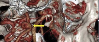
If suspicions arise, doctors refer the patient for angiography - this is an x-ray with contrast, which examines all parts of the aorta. However, if the inner lining of the blood vessels is damaged, this method may not produce results. In addition, the patient is given an ECG to make sure that the cause of the pain is not myocardial infarction. The most reliable method (and the most expensive) is computed tomography.
The only treatment option is prosthetic surgery using a stent graft. Unfortunately, it will not be possible to “glue” a ruptured vessel together with medication.
Definition
A thoracic aortic aneurysm is a condition that occurs when the walls of the top of your body's main artery (aorta) become weakened and bulging.
A thoracic aortic aneurysm is a weakened and bulging area at the top of the aorta, the main blood vessel that carries blood to different parts of your body. The aorta, which is approximately the thickness of a garden hose, runs from the heart through the center of the chest and abdomen. Aneurysms form anywhere along the aorta, which runs from the heart through the abdomen. When they occur in the upper part of the aorta, they are called thoracic aortic aneurysms. Most often, aneurysms form in the lower part of the aorta and are called abdominal aortic aneurysms. In rare cases, an aneurysm develops in both the upper and lower parts of the aorta. This type of aneurysm is called a thoracoabdominal aneurysm.
Because the aorta carries blood to various parts of the body, rupture can cause life-threatening bleeding. Most small, slow-growing thoracic aortic aneurysms do not rupture, but large, rapidly growing aneurysms are at very high risk of rupture. Depending on the size and growth rate of the thoracic aortic aneurysm, treatment may range from watchful waiting to emergency surgery. If a thoracic aortic aneurysm is identified, doctors will closely monitor its condition to plan surgery if necessary. Emergency surgery for a ruptured thoracic aortic aneurysm is very risky.
Search and don't give up
A dissecting aneurysm (longitudinal rupture of the aorta) is practically asymptomatic. At the time of rupture of the vessel, the patient experiences severe “tearing” pain in the chest, which is often mistaken for symptoms of another cardiovascular disease, such as myocardial infarction. If prompt surgical measures are not taken, death after aortic rupture can occur within 2 hours.
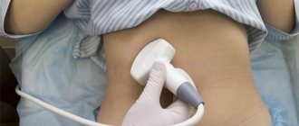
In order not to lead to such a situation, you need to undergo examination on time. An ultrasound examination can detect an aneurysm even in cases where there is no dangerous dilation of the vessel yet, but it is about to appear. X-ray is very informative for an abdominal aneurysm, since it is clearly visible due to the deposition of calcium on the walls of the vessel. More detailed studies - computer and magnetic resonance imaging - are carried out after the ultrasound method. They allow you to determine the strategy for further treatment. It is believed that if the diameter of the aneurysm is less than 5 cm, the risk of rupture of the vessel is low. With larger sizes the risk increases.
Symptoms
Aortic aneurysms grow slowly and are usually asymptomatic, making them difficult to detect. Some aneurysms are small and do not grow larger over time. Other aortic aneurysms grow slowly, increasing in size at about 1.2 cm per year. Others grow at a faster rate, increasing the risk of rupture. The rate of aneurysm growth is difficult to predict.
As the aneurysm grows, people notice:
- Soreness or pain in the abdomen or chest;
- Backache
Aortic rupture: causes, symptoms, how to avoid death, surgery, prognosis
Aortic rupture is a severe vascular pathology, one of the ten most life-threatening conditions. The mortality rate from aortic rupture reaches 90%, and even timely surgical intervention is not always successful.
According to statistics, over the past few decades, the frequency of ruptures of the main vessel of the human body has increased 7 times. Saving a patient with this condition is disproportionately more difficult than with many other potentially fatal diseases - myocardial infarction, cerebral hemorrhage, etc. While it is quite possible to diagnose changes in the aorta that could lead to rupture, modern medicine is still powerless to prevent them.
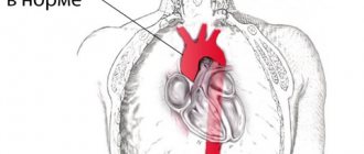
The aorta is the largest and, without exaggeration, the main vessel of the human body, which, delivering blood to all internal organs without exception, experiences a huge load, and the volume of blood flowing through the aorta every minute is quite large. Working continuously and with great stress, the aorta is subject to various kinds of pathological changes, but the real problem of recent decades has been atherosclerosis, which selects the aorta as the main target for damage.
When talking about aortic rupture, we usually mean a violation of the integrity of the vessel against the background of an aneurysm, the cause of which is atherosclerosis, degenerative changes, and inflammatory processes. In this regard, the greatest attention will be paid to the problem of a ruptured aneurysm of this vessel.
Aortic rupture is a severe vascular pathology, one of the ten most life-threatening conditions. The mortality rate from aortic rupture reaches 90%, and even timely surgical intervention is not always successful.
Causes
The exact causes of thoracic aortic aneurysm are unknown, but factors that contribute to its development include:
- Marfan syndrome is a genetic disorder that affects connective tissue in the body. People born with Marfan syndrome are especially at risk of developing a thoracic aortic aneurysm. People with Marfan syndrome have weakened aortic walls, which makes them more susceptible to developing an aneurysm. People with this syndrome have specific physical characteristics: tall height, very long arms, chest deformities, and vision problems.
- Other connective tissue diseases. Ehlers-Danlos syndrome contributes to the development of thoracic aortic aneurysm. In Ehlers-Danlos syndrome, the skin, joints and connective tissue become very fragile and the skin stretches easily.
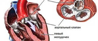
- Heart valve damage. Sometimes people with damage to the heart's aortic valve (the valve through which blood flows out of the heart) are at increased risk of developing a thoracic aortic aneurysm. This is especially true for people with bicuspid aortic valve, where the aortic valve has only two leaflets instead of three.
- Previous injuries to the aorta. If there has been previous damage to the aorta, such as a tear in the aortic wall (aortic dissection), the risk of developing a thoracic aortic aneurysm increases dramatically.
- Traumatic injury. Thoracic aortic aneurysms develop after injuries from falls or road traffic accidents.
Structure of blood vessels
Before determining why an aneurysm occurs, you need to clearly understand what the aorta is and what its structure is. This vessel originates from the left ventricle of the heart and is divided into several sections, namely:
- ascending;
- aortic arch;
- left subclavian;
- right brachiocephalic trunk.
It then passes into the abdominal cavity through the aortic opening in the diaphragm so that its contraction does not interfere with blood flow. The walls of the aorta consist of 3 layers. The inner layer consists of flat cells that fit tightly. It is perfectly smooth and ensures the movement of blood in the vessels.
The middle layer is represented by smooth muscle fibers, which contract together with the heart muscle, creating an additional impulse to move blood through the vessels. The outer layer is fibrous tissue that maintains the shape of the vessel and guarantees its normal location. Having determined what the aorta is, you need to understand exactly why it ruptures and what the provoking factors may be.
Risk factors
Risk factors for developing thoracic aortic aneurysm include:
- Age. Pathologies most often occur in people aged 60 years and older.
- Tobacco use. The longer you smoke or chew tobacco, the greater the risk.
- High blood pressure: Hypertension damages blood vessels in the body, increasing the risk of developing an aneurysm.
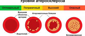
- Plaque deposition in the arteries (aortic atherosclerosis). Atherosclerosis damages the mucous membrane of the blood vessel, increasing the risk of developing aneurysmal dilatation.
- Male gender. Men are more likely than women to develop aortic aneurysms. However, women with aneurysms are at higher risk of rupture than men.
- Race. Aortic aneurysms are more common in white people than in other races.
- Family history. People with a family history of aortic aneurysm are at increased risk of developing one. In people with a family history of aneurysms, they develop at a younger age and are more likely to rupture.
Signs and symptoms of a ruptured aneurysm:
- Sudden, intense, and persistent pain in the abdomen, chest, or back
- Pain that radiates to the back or legs
- Sweating
- Sticky sweat
- Dizziness
- Low blood pressure
- Increased heart rate
- Loss of consciousness
- Dyspnea
- Weakness or paralysis on one side of the body, difficulty speaking, or other signs of a stroke.
Emergency cardiology deals with the treatment of such conditions.
Another complication of aortic aneurysms is the risk of blood clots. Small blood clots can develop in the area of the aortic aneurysm. If a blood clot breaks away from the inside wall of the aneurysm and blocks a blood vessel in another part of the body. This causes pain or blocks the flow of blood to organs and tissues, causing ischemia of the lower extremities, brain, and abdominal organs.
Possible complications
The main complication of the pathology is considered to be rupture of the vessel wall, which leads to life-threatening bleeding. Signs of tissue rupture include:
- sudden sharp pain in the chest area,
- irritation in the leg or back,
- dizziness and weakness,
- lowering blood pressure,
- loss of consciousness and rapid heartbeat,
- partial paralysis
In addition, thrombosis can be a complication of an aneurysm: thrombotic masses can form in the aorta area, which risk breaking off at any time.
Preparing for a doctor's appointment
If you suspect you have a thoracic aortic aneurysm or are concerned about your risk of developing an aneurysm due to your family history, make an appointment with your doctor at a vascular clinic. If an aneurysm is detected early, treatment may be easier and more effective.
Because most thoracic aortic aneurysms are discovered during a routine medical examination or during an examination for another reason, there is no need to make special preparations for your visit to the doctor. If you are being evaluated for an aortic aneurysm, your doctor will likely ask if anyone in your family has had an aortic aneurysm, so be prepared to answer this question. Be prepared to discuss your diet, physical activity and bad habits. If you are not following a diet or exercise routine, be prepared to talk to your doctor about problems you may encounter when starting treatment. Be sure to tell your doctor if you smoke or have quit smoking.
Treatment options
The main treatment method for a patient with this symptom is observation. The treating specialist may recommend a wait-and-see approach; there is no need to be afraid of this. Only with constant enlargement does a thoracic aortic aneurysm run the risk of suddenly rupturing. Regular examination must be carried out at least twice a year. It is important to monitor your blood cholesterol levels and blood pressure, take appropriate medications if recommended, and give up bad habits.
In cases where the size of the aneurysm is constantly growing, surgical intervention is recommended. Planned intervention is much safer than emergency intervention, which is performed when a vessel ruptures. That is why it is important to observe a dangerous pathology and follow all doctor’s recommendations. You can consult with a cardiologist and make an appointment by calling our clinic or using the form on the website.
FIND OUT PRICES
Diagnostic methods
Thoracic aortic aneurysms are often detected during routine medical examinations: chest x-rays, or ultrasounds of the heart or abdominal organs, which are prescribed for other reasons. If your doctor suspects that you have an aortic aneurysm, this can be confirmed by special tests at the heart center. These include:
- X-ray of the chest organs. Your doctor may first notice that you have a thoracic aortic aneurysm when examining chest X-rays. Your doctor may order a chest x-ray as the first test to detect damage to the upper aorta, or may detect that you have a thoracic aortic aneurysm from x-rays taken for another reason.
- Echocardiography. Thoracic aortic aneurysm can be diagnosed by echocardiography, and this method is often used to evaluate family members of patients with aortic aneurysm. Echocardiography is performed when evaluating for an aneurysm in people with Marfan syndrome. This uses sound waves to produce images of the heart in motion in real time. Echocardiography is an informative way to check the blood vessels of the heart; it shows how the chambers and valves of the heart work. In some cases, to get a better look at the aorta, your doctor may recommend a transesophageal echocardiogram, which uses a device inserted into your esophagus to create sound waves inside your body.
- Computed tomography (CT). This painless test allows your doctor to get a clear image of your aorta. During a CT scan, you will lie on a table inside a barbell-shaped device called a gantry. Detectors inside the gantry measure the radiation that passes through your body and convert it into electrical signals. The only disadvantage of using CT in detecting and monitoring aortic aneurysms is radiation exposure, especially in patients who require a large number of repeat examinations, such as those with Marfan syndrome.
- Magnetic resonance angiography (MRA). MRA is another painless imaging modality. Most MRA devices contain a large magnet that is shaped like a steering wheel or tunnel. For this test, you will be asked to lie on a table that slides into a tunnel. The magnet produces signals that vary depending on the type of tissue being magnetically scanned. Your doctor can use these images to help determine if you have an aneurysm.
Replacing parts
Conservative treatment is used only in case of serious contraindications to surgical intervention (old age, presence of concomitant systemic diseases, etc.). The most commonly used surgical methods.
Implantation of a synthetic prosthesis. The artificial vessel has good biocompatibility and can exist in the body for an unlimited time. Carrying out this operation requires the surgeon to have the highest professionalism and mastery of technique. It is most difficult to operate on thoracic and thoraco-abdominal aneurysms, since these rather lengthy and complex surgical procedures are carried out under conditions of artificial circulation. Abdominal surgery is performed for all types of aneurysm, but a less traumatic method of treatment can be used on the abdominal aorta.
Stenting method. By puncture of the femoral artery, a metal stent covered with a special fabric is inserted into the aorta. Opening in the aorta, it strengthens the walls of the vessel and protects it from rupture. After surgery, the patient must maintain normal blood pressure at all times and avoid excessive strain.
Medical observation
For small aneurysms, your doctor may recommend medical monitoring, which includes regular testing to make sure the aneurysm is not getting larger, as well as treatment for underlying conditions that may be contributing to the aneurysm's formation or enlargement.
Your doctor will give you regular tests to determine the size of the aneurysm. Once an aneurysm is diagnosed, you will have an echocardiogram in 6 months, as well as follow-up imaging tests.
If you have high blood pressure or have plaque in your arteries, it is likely that your doctor will prescribe medications to lower your blood pressure and reduce your risk of developing aneurysm complications. Such drugs include:
- Beta blockers. Beta blockers lower blood pressure by slowing your heart rate.
- Angiotensin II receptor blockers. Your doctor may also prescribe these drugs if beta blockers do not lower your blood pressure enough. These medications are recommended for people with Marfan syndrome, even if they do not have high blood pressure.
- Statins. These drugs for the treatment of atherosclerosis of the aorta can lower cholesterol levels, leading to a decrease in plaque deposits in the arteries and a reduced risk of developing aneurysm complications.
If you smoke or chew tobacco, it is important that you quit this bad habit. Tobacco use can worsen the aneurysm.
Aortic rupture: causes, symptoms, prognosis
Aortic rupture is accompanied by a violation of the integrity of the largest artery of the human body and the rapid release of large volumes of blood. This vascular accident is rightfully ranked among the ten most dangerous life-threatening conditions, and it ends in death in 90% of cases. In case of aortic rupture, even a timely operation does not always end successfully, and the probability of the patient’s death remains extremely high.
Observations by cardiologists show that in recent years, the frequency of rupture of this largest vessel has begun to occur several times more often, and saving the patient in these cases is possible much less often than with myocardial infarction, strokes and other vascular pathologies. In most cases, the vascular catastrophe discussed in this article occurs against the background of atherosclerosis, inflammatory or degenerative lesions of the vessel.
Why does aorta rupture? What pathologies and diseases contribute to its rupture? What external factors can affect the likelihood of this pathology? What are the symptoms of this vascular catastrophe? How to identify a threat of aortic rupture? What treatment methods are used for its rupture? What is the prognosis for patient survival after aortic rupture?
The most common causes of aortic rupture are arterial hypertension and atherosclerosis. As a rule, these pathologies lead to the formation of an aortic aneurysm, which, as it progresses, leads to thinning of the vascular wall and its dissection. With severe anxiety and surges in blood pressure caused by other factors (for example, overeating, high ambient temperature, etc.), the artery can rupture, and a violation of its integrity leads to massive blood loss.
Interesting
According to a study by American scientists, residents of the countries of Eastern Europe and the former USSR began to suffer from obesity after the economies of their countries switched to market economy. At the same time, there are now noticeably fewer obese people in Western Europe. This problem most affected Hungary (19%), Russia and Lithuania (18% each).
The main reasons for gaining extra pounds were:
>> psychological stress associated with reforms;
>> change in working hours (the number of people engaged in office work and computer work has sharply increased);
>> expansion of fast food chains;
>> the emergence of numerous stores selling cheap packaged foods (chips, cookies) that are convenient to eat on the go;
>> mass motorization of the population.
Unfortunately, every year it becomes more and more difficult for a city dweller to follow these seemingly simple rules. The air cannot be called fresh, and the food is not so rich in vitamins. In such conditions, the body is constantly in a state of stress.
Main symptoms
If we consider the symptoms, then we should pay attention to the signs of dissection of the vessel walls, which can lead to rupture of the aorta; the reasons for this result can be both congenital and acquired.
Any pain symptoms in the heart area, changes in blood pressure may be symptoms of a dangerous disease
Symptoms of vessel wall delamination include:
- presence of pain. This sign has a pronounced characteristic. In the initial stages of the disease, pain may appear in the chest area or in the shoulder blades; it is often acute. In later stages, pain may migrate to the head and neck area;
- periodic changes in blood pressure readings. In this case, the pressure can decrease and increase;
- dyspnea;
- swelling;
- vomit;
- loss of consciousness, etc.
If we consider in more detail the surgical solution to the issue, then an endoprosthesis is used here. As a rule, such an operation is recommended if the aneurysm is growing at an accelerated pace. It is also worth noting that an increase in the vessel in men to 5.5 cm also requires surgical intervention; as for women, this figure should be less.
Bases and types
Since the aorta is naturally endowed with a strong wall that can withstand high blood pressure and rapid blood supply, spontaneous rupture of integrity does not occur.
Aortic rupture: the causes are not fully understood. However, this is a dangerous situation. Experts suggest a number of circumstances that are precursors to a threatening condition.
The main cause of rupture of the arterial wall is an aneurysm. Other factors become either a trigger for perforation of the vessel wall or a conducive situation for the formation of an aneurysm.
The main reason is aneurysm
When deposits appear on the walls of the artery, one of the reasons is considered to be high blood flow speed, which contributes to the formation of an aneurysm or dissection of the weakened membranes of the aorta.
All circumstances that weaken the lining of the main vessel can cause rupture.
Aortic rupture: what causes or situations leading to a section of the main artery:
- Marfan syndrome, which causes tissues to be much weaker than those of healthy people;
Marfan syndrome
- aneurysm;
- damage and strong blows to the chest area;
- surgical interventions near the heart;
- abnormalities in the aortic valve;
- overweight;
Excess weight
- fungal diseases;
- inflammation: nonspecific arthritis, fungal infections, syphilitic mesaortitis;
- physiological and age-related tissue transformations are not uncommon in men from 40 to 70 years of age;
- unhealthy diet, bad habits;
- atherosclerosis and stenosis of heart valves;
- causing high blood pressure;
- poor heredity, congenital aneurysms;
- pregnancy and childbirth.
According to its location, the aorta runs up from the heart, emerging from the left ventricle. This is the supravalvular ascending aorta. Following this, it bends and goes from the chest to the abdominal cavity. And this is the descending aorta. Shell delamination can occur in both parts.
Detachment in the ascending part of the main artery is not uncommon; this phenomenon is life-threatening and is designated in the medical literature as type A. It is type A that is extremely dangerous and often causes the death of the patient. In such situations, immediate medical attention is required, but the chances of survival are negligible.
The section of integrity in the descending aorta is called type B. If the aorta of the heart ruptures, a person can be saved; usually this pathology is not as life-threatening as type A, and is not so demanding in terms of the timing of specialized care.
According to its location, the aorta runs up from the heart, emerging from the left ventricle. This is the supravalvular ascending aorta. Following this, it bends and goes from the chest to the abdominal cavity. And this is the descending aorta. Shell delamination can occur in both parts.
Surgical interventions
Surgical treatment is recommended for aneurysm sizes of 5.6 cm or more. If you have Marfan syndrome, other connective tissue diseases, or a family history of an aortic aneurysm, your doctor may recommend surgery for even small aneurysms.
Depending on your condition and the location of the thoracic aortic aneurysm, your doctor will recommend:
- Open chest surgery. An operation to open the chest to treat a thoracic aortic aneurysm involves removing the damaged section of the aorta and replacing it with a synthetic tube (prosthesis), which is sewn in place of the removed section. This procedure requires opening the abdomen or chest, takes more than 4 hours and takes several months to fully recover.
- Endovascular intervention. Through a small incision in the groin area, a special stent graft is inserted into the area of the aortic expansion. After which the stent-graft is opened, its position is assessed and secured. The aortic stenting operation is performed in an angiographic operating room, lasts 1 – 1.5 hours and the recovery period takes approximately 1.5-2 weeks.
- Other heart surgeries. If you have a medical condition that contributes to the formation of an aneurysm, such as damage to the heart valves, your doctor will recommend additional surgeries to repair or replace damaged valves to prevent the aneurysm from getting worse.
Cardiac surgeon: how to prevent sudden death from aortic rupture
Gennady Grigorievich, please explain: why does such a disaster happen completely unexpectedly, when a person seemed to feel normal? How high is the risk of such a catastrophe?
Gennady Khubulava
: These are, perhaps, the key words: “I seemed to feel fine,” but in fact, such a catastrophe is in most cases the end of a chronic disease. Aortic ruptures occur as a result of dissection of the aorta changed by a pathological process or an increase in the size of an already existing chronic aneurysm, that is, pathological expansion of the aorta, which a person might not be aware of. The frequency of such ruptures reaches six cases per 100 thousand population per year. It would seem not so much, but look - 6 per 100 thousand, this is already 60 cases per million. For example, in St. Petersburg, where there are five million inhabitants, there are 300 new cases per year - this is a fairly large figure.
How is an aortic aneurysm diagnosed?
Gennady Khubulava:
If we talk about diagnosis, this is most often an accidental finding during examination. That is why doctors insist on regular medical examinations. But it is important to know that if you have any unpleasant sensation in the chest, you need to contact your cardiologist. There are cases of congenital pathology - weakness of connective tissue - the so-called Marfan syndrome. In such patients, aneurysm may occur at a young age, and such patients must be closely monitored.
But, of course, regular examinations are important for all people. Especially if there are risk factors - the person suffers from atherosclerosis, hypertension. Another risk factor is smoking. Well, and of course, old age. The aorta ages just like the rest of the body. This is essentially a tube through which a very large amount of blood passes - about 200 million liters per year - and over time it begins to wear out. Therefore, older patients, especially those suffering from hypertension, atherosclerosis, and coronary heart disease, need more thorough, regular examination and monitoring to prevent acute situations. Medical technologies, and in cardiology, of course, too, are constantly developing and improving. Thus, one of the most important areas in the scientific research of the departments at the S.M. Kirov Military Medical Academy and the I.P. Pavlov First St. Petersburg State Medical University, which I head, we study the strength of the aortic wall, the probabilities of predicting aortic ruptures , we are developing models for organizing the provision of care to patients in acute situations. Modeling these complex processes is impossible without the use of modern information technologies, and in this we work closely with the recently opened Military Innovation Technopolis “Era”. Scientific research put into practice will help prevent many acute vascular accidents.
But you need to understand that maintaining your own health largely depends on the person himself. Patients must take responsibility for their health.
What should you do for this?
Gennady Khubulava:
The simplest thing is that we recommend having a chest x-ray every year. It seems to be the most ordinary, routine study, and, as is often said, not very informative. But, nevertheless, on the fluorogram you can see an increase in the contours of the aorta, and its calcification, and, for example, calcification of the heart valves. And conduct a more in-depth study on such a patient. Echocardiography is usually performed - ultrasound examination of the heart and blood vessels. The third stage, if you need to clarify the diagnosis, is a computed tomography scan. But first of all, we must remember that preventing a disaster is easier than dealing with its consequences.
How is an aortic aneurysm treated?
Gennady Khubulava:
If the increase in the size of the aorta reaches a certain degree, elective surgery is indicated. With a planned operation, the results are much better, since the patient is more thoroughly examined, the operation and everything necessary for it are prepared in advance.
In acute cases - when delamination or rupture occurs unexpectedly - this is a very difficult situation and a big problem. But even here, in many cases, if assistance is provided in a timely manner, there is a chance of saving the patient.
A program for the treatment of acute aortic syndrome has been operating in St. Petersburg for three years now. This is the first program in our country to implement emergency surgical care for patients with acute lesions of the thoracic aorta. During this time, we organized the logistics of providing emergency care to such patients, developed and implemented new organizational and clinical approaches. Both practical doctors and managers and organizers of medical care in our city have worked hard. The results over the three years of work have been improving. But, of course, the most important thing is not to lead to such acute conditions. If the diagnosis is established, surgical treatment is performed as planned. There are many types of operations, but the essence is that the aorta is replaced with various vascular prostheses or stent grafts (special vascular prostheses) are installed endovascularly.