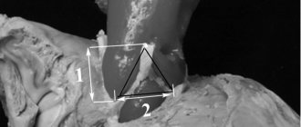
A popliteal artery aneurysm is a pathological expansion of the main artery running from the lower third of the thigh to the upper third of the leg. It is located quite deep in the leg under the knee. The popliteal artery is a continuation of the superficial femoral artery and below the knee it divides into the anterior, posterior tibial arteries and peroneal artery. These arteries supply blood to the leg and foot, so blocking blood flow in the popliteal artery leads to severe circulatory failure in the leg below the knee. The normal diameter of the vessel is about 6-10 mm.
Popliteal aneurysm is a risk factor for sudden acute limb ischemia and subsequent amputation. Unoperated aneurysms lead to leg amputation in 50% of all cases within 3 years.
Popliteal artery aneurysm must be operated on as soon as possible after diagnosis. There is no need to hope that it will “resolve” on its own. The high risk of acute ischemia and good results of planned operations should encourage the patient to consent to surgery. The results of planned interventions are very good.
Treatment technologies at the Innovative Vascular Center
The vascular surgeons of our clinic have significant experience in diagnosing and treating both planned and complicated lesions of the popliteal artery. The main method of treatment in our clinic is autovenous replacement of the popliteal aneurysm. This technology shows better immediate and long-term results. For complicated aneurysms, open surgery allows you to restore the patency of not only the popliteal artery, but also the vessels of the leg. Endovascular interventions for extensions of this localization have very poor results due to the high mobility of the knee joint.
Treatment
If there is a risk of aneurysm rupture or blood clot formation , treatment is carried out only surgically.
There are two methods:
- open access to the aneurysm;
- endovascular surgery.
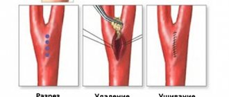
In the first case, the aneurysm wall is excised, followed by shunting. During endovascular intervention, a stent is installed into the lumen of the artery using a catheter.
If the aneurysm is asymptomatic, with a slight increase in size, the doctor prescribes regular monitoring of its condition using ultrasound.
Causes
Popliteal artery aneurysms account for about 1% of all surgical vascular diseases and often occur in both legs. The main reason is the congenital weakness of the artery wall, which contributes to their pathological expansion. The majority of patients (95%) are elderly men with an average age of approximately 71 years. The exact reasons for the development of dilation in the popliteal artery are unknown, but there is a clear connection with atherosclerotic changes in the vessel wall. Sometimes the pathology develops as a result of injuries to the popliteal region, dislocations or fractures. Patients with multiple aneurysms in different arteries should have general tissue weakness. The exact nature of this is still unclear. The tendency of the popliteal artery to pathological expansion is associated with frequent flexion and extension of the vessel due to movements in the knee joint.
Aneurysm of the lower extremities
28.09.2018
Among the structural pathological changes in our blood vessels , I would like to highlight a disease such as aneurysm . At its core, an aneurysm is a significant enlargement of a vessel in a separate area. Quite often, this pathology affects the vessels of our legs . The femoral and popliteal arteries . It is worth noting that the initial stages of aneurysm are practically asymptomatic, which subsequently negatively affects the treatment of this disease. Any disease responds well to treatment in the early stages of its development, aneurysm is no exception. During its development, accompanied by expansion of the affected area of the vessel , femoral artery aneurysm is manifested by swelling in the groin area, cramps in the legs during physical activity, the appearance of ulcers on the toes , and numbness in certain areas of the legs . If left untreated, the symptoms become pronounced and intensify. Soft tissue necrosis is a common occurrence.
It is worth noting that the manifestation of symptoms is accompanied by pain of varying degrees of severity, sometimes pain is observed even at rest. Severe forms of the disease lead to the development of such irreversible complications as gangrene. It is hardly worth explaining that in this case there is a high risk of death.
The development of popliteal artery aneurysm is often caused by trauma. For most professional athletes, this is a fairly common disease. There is also a false form of the disease, the causes of which are bruises, fractures and unsuccessful surgical interventions. It is worth noting that a false aneurysm of the popliteal artery may be a consequence of a previous infectious disease.
Symptoms of the development of a popliteal artery aneurysm are manifested:
- pain in the knee joint;
- a change in the shade of the skin, the skin becomes bluish in color;
- the temperature of the lower extremities differs significantly from the general body temperature, or, more simply, the legs become cold.
Aneurysm of the popliteal artery can lead to such serious complications as paralysis and complete immobilization of the limb. The most optimal treatment option is surgery , which can stop the progressive disease and prevent irreversible consequences. Among the most severe and often irreversible complications of aneurysms of the femoral and popliteal arteries , it is worth highlighting the development of gangrene as a result of a systematic lack of blood supply; rupture of an artery in the affected area, accompanied by heavy bleeding and hemorrhagic shock; the development of thrombosis arising as a result of atherosclerotic changes in the vessel .
Modern technologies make it possible to carry out treatment using a minimally invasive surgical . The surgeon makes a small incision through which the stent is installed. The rehabilitation period after such an operation is considered short. vascular prosthetics began to be used . Its essence lies in excision of the affected area of the vessel and replacing it with a prosthesis. After the operation, normal blood circulation is restored and the danger of artery . Surgery to remove false aneurysm is fundamentally no different from surgery for a true aneurysm .
Published in Cardiology Premium Clinic
Complaints and symptoms
Patients with an aneurysm complain of a feeling of heaviness in the popliteal region, swelling of the foot of the affected limb, and sometimes shooting pains. Most often, such complaints are vague, and the patient may not be aware that he has such a dangerous disease.
With aneurysm thrombosis, a clinical picture of acute ischemia develops: severe pain in the affected limb, changes in color and skin temperature of the foot. Subsequently, disturbances in sensitivity and movement develop. With advanced acute ischemia, rigor of the leg and foot develops; active and passive movements are impossible due to muscle death.
Popliteal artery aneurysm - dangers and effective treatment
Questions and answers
atherosclerosis of the arteries of the lower extremities KHAN stage 2 Good afternoon. Vladimir Grigorievich Kirillov, born in 1954, writes to YOU.
I am disabled group 2 in oncology due to a general disease. Three years ago I began to notice such a phenomenon as... Answer:
All recommendations are correct and should help you when implemented.
Bypass surgery of the lower limb artery
Hello. Please tell me whether repeated operations are performed to bypass the artery in the leg.
Answer:
Of course, if there is a need and conditions for this.
Necrosis of 4 fingers
Hello. My grandfather has necrosis of 4 fingers and swelling of his feet. The photographs show vessels up to the knee. The finger was not removed, explaining that it would go even faster. If you delete it, they said you have to...
Answer:
I advise you to contact competent vascular surgeons dealing with the problem of critical ischemia.
gangrene
where and when can you operate on your leg?? we agree to come from Armenia.. the doctor advised us to contact you.. then I am sending the doctor’s epicrisis
Answer:
Send examination data and photographs of your leg by email.
Celiac stenosis
Good afternoon! After a CT scan with contrast, I was diagnosed with \"Celiac axis stenosis 70%\" and was told that surgery was needed. How serious is this and if surgery is necessary, how urgently?…
Answer:
Any operation has risks.
But stenting the celiac trunk is not very dangerous. An in-person consultation is needed to accurately determine the indications for surgery. Possibly gongrene
I injured my hand, the skin was torn off while drinking on gravel and my hand became swollen in two hours, what should I do?
Answer:
See a surgeon for an appointment.
Hypoplasia of the left vertebral artery
Hello. I have been sick for a long time. Nausea. vomiting. Impaired coordination. unsteadiness. Severe headaches. What examination should I do before someone turns to you for help?
Answer:
Do MSCT of the arteries of the neck and brain with contrast.
Thromboangiitis obliterans
Hello. My mother is 58 years old, disabled group 1. She had both legs amputated below the knees due to thromboangiitis obliterans. We underwent an ultrasound examination of the NK vessels. What additional research needs to be done...
Answer:
What about the stumps now?
Leg amputated
Hello! My brother had his leg amputated in December 2021 due to atherosclerosis, after which he developed gangrene. In January we underwent re-amputation. It is now March, but the wound is not healing. They did a CT scan of the vessels...
Answer:
What to do?
Restore blood flow. Send a link to MSCT of vessels. Gangrene
Hello, my dad had gangrene on his right leg on the big toe, his toe was amputated, the treatment prescribed by the doctor did not help, there was pain, a large crust and there was pus, they applied ointments...
Answer:
It is urgent to perform an ultrasound of the arteries of the extremities and MS CT with contraception; after receiving the examination results, we will be able to offer you the optimal treatment method.
Course and complications
The main risk from a popliteal aneurysm is associated with embolization - blockage of the underlying arteries with pieces of blood clots or occlusion of the aneurysm cavity. Both of these complications can lead to acute ischemia and gangrene of the legs (sudden loss of blood supply). Blood clots (thrombi) gradually form in the cavity of the vessel. When this clot remains attached to the wall of the vessel, it does not pose any danger. If a clot fragment breaks off, it can travel far from the aneurysm and cause blockages in small arteries, preventing blood flow to downstream tissues. A popliteal aneurysm can rupture (rupture), but this is much less common than embolization. In this case, a pulsating hematoma occurs behind the knee. Simultaneously with the rupture, the next stage is thrombosis of the popliteal artery with the development of signs of acute circulatory failure of the limb. Most people develop severe ischemic changes and death of the leg. Only surgery performed within the next 6-12 hours after the complication will help avoid amputation.
Complications
Complications of DVT may include:
- Pulmonary embolism (PE). PE is a potentially life-threatening complication associated with DVT. This occurs when a blood vessel in your lung is blocked by a blood clot that travels to the lung from another part of your body, usually your leg. If you have signs and symptoms of PE, it is important to seek medical help immediately. Sudden shortness of breath, chest pain when inhaling or coughing, rapid breathing, rapid pulse, feeling weak or faint, and coughing up blood may occur with PE.
- Postphlebitic syndrome. Damage to the veins by a blood clot reduces blood flow to the affected areas, causing leg pain and swelling, skin discoloration, and skin ulcers.
- Complications of treatment. Complications may arise from blood thinners used to treat DVT. Bleeding is a side effect of anticoagulants. It is important to have regular blood tests when taking these medications.
Classification and course of the disease
In addition to the classification of aneurysms depending on their location, there are others presented in the table below.
| Because of |
|
| By shape |
|
| By structure |
|
Aneurysms do not have a clear stage; the progression of the disease is judged by the size of the aneurysmal expansion, which varies depending on the location. For example, in the case of an abdominal aortic aneurysm, an expansion diameter of up to 6 cm is considered small, while in the case of aneurysms of the cerebral arteries, a size of more than 2.5 cm is already considered gigantic.
Based on the size, doctors predict the risk of complications and decide on surgical intervention.
Diagnostics
Source: MART PRODUCTION: Pexels
Asymptomatic course and imitation of the clinical picture of other diseases can hide the fact of the presence of an aneurysm until complications develop. Often an aneurysm becomes an incidental diagnostic finding. It is easiest to diagnose a peripheral artery aneurysm symptomatically, since its symptoms are quite obvious.
The doctor can detect large aneurysms of the aorta and peripheral arteries during examination using palpation (feeling) and auscultation (listening). Damage to the arteries of the brain can manifest itself as symptoms of transient ischemic attack, sudden headaches, eye pain, fainting and other symptoms.
A cardiac aneurysm can manifest itself as symptoms of heart failure: shortness of breath, swelling, bluishness of the skin.
The following instrumental diagnostic methods are the most informative for aneurysmal vascular lesions:
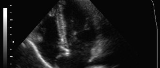
- Ultrasound is a fairly universal technique used to diagnose aneurysms of various locations (except for cerebral vessels in adults). There are several options for ultrasound, used depending on the part of the body being examined and the pathology being sought.
- X-ray examination - allows you to identify aortic aneurysms, as well as the displacement of neighboring organs caused by them.
- CT and MRI are the most accurate diagnostic methods and allow you to visualize aneurysms of any location and size, determine the presence of thrombotic masses in them, and assess the condition of the walls.
- Angiography is a highly informative method that can be used in the diagnosis of any aneurysms. Unlike the above, this procedure is invasive, as it is necessary to deliver a contrast agent through a catheter into the vessel of interest. It is difficult to perform, and therefore is used to clarify cases of questionable diagnosis and controversial indications for surgical intervention.
Symptoms of cardiac aneurysms
The cause of the development of cardiac aneurysms is most often the thinning of the muscle wall due to a heart attack. Having a heart attack weakens the tissue, which often causes the damaged area to bulge. The left ventricle of the heart and the interventricular septum are most susceptible to such pathologies. Symptoms in this case can manifest themselves in the form of pressing pain in the heart area, tingling sensations, and shortness of breath. Heart rhythm disturbances and signs of chronic coronary insufficiency may also be observed. Rupture of an acute aneurysm most often leads to death. In this case, the patient experiences severe pallor, loss of consciousness, coldness of the extremities and hoarse breathing. You can learn about treatment methods for cardiac aneurysms here.
Diagnosis of deep venous thrombosis
Diagnosis of thrombosis is extremely difficult. This is primarily due to the absence of clinical symptoms. According to some data, out of 1000 venous thromboses, only 100 have any clinical manifestations. Of these, 60 patients will develop PE, but only 10 will have clinical signs. It should be recognized that today there is not a single clinical symptom, laboratory or instrumental sign that would indicate with absolute certainty the presence of PE and DVT. Clinical manifestations of thrombosis and ultrasound results can be the basis for the correct diagnosis of venous thrombosis. The clinical picture of deep vein thrombosis consists of a complex of symptoms that characterize a sudden disturbance of venous outflow with preserved arterial blood flow to the limb. Swelling, cyanosis of the limb, bursting pain, local increase in skin temperature, overflow of the saphenous veins, pain along the vascular bundle are characteristic to one degree or another for thrombosis of any localization. Movements in the joints of the limb and sensitivity remain virtually unchanged. General signs, such as low-grade fever, weakness, adynamia, and slight leukocytosis, occur in most patients. The diagnosis of thrombosis largely depends on the location of the lesion, i.e. on the level of distribution of thrombotic masses.
What is an aneurysm?
An aneurysm is a protrusion of the wall of an artery (less commonly, a vein) or heart due to its thinning, stretching or damage.
As a result, a so-called aneurysmal sac appears, which can compress nearby tissue. An aneurysm may be congenital. Moreover, when a child is born, this defect is invisible, and the baby develops completely normally. Aneurysms are also caused by diseases that affect blood vessels: hypertension, atherosclerosis, syphilis (at a late stage), sepsis. The risk of developing this disease increases when a blood vessel is injured or injured and when infected blood clots form. You can live with an aneurysm for years, go about your daily activities, and not experience any symptoms. Meanwhile, the aneurysm can quietly increase in size, threatening to explode at any moment - like a time bomb.