Cardiomegaly or enlarged heart?
Every year, hundreds of thousands of citizens die from cardiovascular pathologies around the world. In most cases, the reason for this is untimely consultation with a doctor and deterioration of cardiac function.
Enlargement of the organ is associated with the development of ventricular hypertrophy, accumulation of metabolic products and neoplastic processes. Cardiomegaly often occurs in healthy people, this includes athletes and pregnant women.
The volume of the heart varies within different limits for each person. If we talk about gender differences, then in men this organ is larger than in women. So for the age category from 20 to 30 years, the approximate heart volume will be the following values:
- women – 580 cm3;
- men - 760 cm3.
This figure also depends on body weight. A diagnosis of cardiomegaly should be made only after a thorough examination, because in some cases a slightly enlarged heart is the norm, which is strictly individual for each person.
Dilatation of the right or left ventricle: causes
Enlargement of the walls of the right or left ventricle is called hypertrophy. In this case, the functioning of the myocardium is disrupted and, as a result, their functional activity worsens. Depending on the location of the depletion of the heart muscle, different etiologies are distinguished.
Right ventricular hypertrophy
Enlargement of the walls of the right ventricle is most often observed in children with congenital defects of intrauterine development. Also, one of the main reasons is associated with an increase in pressure in the pulmonary circulation and the discharge of blood into the right ventricle. In this case, the load on the right ventricle increases.
In adults, the cause of right ventricular hypertrophy is often diseases that interfere with normal breathing. These include the following pathologies:
- rachiocampsis;
- pulmonary vascular diseases (compression, embolism, thrombosis, etc.);
- bronchial asthma;
- tuberculosis;
- bronchiectasis;
- Chronical bronchitis;
- polio, etc.
Left ventricular hypertrophy
Left ventricular hypertrophy is dangerous due to sudden cardiac arrest, causing myocardial infarction and death. Thickening of the walls of the left ventricle can result from the following cardiac pathologies:
- developing atherosclerosis of the aorta;
- hypertonic disease;
- congenital or acquired heart defects;
- obesity.
To prevent the development of such serious diseases, you need to follow preventive measures, which means sticking to a healthy lifestyle and seeing a doctor in order to diagnose all disorders in a timely manner.
Myocarditis in the daily practice of a doctor (part 2)
00:00
Vladimir Trofimovich Ivashkin , academician of the Russian Academy of Medical Sciences, Doctor of Medical Sciences:
– Next – infectious diseases, since we needed to exclude infectious myocarditis. You see that indicators of viral infections (chlamydia, ureaplasma, trichomonas, mycoplasma, herpes simplex viruses, brucella, and so on) were practically negative. The same applies to cytomegalovirus. Blood was taken repeatedly for bacterial culture (culture) - and not once was a positive culture detected. Consequently, we also rejected the infectious nature of heart pathology.
This possibility of excessive sensitivity to certain drugs is sometimes observed in patients with certain genetic polymorphisms of regulatory mechanisms. In this regard, we conducted genetic studies and found that our patient was heterozygous for angiotensin-converting enzyme. She also turned out to be heterozygous for the first type receptor, angiotensin II.
But despite being heterozygous, the presence of angiotensin-converting enzyme levels was essentially unaffected. It turned out to be within normal limits. Therefore, this heterozygosity for angiotensin-converting enzyme and angiotensin type I receptor for angiotensin II essentially remains outside the brackets. We cannot, in one form or another, link these data to the clinical observations that we have in relation to this patient. Perhaps in the future we will be able to answer this question.
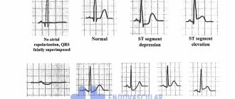
Here is an electrocardiogram from 2010. We do not see electrical alternation here. But what attracts attention is a very pronounced increase in the electrical activity of the left and right parts of the heart. Right leg block. But, what is important: after such a long time, the ST segment elevation in precordial leads V3, V4, V5 remains. This ST segment elevation forces us to continue to pursue our patient's differential diagnosis of what is causing her cardiac decompensation to be so severe.
The left picture shows a chest x-ray from 2008. On the right is an x-ray from 2010. You see some differences. The fluid content in the pleural cavities decreased. The boundaries of the aperture have dropped somewhat. Root stagnation has decreased. However, the heart remains large in diameter. The silhouette of the pulmonary artery and right atrium bulges. Consequently, all radiological signs of congestive heart failure remain. Kerley lines 2 are clearly visible. Our patient has signs of congestive interstitial edema.
04:19
A computed tomography scan of the chest (shown by arrows) shows a fairly significant protrusion in the pleural cavities and large chambers of the heart. Here, using the left atrium as an example, the large left atrium.
During computed tomography of the chest, the patient is positioned on her stomach. She could not lie on her back because she was choking with a cough and did not have enough air. We see signs confirming the presence of interstitial edema, thickening of the interlobular peribronchivascular interstitium. There is no evidence of infiltration of lung tissue.
On an echocardiogram in October 2010, we see signs of enlargement of the chambers of the heart, ventricles, and atria. Left ventricular ejection fraction – 22%. Systolic pressure in the pulmonary artery is 65 millimeters Hg. Art. Experts note an increase in the degree of dilatation of the left atrium, pulmonary hypertension and pressure in the right atrium.
Ultrasound examination of the abdominal cavity confirms signs of right heart decompensation. The portal vein is slightly dilated. And the inferior vena cava is expanded to 32 millimeters. The hepatic veins are dilated to 14 millimeters. No free fluid is detected in the abdominal cavity. Consequently, the presence of right heart decompensation is confirmed.
(Slide show).
In order to finally resolve the issue of the nature of the changes in the myocardium that we encountered, a series of studies were carried out. In particular, the patient underwent perfusion tomoscintigraphy. You see the results of the examination of the apex and anterior wall.
Experts note a massive zone of hypoaccumulation of the indicator, indicating a perfusion defect in the anteroseptal-posterior localization. A conclusion is made about the presence of a sign of large-focal myocardial damage. Essentially, these changes coincide with those changes in the electrocardiogram that we saw in previous electrocardiographic pictures.
07:08
The patient underwent multislice computed tomographic angiography. She put us in a difficult position because it was noted that there was no contrast enhancement in the distal segment of the right coronary artery. The question arose again about the possibility of myocardial infarction suffered by our patient. This required either direct confirmation or denial of this possibility.
(Slide show).
Here is a continuation of this research. In the area of the apex there is a suspicion of the presence of a blood clot. Here we have projected on a diagram the data that we obtained using electrocardiography, echocardiography, scintigraphy and multislice computed tomography. It turns out that the apex, septum and lateral wall of the left ventricle are hypoperfused, that is, damaged.
(Slide show).
The cardiac surgeon consulted our patient and recommended probing of the heart cavities and coronary angiography. The patient underwent coronary angiography. You see that the left type of blood supply to the heart is marked. The anterior interventricular branch is not changed. Noteworthy is the small number of diagonal and septal branches. The trunk of the left coronary artery is not changed. The circumflex branch is also unchanged. Normal picture of the coronary vessels.
(Slide show).
Here is the right coronary artery. You remember that during multislice computed tomography, there was a suspicion of occlusion of the right coronary artery in its distal part. When performing coronary angiography, you see that the coronary artery has smooth contours. Essentially, it is not changed along its entire length, including in the distal sections.
Consequently, we rejected the thromboischemic nature, that is, simply myocardial infarction in this patient. With transesophageal echocardiography, attention is drawn to the hyperechogenicity of the myocardium of the interventricular septum, as well as the pronounced trabecularity of the posterolateral wall of the left ventricle, which allows us to think about myocardial non-compaction syndrome with its infiltration, that is, the possibility of sarcoidosis or amyloidosis. Although, of course, the clinical picture itself does not coincide with the assumption of sarcoid granulomatous or amyloid myocardial damage.
10:17
(Slide show).
The patient underwent magnetic resonance imaging of the heart. It was noted that there is akinesis of the anterior and inferior walls of the left ventricle in the apical and middle segments, the interventricular septum, and the apical segment. Hypokinesis of the interventricular septum in the middle segment, basal segments of the anterior and inferior walls. Increased trabecularity of the apical segment of the lateral wall is noted (without criteria for myocardial non-compactness).
Consequently, we can largely (given the clinical picture) ignore amyloidosis and sarcoidosis. And the data obtained coincide with previous studies on massive myocardial damage in our patient.
Thus, no evidence of space-occupying lesions of the heart or myocardial non-compaction syndrome has been identified. The magnetic resonance image, as experts note, may correspond to myocarditis affecting the left and right ventricles.
(Slide show).
Let's return to the differential diagnosis table. So, we have to guess based on the clinical data that we have. This table, of course, does not completely cover all the symptoms of myocarditis. Only those data that were most clearly and clearly identified in our patient are included here.
The first is ST segment elevation. In what forms of myocarditis is ST segment elevation usually observed? This is either acute hypersensitive eosinophilic myocarditis, or acute (or subacute) necrotizing eosinophilic myocarditis.
What next should we pay attention to? In what forms of myocarditis can we encounter severe myocardial necrosis? In fact, with all forms of myocarditis (infectious, autoimmune, giant cell), very small areas of myocardial necrosis are possible.
But such massive myocardial necrosis, which we assume in our patient, can occur either in acute hypersensitive eosinophilic myocarditis or in acute necrotizing eosinophilic myocarditis.
But if we talk about these two forms, then in acute hypersensitive eosinophilic myocarditis the main clinical picture is rhythm disturbance. Arrhythmia of various kinds characterizes acute hypersensitive eosinophilic myocarditis, occurring with ST elevation. Whereas acute necrotizing myocarditis (or subacute necrotizing myocarditis) is characterized by massive myocardial damage with the development of congestive heart failure.
13:45
Now, if we turn to the rate of myocardial development that characterizes the development of all these adverse consequences, such a subacute course is characteristic of idiopathic giant cell myocarditis and subacute necrotizing myocarditis. In idiopathic giant cell myocarditis, the main clinical form is still severe rhythm disturbances. They determine the course and prognosis of the disease. Whereas in subacute necrotizing myocarditis we are talking about rapid development, the formation of congestive heart failure.
Therefore, based on these criteria (plus a differential diagnosis involving numerous instrumental studies), we finally settled on the possibility that our patient had subacute necrotizing eosinophilic myocarditis, secondary dilated cardiomyopathy, and pulmonary hypertension. Relative insufficiency of the mitral and tricuspid valves. Relatively minor rhythm and conduction disturbances, incomplete blockade of the right leg, and so on. In this case, in our opinion, we were talking about subacute necrotizing myocarditis.
(Slide show).
Naturally, such an unfavorable trend in the decline of contractile function forced us to consult and invite specialists from the Research Institute of Transplantology and Artificial Organs named after Academician Shumakov. We agreed with the director of the Institute, Academician Gauthier. He referred specialists, and the patient was transferred to the Transplantation Institute.
There she underwent a heart transplant and was diagnosed. Postmyocardial cardiosclerosis with dilatation of the heart cavities. Relative insufficiency of the mitral and tricuspid valves, and so on. Our ideas that she suffered from myocarditis coincided.
Unfortunately, we were unable to obtain the results of a histological study that would confirm or reject our idea of the nature of myocarditis. But the mere fact of the coincidence of our clinical diagnosis and surgical diagnosis, plus the presence of slight eosinophilia in peripheral blood and a slight increase in immunoglobulin E, still confirms us in the assumption of the presence of subacute destructive necrotizing myocarditis.
17:08
Further, after heart transplantation, the patient was naturally recommended the necessary medications, which are indicated here.
A year later she is back in our clinic. We see that she feels great. She is still so graceful - she weighs 55 kg, height - 168. Respiration rate - 17. Normal blood pressure. Some tendency to tachycardia is typical.
Here is her electrocardiogram, actually, the cardiogram of the transplanted heart. We see a completely different picture. The echocardiogram shows everything is good and favorable. The sizes of the heart chambers correspond and are not bad. They will tend to the atrium to shrink. Contractile function – ejection fraction of almost 60%.
Most often, surgeons perform transplantation for the following pathologies:
1. Coronary heart disease.
2. Dilated cardiomyopathy.
3. Acquired heart defects.
4. Congenital heart defects.
5. Heart retransplantation.
6. Other pathologies.
Our patient falls into the category of dilated cardiomyopathy as a result of severe necrotizing myocarditis. This is one of the most common forms of indication.
What specific indications for transplantation did our patient have? The drug-refractory end-stage of chronic heart failure in our patient has, of course, already formed. Left ventricular ejection fraction is less than 20%. In our patient it is 15%.
End-diastolic pressure in the left ventricle is more than 25. Our patient also had high end-diastolic pressure. Her cardiac index was less than 2 liters per minute per square meter. Consequently, her heart transplant was performed on time.
What is the forecast? Our patient has been living for a year and a half. The prognosis is good. One-year survival rate - she was included in this group (86%). Three-year – 77%, five-year – 69%. We will hope that our patient will always be in these successful groups. We wish her health and prosperity in her future life. Thank you for your attention.
20:25
Oksana Drapkina : Thank you very much, Vladimir Trofimovich. Questions are already coming in.
Question: Why was this woman not given a myocardial biopsy?
Vladimir Ivashkin : Myocardial biopsy is not an easy procedure. It is advisable to perform it when the condition of the patient allows this procedure.
Our patient experienced progressive cardiac decompensation. Therefore, the large volume of conservative diagnostic studies that have been carried out has convinced us that we are right. We are talking about myocarditis, and not about any other forms. Therefore, in agreement with the transplantologists, we referred the patient. They accepted her, and, you see, everything went well.
I hope this patient has a good life prognosis. First of all, she is a young woman. Secondly, she has no risk factors. You see, she is an elegant woman with normal blood pressure. She has an absolutely clear coronary angiogram (as you looked at), there are no associated diseases. Nothing clouds her future. Let's hope everything goes well.
Oksana Drapkina : Thank you very much, Vladimir Trofimovich. I think there will be more questions. We have finished the section “Lectures, master classes, clinical analysis”.
Causes of cardiomegaly
Most often, an enlarged heart in diameter is diagnosed in adults. Predisposing factors that contribute to the expansion of the boundaries of the shadow of the ventricles and atria are quite diverse, in most cases this is associated with cardiovascular pathologies. So, the etiology of the appearance of cardiomegaly includes the following reasons:
- excessive exercise;
- pregnancy;
- idiopathic cardiomyopathy;
- heart defects;
- anemia in severe forms;
- infectious diseases where the target organ is the heart muscle;
- complications after viral diseases;
- myocardial ischemia or infarction;
- inflammatory processes in the heart;
- severe stress loads;
- excessive alcohol consumption, drug addiction, smoking;
- kidney disease and renal failure;
- rheumatic carditis and endocarditis;
- hypertension, etc.
If an enlargement of the heart muscle is detected, the doctor prescribes the necessary diagnostics and treatment.
Clinical manifestations
When the heart expands across the diameter or in other parts, the patient may experience unpleasant symptoms. This includes the following clinical severity:
- increased fatigue;
- shortness of breath at rest or with minor physical exertion;
- increased blood pressure;
- the appearance of pain in the heart area;
- formation of edema in the lower extremities;
- headaches and dizziness;
- short-term loss of consciousness.
Other signs characteristic of a particular cardiac pathology, if present, may also be added.
During treatment, it is important to identify the focus, which means to determine the disease or disorder that triggered the occurrence of heart enlargement. As soon as this is diagnosed, treatment is prescribed aimed at eliminating this pathology.
As an auxiliary therapy, medications are prescribed, the purpose of which is to reduce the obstacle to normal blood outflow while simultaneously unloading the increased work of the ventricles. This will prevent the risk of complications such as myocardial infarction, angina, shortness of breath and arrhythmia.
If therapeutic actions are ineffective, the doctor may prescribe surgery to improve blood flow. However, they resort to it only in extreme cases.
Symptoms of the disease
Manifestations of cardiomegaly are purely individual. In some cases, symptoms do not occur at all, and a person learns about the expansion of the organ’s boundaries by chance, for example, during fluorography or a doctor’s examination. If the pathology makes itself felt, its manifestations are not much different from the symptoms of diseases of the cardiovascular system. But there are cases when the pathological process develops so rapidly that a “bull’s heart” quickly forms.
Signs include:
- Pain behind the sternum.
- Dizziness, loss of consciousness for no reason.
- Fast fatiguability.
- Shortness of breath with slight exertion.
- Arrhythmia (change in rhythm).
- The appearance of edema (especially in the lower extremities in the evening).
- Cough not associated with respiratory diseases.
Expanding the size of such an important organ of the cardiovascular system entails very dangerous consequences. If the heart rhythm is disturbed, there is a risk of cardiac arrest. The occurrence of noise should be monitored by a doctor, because it indicates a change in the structure of the valves. An enlarged heart affects the quality of the entire cardiovascular system. Over time, there will come a point when the organ cannot pump the required amount of blood, so the risk of developing heart failure increases over the years. If the problem is ignored, then you can get a serious complication - “bull heart” (an increase in the size of the organ to critical).
General recommendations
In addition to treatment recommendations, it is imperative to follow preventive measures. For heart problems, it is very useful to follow these tips:
- You should avoid drinking alcoholic beverages, which have a toxic effect on the myocardium (heart muscle).
- In order to prevent the deposition of cholesterol plaques on the walls of blood vessels, foods high in cholesterol should be excluded from the daily diet. It is advisable to consume fish, olive, flaxseed, corn and soybean oil at least 2 times a week.
- To strengthen and maintain the heart muscle in normal working condition, it is useful to include viburnum, cranberries, cabbage, eggplants, peaches, dried apricots, apples, pomegranates, walnuts, melons, etc. in the daily diet.
- It is necessary to reduce salt intake to at least 2 grams. per day, especially for patients with increased swelling.
- If obesity is recorded, it is necessary to create a proper balanced diet aimed at eliminating extra pounds.
- Sleep at least 8 hours, do not become physically and emotionally overtired.
- Walk outdoors more often.
Enlargement of the heart is not a diagnosis, but only a temporary condition of the heart muscle. With correct and timely actions, you can get rid of this disorder and significantly alleviate your condition.
Symptoms, diagnosis and treatment for cardiomegaly
Have you been struggling with HYPERTENSION for many years without success?
Head of the Institute: “You will be amazed at how easy it is to cure hypertension by taking it every day.
The heart is one of the most important organs of the human body. One of the most dangerous symptoms of diseases that affect not only the cardiovascular system, but also other organs, is an enlarged heart (cardiomegaly). This can manifest itself in different ways: in one case, the patient has symptoms characteristic of diseases of the heart muscle, in another, he lives for a long time and does not even suspect the presence of pathology.
Expansion of the heart in diameter
Causes of heart enlargement
● The weight of a man's heart weighs approximately 330 grams, for women it is slightly less - 253 grams. The increase in size of the heart in both men and women occurs either due to the expansion of its cavities, or due to the growth of the heart muscle (myocardial hypertrophy). Today there is a whole list of diseases that contribute to the expansion (or dilatation) of the heart. One of the most classic diseases is hypertension. The mechanism of heart expansion in hypertension is as follows: narrowing of the lumen of blood vessels leads to increased pressure on the heart, an additional load is placed on it to pump blood, the heart muscles become more tense, and therefore increase in size.
● Diseases that lead to enlargement of the heart include inflammation of the heart muscle - cardiopathy. This happens as a complication after untreated scarlet fever or tonsillitis. These diseases deplete the muscles of the heart, they become flabby, and the cavities of the heart (its ventricles) expand. Enlargement of the heart (dilatation) is manifested by the following clinical symptoms: edema, mainly in the legs, shortness of breath during exercise, palpitations (tachycardia), cardiac arrhythmias (arrhythmia).
Causes of an enlarged heart
The weight of an average man’s heart is 332 grams, a woman’s – 253. It is considered normal if the weight of the organ varies within these limits.
As for the sizes, they are usually correlated with a person’s fist. For an organ to function normally, it is very important that all its parts (atria, ventricles) are normal, or more precisely, the thickness of their walls, length and width as a whole.
What to do if fluorography (x-ray, ultrasound) showed that the heart is enlarged and dilated?
How dangerous is it to literally have a big heart? And as a result of what can the organ become larger? Let's figure it out in order.
The most important reasons why the heart is larger than normal in a fluorography image include:
- great physical activity;
- diseases.
In people who engage in heavy physical labor every day, as well as in professional athletes, the heart also works harder: it is forced to beat more often and pump blood faster.
This leads to the fact that there are often more heart muscle cells and they grow. As a result, the weight of the organ and its size increase.
If physical activity in the future is moderate, an enlarged heart for this reason does not pose a health risk.
If a person subjects his body to excessive stress for a long time, then it is possible to develop a pathology such as a hypertrophied heart, which is already fraught with serious complications and even life-threatening.
The reason that the heart is enlarged in size can be diseases of the cardiovascular system (coronary diseases: for example, hypertension, coronary disease) and the heart itself (viral, inflammatory diseases), as well as heart defects.
So, if there is a defect and the organ is unable to function normally in order to properly supply the entire body with blood, the organ can enlarge.
The heart is expanded in diameter
I ask you, if it is possible to provide an explanation of the medical term “the heart is expanded in diameter,” what does it mean? This is written on the results of my husband’s chest fluorography. He is 54 years old, second-degree hypertension, coronary heart disease, varicose veins in the legs, overweight - this is an incomplete list of diseases accompanying his life.
I ask you to give recommendations on how to improve his health. What is the reason for the enlargement in the heart, is it life-threatening, and if so, how to protect your husband from possible consequences?
Sincerely, Lyudmila Reutskaya
Unfortunately, everything you wrote about your husband’s health condition is very dangerous. The heart enlarges from constant stress caused by high blood pressure and excess weight. At the same time, it cannot adequately perform its function, working as a pump, and tries to cope with this by increasing muscle mass (myocardium). But at the same time, the cavities of the heart expand, the valves stretch, the hole between the ventricles and the atrium becomes larger, and so-called pseudo-defects develop, in which blood flows from the ventricle into the atrium. This leads to heart failure and various heart rhythm disturbances. It is not the diagnosis “enlarged heart” itself that is dangerous, but the fact that these changes can lead to myocardial infarction or cerebral stroke.
Coronary diseases
Hypertension is the most common cause of heart enlargement.
This is explained by the fact that due to increased blood pressure, the organ is forced to pump large volumes of it and work in enhanced mode.
This causes the heart muscles to enlarge and the organ itself to expand.
If a person has ischemia, the heart muscle cells constantly do not receive enough nutrients, as a result of which they degenerate, and connective tissue appears in their place.
The latter, unlike muscle tissue, is not capable of contraction; as a result, the organ cavities become deformed and increase in size.
What to do if an x-ray showed that the organ is enlarged, and the cause of this phenomenon is a disease of the cardiovascular system?
The answer to this question is simple and obvious - treat the root cause and return the organ to normal limits.
If a patient is diagnosed with hypertension, he is usually prescribed pharmaceuticals that lower blood pressure. The latter helps restore the normal size of the organ.
A patient with hypertension or coronary artery disease who has been diagnosed with an enlarged heart must take medications.
The fact is that despite the increased size of the organ, a large heart performs its most important function - pumping blood - much worse, which means that human organs and systems do not receive the nutrients they need - heart failure develops, and the whole body suffers.
That is, returning the organ to its normal size helps prevent heart failure, which in some cases can simply save a person’s life.
Dilatation of the right or left ventricle: causes
Synchronous cardiac hypertrophy to the right and left is an indicator of ventricular expansion.
The main reasons that cause changes in the boundaries of the organ are chronic diseases, heart diseases, drug and alcohol intoxication. The main provoking factor in the formation of enlargement of the right ventricle of the heart is excessive tension. It occurs in the process of increasing blood pressure in the pulmonary circulation, during the discharge of blood into the right ventricle in congenital heart disease.
In a child, the pathology is associated with a heart defect. There is a partial ejection of blood from the left ventricle through the hole in the septum into the right ventricle. As a result, it needs to pump out a much larger amount of blood, and the walls begin to thicken and thicken.
In adults, the organ can be enlarged due to the so-called “pulmonary heart”. A similar pathology appears in diseases that interfere with proper breathing. The pressure rises, the right ventricle becomes overloaded and begins to enlarge.
Changes in heart function are also affected by the activation of neurons located in the hypothalamus. There is a close connection between it and the pituitary gland. Changes in their functioning can lead to cardiomegaly.
The expansion of the left ventricle is formed by constant tension on the heart, which forces it to function with increased intensity. The reasons that can make it work at such a rhythm are the following:
- High blood pressure. Increases the risk of pathology formation.
- Heart defects:
- 1 ventricle instead of 2;
- aortic stenosis;
- defect of the septum between the 2 ventricles;
- pulmonary atresia.
- Idiopathic hypertrophy. Thickening of the heart muscle or some of its parts. For a long time it does not provoke disruptions in myocardial contraction.
- Excessive physical activity. Long periods of exercise force the heart to get used to greater physical stress.
- Diabetes. Coronary disorders provoke ischemia. This leads to the appearance of cardiomegaly, and if the patient has high blood pressure, the likelihood of dilation increases.
- Drinking alcohol. Regular use leads to alcoholic cardiomyopathy, and subsequently to ventricular dilatation.
Non-coronary diseases
Another fairly common cause of an enlarged heart is inflammatory processes affecting muscle tissue (carditis), primarily rheumatic carditis.
So, if a person has suffered a serious infection such as tonsillitis or scarlet fever, complications (rheumatism) can also affect the most important organ that transports blood.
In this case, the muscle loses its elasticity, and the ventricles are overstretched, as a result of which the size of the organ can increase several times, and its functionality, accordingly, will decrease several times.
In this regard, timely treatment of rheumatic carditis is very important. To date, drugs have been developed to completely eliminate streptococcal infections and prevent overstretching of the heart.
If therapy is not followed, the person may die. In addition, being a carrier of streptococcus, the patient infects others.
Endocarditis is an inflammatory disease that affects the internal cavity of the heart and its valves.
Endocarditis in an advanced stage causes expansion of the organ, loss of muscle elasticity and ability to contract. The disease requires immediate treatment.
Myocarditis is a consequence of viral infections, accompanied by arrhythmia and shortness of breath, and heart failure may occur. Video:
In this regard, a patient with myocarditis needs immediate medical attention and supportive care.
Chronic alcohol consumption can cause cardiomyopathy and cardiac dystrophy, as a result of which the heart cavities expand and the heartbeat rhythm changes significantly.
Also, patients with alcoholism, as a rule, have high blood pressure - another factor that contributes to the modification of the heart muscle.
If a person recovers from alcoholism and stops drinking alcohol, and if he has hypertension, takes blood pressure-lowering medications, after some time the organ will restore its normal size.
Thus, if a fluorography image reveals an increase in the size of the heart, you should immediately contact a specialist, find out the cause of the pathological changes and, if necessary, begin therapy: the problem is solvable in most cases.
Right ventricular hypertrophy
The right ventricle allows blood to pass through and pushes it deep into the vessels connecting to the lungs. It is enriched with oxygen. Due to the fact that the right side of the heart and lungs are connected, various difficulties with the respiratory system lead to cardiomegaly.
The right ventricle is 3 times smaller in volume than the left, therefore the electrical activity of the latter is higher. The increase can be pronounced when the mass of the right one exceeds the left one. With moderate expansion, the sizes of the ventricles do not vary. Insignificant excitement is noted. At the initial stage of the disease, symptoms are mixed or absent at all. If there is a tendency towards stable expansion, symptoms are expressed in difficulty breathing, a feeling of heaviness in the chest, and the appearance of painful discomfort.
In addition to these signs, the patient experiences fluttering or freezing and delayed heartbeat. Possible dizziness and unconsciousness.
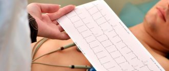
What is cardiac radiography?
X-ray of the heart is a fairly old research method and is to a certain extent inferior to some modern diagnostic procedures. However, to this day, radiography is quite informative. It allows you to quickly and accurately find out the condition of the heart chambers and great vessels. You can also obtain data on the contractility of the organ. Heart disease can be detected without radiography; it is often prescribed as an adjunct. X-rays of the heart are possible in both public and private clinics. This diagnostic method is very common.
Left ventricular hypertrophy
This pathology is a typical heart lesion for those suffering from hypertension. Dilatation of the left ventricle is considered a rather unsafe phenomenon, which is characterized by high mortality. This process involves structural adaptation of the organ regarding metabolism and changes that occur in hemodynamic data.
Cardiomegaly causes significant thickening of the walls. Often it leads to a significant change in the septum, which is located between the 2 ventricles. Due to the expansion that occurs, the elasticity of the walls is lost. Thickening can be either uniform or in certain areas of localization. This specificity directly affects the course of the disease.
Clinical manifestations
To understand what an enlarged heart means, it is necessary to identify its manifestations. The most common symptom indicating changes inside the left ventricle is angina. It develops against the background of compression of blood vessels that provide nutrition to the myocardium. As a result, its expansion in size and its consumption of a significant amount of oxygen in parallel with nutrients are noted. In addition to these symptoms, atrial fibrillation appears, signs of atrial flutter and starvation of the heart muscle are observed. Often a pathology occurs during which the organ freezes for 3-5 seconds and the beat is not visible, which leads to an unconscious state. In certain situations, shortness of breath may indicate the presence of cardiomegaly. The following symptoms are considered additional:
- high blood pressure;
- unstable blood pressure;
- headache;
- disturbances in heart rhythm;
- disturbed sleep;
- poor condition and general malaise;
- painful discomfort in the heart area;
- painful discomfort in the chest.
Dilatation of the right ventricle is not accompanied by any complaints. Only its causes and adverse effects can be demonstrated in the laboratory:
- dilatation of the right ventricle;
- disturbances in heart rhythm;
General recommendations
In addition to medical prescriptions, preventive measures must also be followed. During heart disorders, the following recommendations will be extremely useful:
- Avoid drinking alcohol, which has a toxic effect on the heart muscle.
- Exclusion from the daily menu of products with high cholesterol content. This helps prevent the deposition of cholesterol plaques on the vascular walls. It is optimal to add fish, olive, flaxseed, corn and soybean oil to your diet at least twice a week.
- Inclusion in the menu of such products as viburnum, cranberries, cabbage, eggplants, apples, pomegranates, etc. They will help strengthen and maintain the myocardium in proper condition.
- Salt intake should be limited to 2 g per day, in particular for patients with high swelling.
- When obesity is diagnosed, a proper balanced diet should be formulated that aims to eliminate those extra pounds.
- The duration of sleep should be at least 8 hours, avoiding physical and psycho-emotional fatigue.
- Frequent walks in the fresh air.
Cardiac enlargement is not a diagnosis, but only a temporary condition of the myocardium. With proper and timely treatment of this pathology, it is possible to eliminate the disease and significantly improve the patient’s condition.