Publications in the media
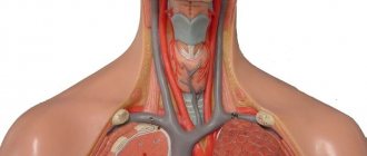
Resuscitation or intensive care is impossible without constant transfusion therapy and taking blood samples for laboratory tests, often emergency. Under these conditions, it is necessary to have a constantly functioning venous access, providing both the infusion of any solution with optimal speed and efficiency, as well as the possibility of laboratory diagnostics and monitoring. Central venous catheterization serves this purpose. In resuscitation practice, the subclavian vein, external and internal jugular veins are most often punctured and catheterized, less often - the femoral and even less often - the ulnar vein (peripheral veins collapse and become empty during hypovolemia, and therefore are poorly contoured).
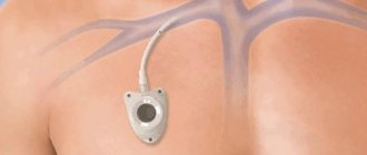
• Subclavian vein •• Indications: shock of any etiology, because Due to anatomical features, this vein does not collapse during hypovolemia. In addition, when using the subclavian vein, good comfort is provided for the patient and the possibility of careful care even with a long stay of the catheter in the vein (for several months) •• Disadvantages of catheterization of the subclavian vein - a high risk of developing pneumothorax, the possibility of injury to the subclavian artery and difficulties when necessary to stop bleeding (impossibility of compression).
• External jugular vein •• Indications ••• Access to this vein is preferable in the elderly, it is well contoured even in obese patients ••• Less danger of developing pneumothorax ••• Its rapid effective compression is possible in case of hemorrhagic complications due to coagulation system disorders •• Inconveniences - the impossibility of its long-term use due to discomfort for the patient, difficulties in care, and relatively unreliable fixation.
• Internal jugular vein •• Characterized by rare complications, especially pneumothorax, good control in case of hemorrhagic complications, high rate of successful catheterizations even during cardiopulmonary resuscitation •• Disadvantages ••• Possibility of injury to the thoracic lymphatic duct or carotid artery during catheterization on the left •• •• The impossibility of its use in intracranial hypertension and during hemodialysis and tracheostomy ••• This vein can be collapsed with deep shock or hypovolemia.
Execution technique. The patient's position is lying on his back, the head end of the table is lowered, a cushion is placed under the patient's back, the patient's head is turned in the direction opposite to the puncture site. The patient's arm on the puncture side is brought to the body, sharply supinated, the assistant pulls it in the caudal direction. The patient may be positioned according to Trendelenburg. There are two operational approaches for puncture catheterization of the subclavian vein.
• Subclavian access •• A 0.5% procaine solution is injected intradermally with a syringe with a thin needle to create a “lemon peel” at a point located 1 cm below the clavicle on the line separating the middle and inner third of the clavicle. The needle is advanced medially towards the upper edge of the sternoclavicular joint, continuously applying procaine solution. The needle is passed under the collarbone and the rest of the procaine is injected there. The needle is removed •• Using a thick sharp needle, limiting the depth of its insertion with the index finger, the skin is pierced to a depth of 1–1.5 cm at the location of the “lemon peel”. The needle is removed •• A syringe with a capacity of 20 ml is filled up to half with 0.9% sodium chloride solution, and a not very sharp (to avoid puncture of the artery) needle 7–10 cm long with a bluntly beveled end is put on. The direction of the bevel should be marked on the cannula. When inserting the needle, its bevel should be oriented in the caudal-medial direction. The needle is inserted into a puncture previously made with a sharp needle (see above), and the depth of possible needle insertion should be limited to the index finger (no more than 2 cm). The needle is advanced medially towards the upper edge of the sternoclavicular joint, periodically pulling the plunger back, checking the flow of blood into the syringe. If unsuccessful, the needle is pushed back without removing it completely, and the attempt is repeated, changing the direction of advancement by several degrees. As soon as blood appears in the syringe, part of it is injected back into the vein and again sucked into the syringe, trying to obtain a reliable reverse blood flow. If a positive result is obtained, ask the patient to hold his breath and remove the syringe from the needle, squeezing its hole with a finger. •• A conductor is inserted into the needle with light screwing movements halfway; its length is slightly more than two times the length of the catheter. The patient is again asked to hold his breath, the guide is removed, closing the catheter hole with a finger, then a rubber stopper is put on the latter. After this, the patient is allowed to breathe. If the patient is unconscious, all manipulations associated with depressurization of the lumen of the needle or catheter located in the subclavian vein are performed during exhalation •• The catheter is connected to the infusion system and fixed to the skin with a single silk suture. Apply an aseptic dressing.
• Supraclavicular approach •• Position of the patient - as with subclavian access •• The doctor pierces the skin in the area of the angle formed by the lateral leg of the sternocleidomastoid muscle and the upper edge of the clavicle, moving 0.5 cm upward from the clavicle. After puncturing the skin, the needle is directed at an angle of 45° to the sagittal and 15° to the frontal plane of the clavicle and the vein is pierced. Further actions are the same as for infraclavicular access.
• After each infusion, 0.1 ml of heparin is injected into the catheter (the so-called “heparin lock”), so that the catheter can be used for several weeks without complications.
• If thrombosis, phlebitis or catheter sepsis is suspected, the catheter must be removed, followed by appropriate treatment measures.
Intravenous (venous) catheter
Vascular catheters are designed to provide access to central and peripheral veins.
These catheters should not be confused with cavity catheters, such as the Foley catheter, Nelaton catheter and others. Indications for placing a peripheral venous catheter.
1. Emergency administration of infusion solutions with the prospect of using the created venous access in the hospital. Most often this is done in the confines of an ambulance.
2. In the hospital, peripheral catheters are placed during long-term intravenous therapy.
3. Intravenous anesthesia during surgery.
4. Install a catheter for women in labor
5. Frequent collection of venous blood for tests.
6. Blood transfusion.
7. In case of parenteral (intravenous) nutrition of the patient.
8. Temporary measure before insertion of a central catheter.
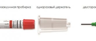
Description of the peripheral venous catheter
Externally, they are a packaged disposable product for long-term intravenous infusions.
A – peripheral intravenous catheter with a port. B – peripheral intravenous catheter without port. A standard intravenous peripheral catheter consists of: - a polymer tube connected to a polymer cannula (1), - a metal needle tube connected to the cannula, - a polymer plug (2). — port (3) optional.
Classification of peripheral venous catheters
There are the following subtypes of intravenous catheters:
1. By the presence of an additional port.
- with port; - without port;
Peripheral catheter with an additional port.
There is a variant of an intravenous catheter with an additional port.
The port is located on top of the catheter. The catheter port allows bolus (quick) administration of medications or flushing of the catheter with heparin and saline. solution. Most modern peripheral catheters have an additional port. The port is closed with a plastic cap to ensure sterility Example of an intravenous peripheral catheter with an additional port
2. By the presence of wings:
— peripheral catheter with wings; - peripheral catheter without wings;
The wings may have holes for suturing to the patient's skin.
3. Based on the presence of a safe device at the peripheral catheter:
— peripheral catheters are safe;
- standard peripheral catheters
Peripheral catheters are safe.
Currently, peripheral safety catheters are becoming available from a number of manufacturers. Most of them implement a safe device due to the presence of a special capsule, or “bullet,” which, after catheterization, is automatically put on the needle. This eliminates the risk of needle injury to medical personnel. In this case, the technique for placing a peripheral safety catheter is no different from the standard technique. The price for peripheral safe catheters is significantly higher than for catheters that do not have such a device. You can buy a peripheral catheter
on our website.
4. According to the presence and number of X-ray stripes on the wings of the catheter:
- without x-ray contrast stripes;
- 2 stripes;
- 4 stripes;
- 6 stripes.
5. Intravenous catheters sizes by color
In accordance with GOST ISO 10555-5-2012, the sizes of catheters are determined. The size of the peripheral catheter is determined by the gauge. Intravenous catheters differ in color. For convenience, each peripheral catheter size is color coded. This allows the medical professional to quickly select the appropriate catheter size for a specific procedure. The table below shows intravenous catheters with sizes by color. Each tube outer diameter has a specific color code.
This table is not identical to GOST R ISO 6009-2013.
You can select and buy an intravenous catheter in our catalog. Delivery is carried out throughout the Russian Federation by SDEK. To the catalogue.
6. Needle butterfly
It is a special type of peripheral catheter. The butterfly needle is a needle integrated with wings with a tube. The length of the butterfly needle tube differs depending on the model. Unlike the classic peripheral catheter, the butterfly needle is intended for short-term insertion. Also suitable for blood collection. One of the key advantages of the butterfly needle is the small diameter of the needle. Butterfly needles are placed for no more than 24 hours.
Scope of application of butterfly needle:
1. Resuscitation and gerontology for patients with “collapsed” veins
2. Neonatology (maternity hospitals and specialized departments of hospitals). Designed for administration and collection of blood to newborns and young children.
3. Blood collection from severe patients.
Safe butterfly needles
There are butterfly needles with a safety device, this is the so-called safety butterfly needle
After the infusion is carried out using a safe butterfly needle, the medical staff removes the needle from the vein and presses a special button on the body of the safe butterfly needle. As a result, the needle goes into a special cylinder, eliminating the risk of infection of personnel. Safe butterfly needles are most in demand when working with urgent patients and with known infected patients
GOST catheters
The main GOST for catheters is GOST ISO 10555-5-2012. This guest describes:
- catheter structure;
- catheter color coding;
- requirements for the needle, catheter cannula;
- flow rate;
- strength of the connection between the cannula and the tube;
- information on individual packaging.
The material was prepared using the following sources:
- GOST ISO 10555-5-2012 Single-use sterile intravascular catheters. Part 5. Peripheral catheters with an internal needle;
- GOST R ISO 6009-2013 Single-use injection needles. Color coding;
- GOST R ISO 11070-2010 Single-use sterile introducers. Technical requirements and test methods.