The mean electrical axis of the QRS complex is the basic measurement required for every electrocardiogram. In most healthy individuals it is between -30° and +100°. An angle of -30° or more negative is described as axis deviation to the left , and an angle of +100° or more positive is described as axis deviation to the right . In other words, axis deviation to the left is an altered position of the average electrical axis of the QRS complex in people with a horizontal position of the electrical axis of the heart. Axis deviation to the right is an altered position of the average electrical axis of the QRS complex in people with a vertical position of the electrical axis of the heart.
The position of the average electrical axis of the QRS complex depends on the anatomical position of the heart and the direction of propagation of the impulse through the ventricles (direction of ventricular depolarization).
General information about pathology
Cardiologists see the chest as a three-dimensional coordinate system that contains the heart. Each contraction is accompanied by a number of bioelectrical changes, which change the direction of the cardiac axis.
What is the electrical axis of the heart
The electrical axis of the heart describes the functioning of the organ based on its size and functioning characteristics. It is almost identical to the axis of the organ’s location in the body. The parameter shows the electrical processes that bring the heart muscle to work.
It is checked using an electrocardiogram. Sensors are attached to various areas of the chest and take readings. After which, on the transcript, the doctor determines the axes and sees whether there are any deviations from the norm.
Normal electrical axis of the heart
Normally, the axis of the heart should almost completely coincide with the axis of the location of the organ in the chest. The cardiac muscle is located semi-vertically. The lower part looks down and to the left. Accordingly, the EOS should also be tilted down and to the left.
The alpha angle normally ranges from 0° to 90°.
Vertical position of the EOS
In people with a thin build, tall stature and long limbs, the heart axis may be vertical. The alpha angle will normally range from +70° to + 90°. It is also normal during exhalation and when a person gets up from a horizontal position.
Horizontal position of the EOS
The horizontal position of the EOS is typical for stocky and plump people with short stature. The alpha angle norm will correspond to the range from 0 to +30°.
It also appears when a person lies down and the internal organs put pressure on the diaphragm and at the moment of full inhalation.
What does a sharp deviation mean?
Any deviations more or less from the norm indicate disturbances in the functioning of the heart muscle. A minus angle indicates that the EOS is excessively tilted to the left. Values above +90° indicate a tilt to the right.
Often these values are not indicated in the ECG interpretation. Doctors in conclusion simply write about axis deviation to the left or right.
A sharp deviation in EOS is not a diagnosis in itself, but can be a symptom of problems such as:
- left ventricular hypertrophy;
- disturbances in the functioning of the left ventricular valves, due to which the volume of blood in the department increases;
- failures in electrical conductivity;
- cardiac blockades.
Pathological deviations of the axis: what do they mean and what are the consequences?
The situation itself cannot serve as a basis for making a specific diagnosis, only indicating the presence of electrical disturbances. No cardiologist will convince you of the presence of pathology only based on EOS. To establish the fact of the disease, it is necessary to support the examination conclusion with the correct clinical interview and additional diagnostic measures.
The position of the EOS is influenced by a number of factors:
- congenital heart defects;
- secondary changes in the anatomical relationships between the right and left parts of the heart;
- abnormal location of organs in the chest cavity (dextrocardia, vicarious emphysema after lobectomy);
- chest deformation (kyphosis, scoliosis, carinatum or funnel-shaped curvature);
- disruptions in the conduction system of the organ (especially in the Hiss bundles), which cause heartbeat disturbances;
- cardiomyopathies of various origins;
- long history of hypertension and coronary heart disease (CHD);
- chronic heart failure;
- respiratory diseases with an obstructive component (COPD, bronchial asthma, emphysema);
- decompensated liver failure (ascites, flatulence).
What diseases cause disturbances?
Deviation of the electrical axis of the heart to the left (levogram) (angle α from 0 to -300) has several reasons:
- Hypertrophy of the left half of the heart. Angle α is directly proportional to the degree of growth of LV mass. The pathology develops with idiopathic cardiomyopathy, arterial hypertension, excessive stress (“athletic heart”), coronary artery disease, and cardiosclerosis.
- Myocardial infarction (with necrosis along the posterior wall).
- Pathology of intracardiac conduction. Most often this is a blockade of the left leg or the anterior superior branch of the Hiss bundle.
- Ventricular tachycardia.
- Valvular heart defects.
- Myocarditis.
There is also a sharp deviation of the EOS to the left when the angle α > -300.
Deviation of the electrical axis of the heart to the right (rightogram) (angle α > +900) is observed when:
- Failures in the conduction of nerve impulses along the fibers of the Hiss bundle.
- Pulmonary stenosis (when pressure in the right ventricle increases).
- IBS.
- Myocardial infarction of the right sections.
- Cardiorespiratory diseases that have formed a “pulmonary heart” (in this case, the LV does not function fully and overload of the right ventricle occurs).
- Thromboembolism of the branches of the pulmonary artery (due to blockage, gas exchange in the lungs is disrupted, the vessels of the pulmonary circulation narrow and overload of the pancreas occurs).
- Mitral valve stenosis (after rheumatic fever). The fusion of the leaflets prevents the complete expulsion of blood from the left atrium, which causes pulmonary hypertension and overloads the RV.
A sharp deviation of the EOS to the right is observed at the angle α = +1200.
It is worth remembering that none of the above diseases can be diagnosed based solely on the position of the EOS. This parameter is only an auxiliary criterion in identifying any pathological process.
Symptoms and manifestations
A cardiogram allows you to detect a sharp deviation of the electrical axis of the heart to the left. But besides it, there are a number of manifestations that may indicate a deviation.
Manifestations on the ECG
An electrocardiogram is a recording of changes in potential differences during the process of excitation and relaxation of the atria and ventricles, caused by the electromotive force of the heart (EMF) from the surface of the human body.
Impulses on the cardiogram are displayed in the form of teeth of various sizes:
positive – P, R, T – directed upward relative to the isoline;
negative – Q, S.
During an ECG, the impulse is placed in three planes:
I – left-right hand;
II – left leg – right hand;
III – left leg – left arm.
With a levogram, the following picture can be observed: RI>RII>RIII, SIII>RIII.
In children
In children, left-sided axis deviation is most often associated with congenital heart defects. The alpha angle can be up to -90°.
When the position of the ventricles changes, the axis deviates by an angle from 0 to -20°. With changes in the atria, the axis deviates to -60°.
Clinical manifestations
Axis deviation in itself has no manifestations. Since it is one of the symptoms of other diseases, symptoms may vary.
With coronary heart disease, pressing pain appears behind the sternum. It intensifies with stress or physical activity and goes away in a calm state.
Heart defects of varying degrees may be accompanied by shortness of breath, swelling, and the appearance of a wet cough when lying on the back. If the defect is mild, there may be no symptoms at all, and EOS declines gradually.
With hypertension, the patient has increased blood pressure, which is accompanied by headaches and weakness.
In children
A child in the neonatal period has a sharp deviation of the axis to the right, this is the norm. If such a disorder is observed in an adult, he will develop symptoms of right ventricular hypertrophy.
In a child, this is due to the fact that the right side of the heart has more mass than the left side. By the age of one year, this condition is normalized, and the organ should take a vertical position in the chest. During this period, it can rotate around its axis in different directions.
The left ventricle then gains mass and is no longer attached to the chest. At the age of 6-7 years, the organ acquires the correct semi-vertical position.
Reasons for shift to the left
The reasons for the displacement of the EOS to the left side can be caused by both heart disease and physiological conditions.
Heart diseases
The reasons for a pronounced deviation of the electrical axis to the left can be:
- Increased pressure in blood vessels. Occurs due to disruption of the kidneys or hormonal system, as well as hypertension.
- Heart defects of various nature. Improper functioning of the valves leads to stagnation of blood in the ventricles, between the atria or in the vessels. As a result, the chambers enlarge and muscle growth begins.
- Cardiac ischemia. Due to insufficient blood flow in the heart muscle, the connective fibers in the walls increase. The organ becomes heavier and deviates.
- Cardiomyopathy. In this disease, the heart chambers and muscle walls become enlarged due to structural abnormalities.
- Myocarditis. The walls of the heart become inflamed. Not all doctors can diagnose the disease. To identify it, a special study is required.
- Myocardial infarction localized in the left ventricle.
Physiological conditions
If the EOS is slightly deviated, this can be considered a variant of the norm. This deviation can occur in athletes, as well as in people with an asthenic body type.
Axis shifts can be associated with deep inhalation and exhalation, when the diaphragm greatly expands and contracts, resulting in pressure on the internal organs. In a horizontal position, the axis also tilts to the left.
In what cases is EOS deviated in children?
In newborns, the deviation of the electrical axis of the heart to the right is considered normal. This is due to the fact that the right ventricle in childhood is heavier than the left. In the second year of life, the EOS begins to level out. And by the age of six, the heart occupies the correct position in the chest.
What does a sharp deviation mean?
Since the deflection angle can vary, the process steps also vary. Changing an academic degree is a gradual process. The more the size of the defect increases, the more the indicator deviates from the norm. If the deviation is from -450 to -900 degrees from the norm, the organ is said to be sharply shifted to the left.
In adults
A shift in the position of the heart in the chest may indicate an ECG disorder if the person is in good condition and no other health problems are detected.
Typically seen in people who exercise regularly and athletes.
The obvious deviation is not accidental; it is a sign of pathology in adults. There may be clogs that accumulate over several years.
Diagnostics
Deviation of the heart axis to the left is diagnosed first of all by performing an electrocardiogram. A cardiologist determines the angle of deviation in several ways.
Visual definition
Visual comparison of the R and S waves is a simple and clear way. However, it does not detect deviation accurately. When the EOS deviates to the left, you can see a picture of RI - SIII on the cardiogram, which means the R-type of the ventricular complex in the first lead and the S-type in the third.
Alpha angle offset detection
Calculating the alpha angle allows you to accurately determine the degree of deviation. Diagnostic doctors find the algebraic sum of the teeth in the first and third standard leads. They calculate the height of the teeth in mm, then add the resulting values. The final result is substituted into the table according to Diede and the alpha angle is calculated.
Sinus rhythm on ECG
Sinus rhythm characterizes oscillations that arise from impulses in a certain node and diverge through the atrium and ventricle. As a result, the heart muscle contracts. Sinus rhythm disturbances indicate arrhythmia or blockade.
The importance of the cardiac conduction system for determining EOS
The conduction system of the heart is necessary for the autonomous generation of electrical impulses and their distribution to all parts of the muscle. However, if the left ventricle is enlarged, it takes longer for electrical impulses to reach it. Consequently, the normal ventricle depolarizes much earlier than the hypertrophied ventricle, which remains electropositive. Due to this, a powerful vector is formed directed towards the hypertrophied ventricle, and the electrical axis of the heart is deviated towards the hypertrophied ventricle.
Eos is deviated to the left: what does it mean, causes and treatment
Scheme for determining the electrical axis of the heart
How to learn to determine EOS using an electrocardiogram? First, a little theory.
Let's imagine the Einthoven triangle with the axes of the leads, and also supplement it with a circle that passes through all the axes, and indicate degrees or a coordinate system on the circles: along the line of the first lead -0 and +180, above the line of the first lead there will be negative degrees, in increments at -30, and positive degrees are projected down, in increments of +30.
Let's consider another concept necessary to determine the position of the EOS - the alpha angle (2 Practical principles for determining
Normal position of the EOS
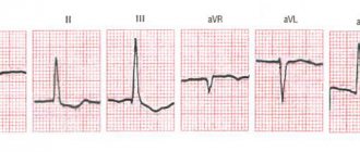
In front of you is a captured cardiogram. So, let's proceed to the practical determination of the position of the heart axis. We look carefully at the QRS complex in the leads:
- With a normal axis, the R wave in the second lead is larger than the R wave in the first lead, and the R wave in the first lead is larger than the R wave in the third: R II>RI>RIII;
- The deviation of the EOS to the left on the cardiogram looks like this: the largest R wave is in the first lead, a little smaller in the second, and the smallest in the third: RI>RII>RIII;
- A rotation of the EOS to the right or a shift of the heart axis to the right on the cardiogram appears as the largest R in the third lead, somewhat smaller in the second, the smallest in the first: R III>RII>RI.
Alpha Angle Definition
But it is not always easy to visually determine the height of the teeth; sometimes they can be approximately the same size. What to do? After all, the eye can fail... For maximum accuracy, the alpha angle is measured. Here's how they do it:
- We find QRS complexes in leads I and III;
- We sum up the height of the teeth in the first lead;
- Let's sum up the height in the third lead;
Important point! It should be remembered when summing that if a tooth is directed downward from the isoline, its height in mm will be with a “-” sign, if - with a “+” sign - We substitute the found two sums into a special table, find the place where the data intersects, which corresponds to a certain radius with degrees of the alpha angle. Knowing the norms of the alpha angle, it is easy to determine the position of the EOS.
3Why do I use a pencil to diagnose or when I don’t need to look for the alpha angle?
Visual determination of the alpha angle
There is another method, the simplest and most beloved by students, of determining the position of the EOS using a pencil. It is not effective in all cases, but sometimes it simplifies the determination of the cardiac axis, makes it possible to determine whether it is normal or whether there is a displacement. So, with the non-writing part, we apply the pencil to the corner of the cardiogram near the first lead, then in leads I, II, III we find the highest R.
We direct the opposite pointed part of the pencil to the R wave in the lead where it is maximum.
If the non-writing part of the pencil is in the upper right corner, but the pointed tip of the writing part is in the lower left, then this position indicates the normal position of the heart axis.
If the pencil is located almost horizontally, we can assume a shift of the axis to the left or its horizontal position, and if the pencil takes a position closer to vertical, then the EOS is deviated to the right.
4Why determine this parameter?
Limits of deviation of the electrical axis of the heart
Issues related to the electrical axis of the heart are discussed in detail in almost all books on ECG; the direction of the electrical axis of the heart is an important parameter that needs to be determined.
But in practice, it is of little help in diagnosing most heart diseases, of which there are more than a hundred.
Decoding the direction of the axis turns out to be really useful for diagnosing 4 main conditions:
- Blockade of the anterosuperior branch of the left bundle branch;
- Right ventricular hypertrophy. A characteristic sign of its increase is the deviation of the axis to the right. But if left ventricular hypertrophy is suspected, a displacement of the heart axis is not at all necessary and the determination of this parameter does not help much in its diagnosis;
- Ventricular tachycardia. Some of its forms are characterized by deviation of the EOS to the left or its uncertain position; in some cases, a turn to the right occurs;
- Block of the posterosuperior branch of the left bundle branch.
5What is the normal EOS?
EOS position options
In healthy people, the following descriptions of EOS take place: normal, semi-vertical, vertical, semi-horizontal, horizontal. Normally, as a rule, the electrical axis of the heart in persons over 40 years of age is located at an angle of -30 to +90, in persons under 40 years of age - from 0 to +105. In healthy children, the axis can deviate up to +110.
For most healthy people, the indicator ranges from +30 to +75. In thin, asthenic individuals, the diaphragm is low, the EOS is more often deviated to the right, and the heart occupies a more vertical position. In obese people, hypersthenics, on the contrary, the heart lies more horizontally, and there is a deviation to the left.
In normosthenics, the heart occupies an intermediate position.
6Normal in children
ECG of a newborn
In newborns and infants, there is a pronounced deviation of the EOS to the right on the electrocardiogram; by the age of one year, in most children, the EOS moves to a vertical position.
This is explained physiologically: the right parts of the heart are somewhat predominant over the left ones both in mass and in electrical activity, and changes in the position of the heart can also be observed - rotations around its axes.
By the age of two, many children still have a vertical axis, but in 30% it becomes normal.
The transition to the normal position is associated with an increase in the mass of the left ventricle and cardiac rotation, during which the fit of the left ventricle to the chest decreases. In preschool children and schoolchildren, normal EOS prevails; the vertical electrical axis of the heart may be more common, and less often the horizontal electrical axis of the heart. Summarizing the above, the norm in children is considered to be:
- during the newborn period, EOS deviation is from +90 to +170
- 1-3 years - vertical EOS
- school age, adolescence - half of the children have a normal axis position.
7 Reasons for EOS deviation to the left
Left ventricular hypertrophy
A deviation of the EOS at an angle from -15 to -30 is sometimes called a slight deviation to the left, and if the angle is from -45 to -90, they speak of a significant deviation to the left. What are the main causes of this condition? Let's take a closer look at them.
- Variant of the norm;
- GSV of the left bundle branch;
- Left bundle branch block;
- Left ventricular hypertrophy;
- Positional changes associated with the horizontal position of the heart;
- Some forms of ventricular tachycardia;
- Malformations of endocardial cushions.
Additional diagnostic methods - a set of examinations
Based on the indications of axis deviation to the left, it is impossible to reliably determine what changes are occurring in the heart muscle. Therefore, cardiologists prescribe a number of additional examinations.
- Ultrasound. Allows you to determine how enlarged the ventricles are and how their contractile function is impaired.
- Electrocardiogram with exercise (walking on a treadmill or riding an exercise bike). Allows you to detect coronary heart disease.
- Chest X-ray. If the tissue is significantly enlarged, the shadow of the heart will be larger.
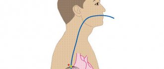
- 24-hour Holter ECG monitoring. Performed in case of significant cardiac dysfunction src=”https://infoserdce.com/wp-content/uploads/2021/08/foto-7.jpg” class=”aligncenter” width=”375″ height=”510″[ /img]
- KAG. Also necessary for diagnosing coronary artery disease.
- Echocardioscopy. Allows you to fully assess the condition of all components of the heart muscle.
Additional diagnostics
To find out the reasons for the EOS deviation, the ECG is analyzed in detail. They may also assign:
- EchoCG (ultrasound of the heart) - to identify possible organ defects.
- Stress echocardiography – ultrasound of the heart under stress – for diagnosing ischemia.
- Angiography of the coronary vessels - their examination to identify blood clots and atherosclerotic plaques.
- Holter monitoring – recording an ECG using a portable device throughout the day.
After a detailed examination, appropriate therapy is prescribed.
Treatment
Since deviation of the EOS to the left is not a disease, it is necessary to understand the reasons for its occurrence. And only after a thorough diagnosis, doctors prescribe treatment for the identified disease.
Coronary heart disease, cardiopathy and heart failure are treated with medications. In addition, you need to follow a diet and daily routine, avoid stress and give up bad habits.
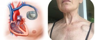
Congenital and acquired heart defects often require surgical intervention. In particularly difficult cases, a pacemaker is installed. The device sends signals to the heart muscle, thereby causing it to contract.
If the axis deviation occurs abruptly, the patient may have a heart attack. This condition requires urgent hospitalization and threatens cardiac arrest.
If EOS deviation is asymptomatic, there is no need to worry. However, it is necessary to constantly monitor the work of the heart and do an ECG at least once a year. Such prevention will make it possible to identify serious diseases in time and begin their treatment.
Possible consequences and complications
The main threat to the body is not the deviation of the axis to the left, but the diseases that accompany it. Enlargement of the left ventricle can cause the following consequences:
- severe heart failure;
- angina pectoris;
- arrhythmia;
- death due to cardiac arrest.
All these consequences are interconnected and follow from one another. Therefore, if the ECG revealed a deviation of the heart axis to the left, it is necessary to undergo additional examinations. They will allow you to identify the true cause of the changes and prescribe the necessary treatment in time.
When additional examination is required
Standard EOC values are about the same for everyone, but a tall person may have a slightly different heart size and location, although they will not get sick. Therefore, if any abnormalities are detected during the initial examination, additional tests are necessary.
Changes in this parameter are also normal for athletes.
Because they exert a lot of effort during constant training, their heart pumps a large amount of fluid, causing the cavities to stretch. They may have horizontal deviations when the organ position is from -15 to +30.
If during the examination a person takes a deep breath or changes his body position, even if it is correct, a deviation of the healthy heart to the left is detected.
Diseases accompanied by levogram
Most patients are trying to understand what it means when the electrical axis of the heart is deviated to the left and what diseases this is associated with. The most common cause is hypertension. When blood pressure is constantly elevated, the load on the left chambers of the heart muscle increases. Which leads to an enlargement of the ventricle and atrium. Accordingly, the electrical axis of the heart shifts to the left.
Acquired heart defects associated with the appearance of atherosclerotic plaques also lead to an increase in the hemodynamic load on the left ventricle. This occurs due to damage to the aortic and mitral valve.
Myocarditis leads to inflammation of the walls of the heart muscle. Accordingly, the tissues increase in volume and hypertrophy occurs. The disease can only be determined in special cardiology centers by undergoing a scintigraphic examination.
Author of the article: Yulia Dmitrieva (Sych) - In 2014, she graduated with honors from Saratov State Medical University named after V. I. Razumovsky. Currently working as a cardiologist at the 8th City Clinical Hospital in the 1st clinic.
The electrical axis of the heart (EOS) is deviated to the left (levogram): what does this mean?
If the electrical axis of the heart (EOS) is deviated to the left (levogram) or to the right (pravogram), then this may indicate disturbances in the functioning of this organ. Let's look at why this can happen, when it is dangerous and when it is not, and how this condition is treated.
How is offset determined?
The position of this axis is determined by electrocardiography, after analyzing an electrocardiogram from several leads.
To detect changes in the normal location of the axis, 2 methods can be used.
Alpha angle deviation
This technique is most often used by diagnosticians. Normally, the EOS completely coincides with the anatomical axis (the heart is located semi-vertically, and the lower end deviates down and slightly to the left). Its location is determined by the alpha angle formed from 2 straight lines (1 abduction axis and the EOS vector line).
To identify the angle, the sum of the S, R and Q waves in 3 and 1 standard leads is calculated. The positive and negative value of each tooth must be taken into account.
Diede's table is then used. Having entered the result, the doctor determines the criteria for the alpha angle.
Here's what it looks like:
Click on the image to enlarge Normally, this angle should be from – 29° to + 89°. Significant (pronounced) left-sided displacement of the axis is a sign of pathological disorders. When it changes to -30°, we are talking about a left-sided deviation, and with values from +90° to +180° - a right-sided deviation.
A left-side deviation of the angle from – 30° to – 44° is considered small (insignificant), but if it is shifted from – 45° to – 90°, this is considered significant and in most cases accompanies cardiac pathologies.
Visual definition
This technique for determining the displacement of the heart axis is most often used by therapists and cardiologists. After an ECG, the doctor compares the magnitude of the S and R waves in leads 1 and 3. If within one of them the value of R is greater than S, we are talking about a ventricular complex (R-type). Otherwise, the complex belongs to the S-type.
When the axis deviates to the left, tooth RI - SIII. This means that the ventricular complex is R-type in lead 1, and S-type in lead 3.
Standard lead of QRS waves in different positions of the EOS (a, b – right-sided displacement; c – normal position of the axis; d, e – left-sided displacement)
The main tool for determining deviations of the EOS to the left is electrocardiography, in which this can be seen visually, however, a number of auxiliary studies are necessary to confirm the result.
Additional diagnostic methods
After an ECG is performed, its results are carefully studied to identify the cause of the pathological condition. In most cases, a repeat cardiogram is prescribed, which is necessary to eliminate technical errors (incorrect application of electrodes, device malfunction, etc.).
In addition, the following studies are recommended:
- Holter monitoring - if the doctor diagnoses a conduction disorder or arrhythmia using an ECG, then 24-hour monitoring of cardiac activity (24-hour ECG) is performed, which makes it possible to more accurately determine the area of the heart with a conduction disorder.
- Ultrasound of the heart - this study is aimed at obtaining more information about cardiac output, blood flow, and the condition of the heart chambers. If indicated, ultrasound can be supplemented with Doppler ultrasound.
- ABPM (daily blood pressure monitoring) is prescribed for a sharp increase in blood pressure against the background of left ventricular hypertrophy with deviation of the cardiac axis. This examination allows you to find out the stage of hypertension and determine the most appropriate treatment.
- Cardiac surgery consultation is prescribed for any pathologies of the heart and, especially for defects with a tendency to progress.
It must be borne in mind that deviation of the EOS to the left is just an ECG sign indicating diffuse changes in various pathologies, therefore a comprehensive diagnosis must be prescribed.
Reasons for displacement
Changes in heart activity on the electrocardiogram are provoked by many factors.
Let's consider each case in more detail.
Heart diseases
The main reason for the shift to the left of the heart axis is left ventricular hypertrophy. Changes can be provoked by: ischemia (including heart attacks and post-infarction cardiosclerosis), aortic and mitral valve disease, cardiomyopathy, myocardial dystrophy and other diseases.
Changes in the cardiogram are possible with atrial fibrillation, heart defects (acquired and congenital), and left bundle branch block.
Physiological conditions
A slight deviation of EOS on electrocardiography often occurs in completely healthy people, for example, athletes, thin and tall patients.
The electrical axis can shift to the left during deep exhalation, when the diaphragm is high, and when the body position changes (from vertical to horizontal), which is caused by compression of the diaphragm by internal organs. Such shifts are considered quite normal.
In what cases is EOS rejected in children?
In children, EOS can change according to age. For example, newborns are characterized by a right-sided deviation and this is not a pathology. In adolescence, the EOS angle has stable indicators.
Most often in children, left-sided axis deviation (up to –90°) is caused by congenital defects that can be complicated by concomitant cardiovascular anomalies.
This is possible with an open ductus arteriosus, in case of high loads on the left ventricle, which happens with mitral heart defects or coarctation of the aorta.
This picture in a child is possible with a defect of the interventricular septa or with a high position of the diaphragmatic dome.
A shift of the axis to the left (from 0 to –20°) is also possible due to a change in the position of the ventricles. Congenital heart disease with incomplete atrioventricular communication, as well as atrial septal defects, are also accompanied by a change in the axis from –20° to –60°.
Clinical manifestations
EOS displacement is not a disease, so it is not characterized by certain clinical signs. In addition, the pathologies that cause it can also occur with mild symptoms. In this case, deviations of the electrical axis of the heart to the left are often detected only when deciphering the electrocardiogram.
There are certain symptoms specific to certain diseases. For example, with hypoxia of the left ventricle, they are expressed by paroxysmal pain in the chest and surges in blood pressure. Tachycardia and severe headache may occur. With left bundle branch block, fainting and bradycardia are possible.
Treatment
Deviation of the heart axis to the left does not require the use of specific therapy. All measures are aimed at neutralizing the underlying disease, accompanied by displacement of the EOS and disturbance of sinus rhythm. For arterial hypertension, antihypertensive drugs are prescribed; ischemia requires the use of ACE inhibitors, statins, and beta-blockers.
Deviation of the EOS does not pose a threat to the patient’s life, but if the position of the axis changes very sharply, there is a possibility of blockade of the legs of His. If such changes are detected, a mandatory consultation with a cardiologist is required to clarify the diagnosis. This approach makes it possible to timely identify a borderline state in the heart.
Dmitrieva Yulia (Sych) – In 2014, she graduated with honors from Saratov State Medical University named after V. I. Razumovsky. Currently working as a cardiologist at the 8th City Clinical Hospital in the 1st clinic.