Patient complaints can vary from mild malaise and unexpressed chest pain to a fulminant course leading to acute heart failure and sometimes death. Most often, the initial complaints are of the nature of a flu-like syndrome: fever, pain in muscles and joints, sometimes symptoms of damage to the gastrointestinal tract such as gastroenteritis
- chest pain, usually long-term, not associated with physical activity, of various types
- palpitations, almost constant, often interruptions in heart function, various arrhythmias
- shortness of breath, decreased exercise tolerance
- weakness, which may be the leading and almost the only complaint.
Myocarditis - symptoms and treatment
Viral infection plays a key role in the development of active myocarditis.
Phases of myocarditis development:
- Penetration of cardiotropic viruses (Coxsackie, mumps, parvovirus B19, adenovirus, etc.) or other infectious agents into the myocardium. Viruses enter cardiomyocytes, the muscle cells of the heart, multiply and cause their death. As a result of the direct action of viruses, the immune system is activated. The first phase of the disease, with an adequate immune response, ends with victory over the viruses or transition to the second phase.
- Autoimmune reactions occur, cardiomyocytes are destroyed under the influence of immune cells.
- Remodeling (modification) of the myocardium occurs—structural changes in the heart muscle [8].
Depending on the characteristics of the body’s defense mechanisms and the characteristics of the pathogen, one or another phase of the disease may predominate.
Normal immune reactivity helps eliminate the virus and allows the damaged myocardium to heal. However, immune disorders can lead to an imbalance: the body either fails to eliminate the virus, or favorable conditions are created for the cells of the immune system to remain in the heart muscle for a long time. Cells of the immune system secrete cytokine proteins that destroy cardiomyocytes.
In the mechanism of development of viral myocarditis, key factors are identified: the direct damaging effect of viruses, which leads to acute and chronic autoimmune reactions, and subsequent cardiac remodeling.
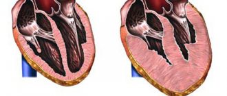
It has been established that remodeling of the heart muscle is the main link in the pathogenesis of the development of heart failure in myocarditis. In this process, the left ventricle is first affected, then the right parts of the heart are involved. Changes in cardiac geometry occur in the initial stages of myocarditis and progress with chronic inflammation [2].
In severe cases of the disease, the myocardium is damaged as a result of apoptosis of cardiomyocytes - programmed cell death. The more cardiomyocytes that show signs of apoptosis, the higher the risk of developing fatal heart failure in acute myocarditis [6].
Circulatory disorders with viral myocarditis go through three phases:
The first phase is hyperdynamic. Develops within 1-3 days after infection. During this phase, myocardial contractility increases, the volume of blood that the heart pumps per minute (minute volume) increases, as the activity of the sympathetic division of the autonomic nervous system increases. Also in this phase, diastolic dysfunction of the heart muscle develops - a violation of myocardial relaxation. There is slight damage to cardiomyocytes as a result of exposure to viruses and mediators of the immune system. There are no pronounced pathological changes in cardiac cells yet.
The second phase is depressive. Occurs between 4 and 7 days after infection. During this phase, a decrease in contractility, cardiac output, and impaired myocardial relaxation progresses. On the 5th day, dilatation (expansion) of the left ventricle develops. On the 7th day, cardiogenic shock and severe manifestations of venous stagnation may occur. In this phase, the virus continues to damage cardiomyocytes, and active damage to cardiomyocytes by cytokines is also observed. Edema develops, the myocardium becomes saturated with inflammatory cells, which leads to an increase in diastolic dysfunction of the left ventricle (impaired relaxation).
The third phase is recovery. If the course is favorable, recovery begins between 9 and 14 days. Intercellular edema and inflammation are reduced, which leads to a partial restoration of myocardial contractility and the compliance of the left ventricular cavity to stretching [1].
Causes of myocarditis
There are many pathogens that cause the disease.
Most often these are viruses:
- Coxsackie B virus
- adenovirus
- parvovirus B19
- herpes simplex virus
- influenza A virus.
Potential pathogens are also considered:
- echovirus (usually affects the gastrointestinal tract)
- Epstein-Barr virus
- HIV
- rubella virus.
The disease can be caused by bacteria:
- staphylococci
- streptococci
- diphtheria bacilli
- the bacteria that causes Lyme disease are Borrelia;
parasites:
- toxoplasma
- trypanosomes, including those that cause Chagas disease, which is common in Central and South America;
mushrooms:
- candida
- aspergillus
- histoplasma.
Other causes of myocarditis may include:
- medications or drugs that may cause an allergic or toxic reaction. These are penicillin and sulfonamides, some anticonvulsants, cocaine.
- chemicals (carbon monoxide).
- burn disease
- organ transplantation
- rheumatological diseases - systemic lupus erythematosus, Wegener's granulomatosis, Takayasu arteritis, giant cell arteritis.
Sometimes the cause of myocarditis remains unclear.
Consultation and examination with a cardiologist
IMMA medical clinics have modern equipment and their own laboratories to accurately determine myocarditis in the initial stages. The full range of studies includes an initial examination by a qualified cardiologist, clarifying tests, and diagnostic measures using modern technologies. Diagnosis and treatment of myocarditis in children and adults takes place in equipped clinics, trained by experienced personnel, under the guidance of leading cardiologists.
To make an appointment, use the contacts listed on the website. The professional staff of the clinic will provide all possible information support.
Rehabilitation
An important part of recovery is reducing the load on the heart by regulating physical activity. You should not overdo physical activity immediately after recovery. Sometimes it is better to limit physical activity, including walking, for several months. You should consult your doctor about your activity regimen and when you can return to normal activities.
It is also important to visit your doctor regularly after recovery. Usually, before the visit you need to perform an echocardiogram, an electrocardiogram, and a general blood test.
If myocarditis is prolonged or there is severe heart failure, it is important to adhere to a diet with limited salt, liquid, alcohol and smoking.
Who is at risk
The most common cause of myocarditis is viral pathologies of various origins. Therefore, the risk group includes every person who has had herpes, hepatitis B, C, rubella, chicken pox, measles, diphtheria and even influenza. Since most people have been diagnosed with some kind of viral disease, everyone must undergo a preventive examination in order to exclude the presence of inflammatory processes in the heart. This can subsequently save lives.
The risk of developing myocarditis increases in proportion to the presence of infectious pathologies. Listening to your heart is recommended for everyone who has suffered or is a carrier of diseases caused by the following pathogens:
- Bacteria that provoke the development of tuberculosis, chlamydia, staphylococcus.
- Fungal genus Aspergillus fumigatus and Candida. They cause such common diseases as tonsillitis, laryngitis, otitis media, conjunctivitis, candidiasis, etc.
- Parasitic worms - roundworms, echinococci, trichinella, lamblia, toxocariasis.
- Diabetes mellitus in any degree.
- Thyroid diseases.
- Autoimmune pathologies – rheumatoid arthritis, lupus erythematosus, vasculitis.
The risk group includes people whose professional activities involve working in enterprises with hazardous working conditions, where there is a possibility of heavy metal poisoning. Taking certain medications can cause inflammation of the myocardium. For example, antiallergic cephalosporins or antidepressants have an extremely negative effect on cardiac activity. Social factors play an important role for the myocardium. Abuse of alcoholic beverages, smoking and other dangerous and harmful addictions contribute to the development of myocarditis.
Myocarditis code according to ICD-10
I01.2 - Acute rheumatic myocarditis
I40 - Infectious myocarditis
I40.1 - Isolated myocarditis
I41.0 - Myocarditis in bacterial diseases classified elsewhere
I41.1 - Myocarditis in viral diseases classified elsewhere
I41.2 - Myocarditis in other infectious and parasitic diseases classified elsewhere
I41.8 - Myocarditis in other diseases classified elsewhere
I51.4 - Myocarditis, unspecified
Diagnostics
The examination begins with questioning the patient and assessing his condition. The doctor examines the color of the skin, determines the presence of edema, listens to heart sounds, assesses the frequency and rhythm of the pulse, and measures the temperature. Then an instrumental examination is performed:
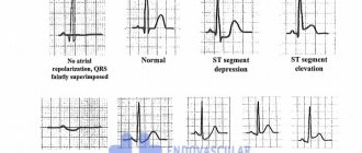
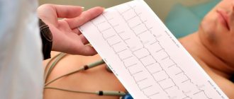
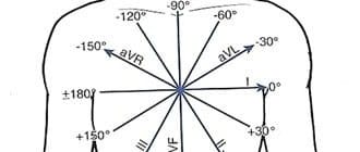
- Electrocardiography (ECG). Allows you to determine arrhythmias and identify disturbances in myocardial contractility. A diagnosis cannot be made on the basis of an ECG alone, since there are no changes characteristic only of myocarditis.
- Chest X-ray. The image can determine the size and shape of the heart, and reveal the accumulation of fluid in the pleural sinuses, which will indicate heart failure.
- Magnetic resonance imaging (MRI). It records the size of the heart, its shape and structure, and changes in muscle motor activity.
- Echocardiography. Allows you to create moving images of a beating heart. During echocardiography, you can determine the thickness of the myocardium, detect expansion of the heart chambers, decreased pumping function, fluid in the pericardial sac, and find out whether there are problems with the valves.
- General blood analysis. Based on the results of a general blood test, you can evaluate the ESR and the level of leukocytes - “inflammatory cells”.
- Biochemical analysis will help determine the levels of enzymes that indicate muscle damage - creatine phosphokinase, troponin. In addition, antibodies to bacterial agents can be detected in the blood, and viruses can be detected using PCR.
- Heart biopsy. This is the most accurate diagnostic method, but due to its complexity it is rarely used.
- MiniRNA profiling. This new method makes it possible to differentiate the fulminant form of myocarditis from the acute form.
Relevance of the problem
The problem of admission to pregnancy and management of women with heart and vascular lesions is becoming increasingly urgent. The desire to move higher on the social ladder, to acquire a financial “safety cushion”, and a later age of marriage have led to an increase in the age of first-time women. Motherhood in adulthood is more often associated with the risk of endocrine and cardiovascular diseases (or CVD).
Also, the high level of development of medicine has become the reason that more and more women with congenital heart defects survive to reproductive age and are allowed to become pregnant, while the increased load on the body becomes the main cause of maternal mortality. Among all CVDs, the greatest risk of adverse outcome of pregnancy and childbirth is borne by:
- congenital heart defects;
- rheumatic heart valve lesions;
- conditions with increased blood pressure.
Cardiomyopathies are extremely rare, but pose the greatest danger to women.
Timely detection of CVD allows for preventive monitoring, hospitalization and timely treatment.
Prevention of myocarditis
There are no specific methods that protect against the disease. However, you can reduce the risk of myocarditis in the following ways:
- Avoid crowds of people during a flu or ARVI epidemic.
- Wash your hands regularly with soap. This will help you avoid getting infectious diseases.
- Avoid risky behavior, e.g. practice protected sex, do not use drugs.
- Protect yourself from ticks. Wearing long sleeves and using repellents containing DEET will help with this.
- Routine vaccination. Rubella and influenza are diseases that can cause myocarditis.