Left ventricular enlargement (or hypertrophy) is the expansion and thickening of the walls of the main pumping chamber of the heart. Hypertrophy can develop in response to some negative factor, for example, high blood pressure or significant physical activity. The enlarged heart muscle loses its elasticity and eventually cannot pump blood with the required force. Enlargement of the left ventricle of the heart most often occurs in people who have uncontrolled high blood pressure. This condition is quite dangerous, as it can ultimately lead to heart attack and stroke. Older people with overweight, hypertension and diabetes are at risk.
Symptoms of the development of an abnormal condition
Dilatation of the left ventricle in most cases develops very slowly. The patient may not experience any unpleasant signs or symptoms, especially in the early stages of the disease. But as hypertrophy develops, the following may occur:
- shortness of breath;
- unexplained fatigue;
- chest pain, especially after exercise;
- sensation of fast, fluttering heartbeats;
- dizziness or fainting.
You should seek medical help if:
- there is a feeling of pain in the chest that lasts longer than a few minutes;
- there are serious breathing difficulties that interfere with daily activities;
- have severe, recurring memory problems;
- there are loss of consciousness;
- shortness of breath combined with rapid heartbeat is troubling.
Degrees of dilatation
Another basis for classification is the degree of pathological abnormalities. Accordingly, they talk about 3 or 4 stages of disease development.
Lightweight
It is formed as a result of a genetic factor, intrauterine defects and acquired conditions.
It is characterized by a complete absence of a clinical picture, which makes diagnosis a matter of chance.
It is possible to identify the initial deformation of the organic plane using echocardiography. It doesn't take much qualification to state a fact.
Determining the root cause falls on the shoulders of the cardiologist and is carried out with the help of a group of measures.
Moderate dilatation
At this stage, the process is diagnosed much more often.
- Myogenic dilatation of the heart
A bright nonspecific picture is typical: shortness of breath, chest pain, arrhythmias. These are common signs of any condition associated with impaired functioning of the heart and blood vessels.
However, the chances of early diagnosis are high, which is good news for the patient.
The prospects for a complete cure are already vague, but with a well-thought-out combination of therapy, the patient will not notice the difference. You can keep the process under complete control.
Marked dilatation of the left atrium
Considered extreme in some national classifications.
It is defined by a vivid clinical picture with a significant decrease in tolerance to physical activity, the inability to adequately work and perform household duties.
Organic defects are gross and are observed not only on the part of the heart; remote systems are also changed.
The prospects for a cure are minimal. At the same time, life expectancy during therapy rarely exceeds 3-4 years.
Reasons for the development of the anomaly
Enlargement of the left ventricle can occur if some unfavorable factor causes the heart to work harder than usual. This means that the heart muscle will need to make several times more contractions in order to pump blood around the body.
Model of the heart with left ventricular hypertrophy
Reasons that can provoke a significant deterioration in heart function:
- High blood pressure (hypertension) is considered the most common cause of thickening of the ventricular wall. More than one third of all patients learn about hypertrophy at the time of diagnosis of arterial hypertension.
- Aortic valve stenosis is a disease that is a narrowing of the flap of muscle tissue that separates the left ventricle from the aorta. Narrowing of the aortic valve causes the heart to contract several times more often in order to pump blood into the aorta.
- Hypertrophic cardiomyopathy is a genetic disease that occurs when the heart muscle becomes abnormally thick and stiff.
- Professional sports. Intense, long-term strength training, as well as irregular performance of endurance exercises, can lead to the fact that the heart is not able to quickly adapt and cope with the additional load. As a result, the left ventricle may swell (enlarge).
Causes of pathology
The myocardium is a muscle that tends to hypertrophy. But if in the case of ordinary muscle tissue this is not dangerous, then with cardiac tissue the opposite is true. Left atrium enlargement is a pathological condition that requires treatment.
Hypertrophy can be true or false. The first type is caused by hyperplasia, that is, the proliferation of certain cells. It is the result of high stress on an organ or system. This species is also called a worker. People who deal with high physical activity are often susceptible to true hypertrophy.
The false type of enlargement is the progressive proliferation of fat cells, which leads to this type of hypertrophy. This form of the disease is most often observed in people with a sedentary lifestyle.
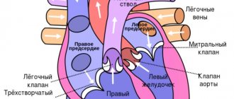
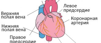
The mechanism of the hypertrophy process is as follows: the human heart has 2 halves (right and left). Each of them is divided into a ventricle and an atrium, which are connected by openings. The lumen of these holes is also equipped with valves. On the left it has two doors, and on the right - three.
Thanks to them, the heart regulates blood circulation through systematic contraction and release of blood portions into nearby vessels. During heavy physical activity, the body's cells' need for oxygen increases, causing the heart to work faster. To do this, you need more muscle tissue, which is formed during such overloads.
Such changes are fraught with consequences, the most unfavorable of which is death. In addition to heavy physical activity, there are also a large number of factors that can lead to hypertrophy. You need to know them and try to avoid them.
Hypertrophy of any part of the heart can be congenital or acquired. This pathology can affect several heart chambers at once, but the left atrium is more often affected. It is the increase in the size of this section that does not act as a separate disease, but is only a symptom that indicates concomitant pathological processes.
There are many reasons for increased thickness of the walls of the left atrium. The main ones can be called:
- excess weight, especially in young people;
- cardiovascular pathologies;
- narrowing of the mitral valve in the cavity of the left atrium;
- high blood pressure;
- insufficiency of the valve apparatus;
- frequent stressful situations and depression;
- acute renal failure;
- diabetes;
- respiratory diseases;
- constant physical activity;
- playing sports;
- the presence of muscular dystrophies;
- presence of bad habits;
- severe genetic inheritance.
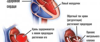
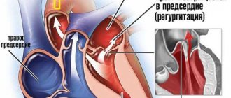
Narrowing of the mitral valve is a heart defect. Its essence lies in stenosis of the fissure, which is a connecting channel for the atrium and ventricle.
This syndrome is often accompanied by severe insufficiency of this valve, which is characterized by the process of regurgitation. Some of the blood flows back from the ventricle into the atrium, and the valve is unable to prevent this.
Sports can also be a cause of this cardiac pathology if the loads do not correspond to the person’s capabilities. With frequent and high tension, the heart muscle cannot cope with the influx of a large amount of blood; it needs additional strength to push it out. As a result, an increase in cardiac muscle tissue can be observed to compensate for this process. Before choosing the type and amount of sports activity, you should first consult with specialists and find out about training and possible regimes.
Ignoring heart problems is very dangerous. To protect yourself from possible malfunctions of the main organ, you need to lead a healthy lifestyle, moderately engage in physical activity and sports, and also consult doctors more often for the timely detection of various concomitant diseases.
What can hypertrophy lead to?
The disease cannot be ignored, because a significant enlargement of the ventricle can greatly change the structure and function of the heart. An enlarged ventricle may weaken and lose elasticity, increasing pressure in the heart. Hypertrophied tissue can also compress blood vessels and restrict blood flow directly to the heart muscle.
On the left is a normal heart, on the right is an enlarged ventricle
As a result of these changes, the following complications may occur:
- complete interruption of blood supply to the heart;
- inability of the heart to pump enough blood around the body (heart failure);
- abnormal heart rhythm (arrhythmia);
- irregular, fast heartbeat (atrial fibrillation);
- insufficient oxygen supply to the heart (coronary heart disease);
- enlargement of the aorta (dilatation of the aortic root);
- stroke;
- unexpected deterioration in cardiac function (sudden cardiac arrest);
- sudden loss of consciousness.
Hypertrophy is fraught with a significant deterioration in heart function
The consequences of hypertrophy can be called catastrophic for health, so if the patient has identified the reasons for the development of the disease, it is necessary to consult a cardiologist.
Development mechanism
LA dilatation is not an independent process.
It is not considered a nosological unit. How does such a state form? Normally, cardiac structures function continuously, acting as a large pump. The release of blood is ensured by alternating synchronous contraction of all chambers of the heart.
Liquid connective tissue moves in one direction: from the upper to the ventricles, no reverse flow is observed.
As a result of congenital genetic defects, acquired sclerosis of active tissues (replacement of affected areas with scars), a long inflammatory period, etc., blood is retained in the left atrium longer than it should be. Or regurgitation is observed (return of liquid connective tissue from the ventricle).
Over a long period of time, the chamber becomes stretched and the normal dimensions of the organ are disrupted.
This leads to a decrease in release into the systemic circulation, and general and local hemodynamics decrease.
Removed tissues and systems suffer from a lack of oxygen and nutrients. The process of development of organ defects and functional failure begins.
Diagnostic methods
Before making a diagnosis, your doctor will take your medical history and perform a thorough physical examination, including taking your blood pressure and testing your heart function. If preliminary studies indicate that the ventricle may indeed be enlarged, a number of additional screening tests are performed.
Electrocardiogram (ECG)
Electrical signals will not confirm ventricular enlargement. But cardiologists can identify some difficulties in the passage of the impulse, which will indicate a violation of the density of the muscle tissue of the heart.
MRI
Images of the heart taken with a special tomograph will directly indicate ventricular hypertrophy.
Diagnostics
The examination of patients is carried out under the supervision of a cardiologist, as necessary, a group of other specialists.
- What is dilatation of the left atrium cavity: causes, symptoms, treatment and prognosis
Approximate list of events:
- Oral questioning of the patient and collection of anamnestic data. It is necessary to establish many factors that would play a role.
- Measurement of blood pressure (possibly an increase or decrease), heart rate (tachycardia is typical, flowing in parallel with various types of arrhythmias).
- Daily monitoring. Registration of blood pressure levels for 24 hours. Used for early diagnosis.
- Electrocardiography. Plays the same role. Shows the degree of deviations from the cardiac structures.
- Echocardiography. Basic technique. It makes it possible to identify organic defects at first glance, determine their degree, and predict complications.
- MRI if there is a suspicion of a tumor process in the heart.
Stress tests are not performed due to the possible cessation of muscle function and sudden death.
Treatment of hypertrophy
Treatment depends on the underlying cause of the enlarged ventricle and may include medications and/or surgery.
Left ventricular enlargement can be controlled by taking tablets
The most common medications prescribed for hypertrophy are the following.
Angiotensin-converting enzyme (ACE) inhibitors
These drugs dilate blood vessels, lower blood pressure, improve blood flow, and help reduce the workload of the heart. Trade names of the drugs: captopril, enalapril and lisinopril. The most common side effect is a persistent, irritating dry cough.
Angiotensin receptor blockers
These drugs are similar to ACE inhibitors, but do not cause a persistent cough.
Beta blockers
Beta blockers help lower your heart rate and normalize your blood pressure. Beta blockers are not usually prescribed as primary treatment for hypertrophy.
Diuretics
Thiazide diuretics help ease blood flow to the heart and lower blood pressure. Trade names: chlorthalidone and hydrochlorothiazide.
Only an experienced cardiologist will select the right type of pills
Surgical treatment consists of repairing or completely replacing the aortic valve.
Atrial flutter - symptoms and treatment
Drug treatment
Drug therapy involves taking drugs that restore sinus rhythm.
The main antiarrhythmic drugs are:
- amiodarone (cordarone);
- sotalol (sotahexal);
- propafenone (ritmonorm);
- verapamil;
- cardiac glycosides.
The combination of these antiarrhythmic drugs increases the effectiveness of treatment.
Amiodarone and sotalol are class III antiarrhythmic drugs.
Amiodarone is most effective in preventing the development of atrial fibrillation, as well as arrhythmia after a heart attack. It has been proven to improve survival in patients with severe circulatory failure. May improve myocardial contractility [5]. Contraindicated in hypo- and hyperthyroidism.
Sotalol, as a means of preventing atrial fibrillation, is less effective than amiodarone. The safest dose of this antiarrhythmic drug for atrial flutter is 120 mg twice a day. But for life-threatening heart rhythm disturbances, the dose of sotalol can reach 640 mg per day. The most common side effects of the drug include symptoms of digestive tract disorders: abdominal pain and diarrhea [5]. Contraindicated in COPD and severe bronchial asthma.
Propafenone is a class I antiarrhythmic drug. Its preventive effect is weaker than that of amiodarone and sotalol. Long-term use of this drug increases the risk of repeated attacks of arrhythmia. It can lead to side effects such as shortness of breath, dizziness, vomiting, changes in taste, constipation, headaches, abdominal pain, blurred vision, ataxia and diarrhea [5].
Verapamil is a class IV antiarrhythmic drug. Its effectiveness is lower than that of amiodarone and sotalol, and fairly high doses are required to prevent flutter attacks. Side effects include a decrease in heart rate to 50 beats per minute, hypotension and progression of heart failure.
Among cardiac glycosides, digoxin is most often used. Typically, this drug is prescribed for a combination of atrial flutter and circulatory failure.
The antiarrhythmic effect of glycosides does not develop immediately: after about two weeks from the start of treatment. Long-term use increases the risk of overdose. Other reasons that increase the risk of overdose include low levels of potassium, magnesium, oxygen and high levels of magnesium in the blood, kidney and liver failure, old age, poor general condition of the body and taking diuretics.
If it is necessary to take diuretics, preference should be given to potassium-sparing diuretics, for example veroshpiron. At the same time as taking diuretics, you need to monitor your heart rhythm [5].
Considering that atrial flutter most often occurs against the background of low potassium levels in the blood, potassium supplements are added to standard drug therapy:
- 4% potassium chloride solution;
- asparkam;
- Panangin.
Among other things, all patients with atrial flutter and fibrillation are advised to take anticoagulants to prevent the formation of blood clots [5][13].
Electropulse therapy
When symptoms of heart failure appear, preference is given to electrical pulse therapy - EIT. It is carried out using a special device - a defibrillator. One of its electrodes is fixed slightly outward from the apex of the heart, the other to the right of the sternum. If atrial flutter is present and the patient is stable, start with a 25 J shock.
After the procedure, the patient must remain in bed during the day. He is prescribed antiarrhythmic and antithrombotic therapy. Atrial function is restored within a few days after the flutter attack has stopped.
If there are no complications, the patient is discharged from the hospital 6-7 days after EIT [5]. In rare cases, complications are possible: systemic embolism, ventricular tachycardia, acute left ventricular failure, ventricular fibrillation, myocardial damage, extrasystole, hypertension, sinus tachycardia and bradycardia.
When atrial flutter is combined with sick sinus syndrome, permanent pacing . This effect on atrial rhythm significantly reduces the risk of developing flutter attacks.
Ablation
With typical atrial flutter, preference is given to ablation - the destruction of pathological electrical foci.
Ablation, which is carried out using high-frequency currents, is called radiofrequency. The destruction of pathological foci using low temperatures, down to −70 °C, is called cryoablation.
In radiofrequency ablation, a catheter is inserted into the atrium through the common femoral or subclavian vein. Using electrical signals sent through the catheter, the doctor finds the arrhythmogenic zone and begins to influence it through the electrode. After this, the doctor re-checks the electrical activity of all chambers of the heart to ensure the effectiveness of the procedure and ends the operation. The effectiveness of this method is 95% [14].
Open heart surgery aimed at isolating the atrium is also possible However, they are difficult to perform and are not performed in patients with severe heart failure.
Prevention of hypertrophy
Lifestyle changes will help not only prevent the development of hypertrophy, but also improve the condition of an already enlarged ventricle. Since hypertrophy is common in people suffering from obesity, maintaining an ideal body mass index will be the best prevention of the disease. It is also worth limiting the amount of salt in your diet to normalize blood pressure. If hypertrophy is suspected, it is recommended to drink alcohol in moderation, and if treatment is prescribed, then it is better to avoid strong drinks altogether.
Despite the fact that one of the reasons for an enlarged ventricle of the heart is heavy physical activity, you should not give up sports. Regular physical exercise, such as walking, Pilates, yoga, will not only do no harm, but, on the contrary, will strengthen the heart. If a diagnosis of hypertrophy has already been made, it is necessary to ask a physiotherapist to select the optimal exercise program. 30 minutes of moderate physical activity will strengthen weakened heart muscle and prevent its enlargement.
A healthy lifestyle and proper nutrition will allow you to forget about problems with the left ventricle for a long time.