Causes of the disease
To understand the features of left ventricular atrophy and determine what it is, you need to look at the anatomy of the heart.
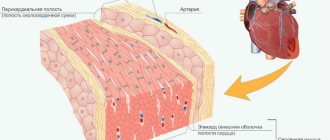
The heart is one of the most important human organs, whose functions include blood supply to the entire body. Its basis is the myocardium, consisting of striated muscle tissue. It allows the heart to contract, causing blood to be pumped between the atria and ventricles. In its structure, the myocardium resembles skeletal muscles, but, unlike it, the heart muscle does not obey the cerebral cortex. Signals are sent to it by the vasomotor center, located in the medulla oblongata.
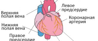
The heart consists of 4 chambers, namely 2 atria and 2 two ventricles. Thanks to their alternating contractions, blood circulates in the small and large circles, providing nutrition to all tissues of the body.
In general, the cardiac cycle looks like this:
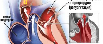
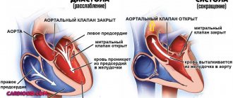
- Atrial systole (contraction). They fill with blood and contract. From the right atrium, blood flow moves into the same ventricle, and the same happens with the left.
- Contraction of the ventricles. Blood entering the left cardiac stomach is sent to the systemic circulation through the aorta. Contraction of the right ventricle leads it to the pulmonary artery (pulmonary circulation).
- Diastole (relaxation). This phase represents the relaxation of all chambers of the heart to fill the atria with blood. There is little oxygen in the bloodstream from the systemic circulation. This blood is called venous and enters the right atrium. The blood flow from the pulmonary circle is rich in oxygen, thanks to the pulmonary vessels. It is called arterial and goes to the left atrium.
The time it takes for the contraction phase to take place is slightly longer than for the relaxation phase. If everything goes well, then the person does not have problems with blood circulation. In the case of atrophy of the left ventricle of the heart, the body does not receive all the substances it needs due to the inability of the affected tissues to fully perform their functions. This disrupts blood flow, causing shortness of breath and swelling. In the congenital form of the pathology, developmental delays are often observed.
Left ventricular atrophy may be congenital or an abnormality acquired over time.
In the first case, the resulting dystrophy is a secondary phenomenon of disorder of myocardial cells. Its development is provoked by congenital heart defects and cardiomyopathies. Anomalies in the structure of the heart muscle mainly affect the connective tissue of the myocardium and the valvular component. Doctors still don’t know much about cardiomyopathy, but they often eliminate the consequences of this disease, among which are left ventricular atrophy.
The acquired form of the disease occurs in approximately 70% of patients. The reasons for its development are mainly the following:
- Viral infections. Among them, influenza and Coxsackie virus prevail. Atrophy of the left cardiac ventricle is a consequence of the effect of infection on the myosymplast. It is a fusion of cells, so it has several nuclei. After a virus attack, the myosymplast remains intact, but failures occur at the genetic level, as a result of which dystrophic changes in the myocardium can begin. The degree of atrophy depends on the number of affected cells.
- Bacterial infections. Doctors distinguish scarlet fever from this group. This is due to the fact that the bacterium that causes the disease is very similar in its gene code to cardiomyocytes, which are the muscle cells of the heart. The immune system, after the heart muscle is damaged by a bacterial infection, perceives its tissues as foreign and attacks them. This process leads to dystrophic changes in the myocardium.
- Lack of nutrition (ischemia) of the myocardium. In adults suffering from left ventricular atrophy, this is the main cause. This is usually associated with chronic ischemia, which gradually leads to dystrophic changes in the heart muscle.
- Intoxication. Long-term exposure to toxins in the body negatively affects the heart muscle. The main culprit of intoxication is alcoholic drinks. In just 2-3 years of their intensive use, the myocardium undergoes significant atrophy.
Kinds
In modern medicine, there are several varieties of this pathology, depending on the reasons for its development:
- Enlarged left ventricle of the heart
- Fat. The disease is provoked by impaired fat metabolism, pathologies of the cardiovascular system, alcohol poisoning, hypoxia, etc.
- Ischemic, occurring during a coronary crisis. This condition can cause heart failure.
- Dishormonal. It is observed when there are hormonal imbalances. Very often occurs in women during menopause.
- Diffuse. Occurs in the presence of an extensive inflammatory process.
- Grainy. The cause of the pathology is protein metabolism disorders.
Left ventricular atrophy often occurs in professional athletes. One of the main factors leading to the development of this disease is regular physical activity. The second reason is an unbalanced diet.
Symptoms of pathology
Atrophy of the left ventricle of the heart can be identified by the clinical picture characteristic of this pathological process:
- Developmental delay. The problem is characteristic of the congenital form of the disease. The patient's tissues do not receive the required amount of necessary substances for full growth, as a result of which developmental retardation is observed.
- Dyspnea. Atrophic changes in the left ventricle disrupt blood flow, which is why the body does not receive the required amount of oxygen. This phenomenon leads to ischemia of internal organs and tissues. To correct the situation, the respiratory center receives a signal to increase the amount of air inhaled. Shortness of breath is a consequence of this entire process and is designed to increase the concentration of oxyhemoglobin in the blood. It is a combination of hemoglobin and air in red blood cells.
- Edema. They represent the second clear sign of acquired ventricular atrophy and indicate heart failure. Edema occurs due to stagnation of venous blood, since the heart cannot fully perform its functions. Initially, the problem begins in the lower extremities. As swelling progresses upward, it can be concluded that heart failure is worsening.
- Irregularities in heart rhythm. Chronic lack of nutrition leads to the death of cardiomyocytes. They perform not only the functions assigned to them, but also regulate the heart rhythm. With atrophic changes in the myocardium, the number of cardiomyocytes is reduced, which leads to disruptions in the process of heart contraction.
How is atrophy different from hypertrophy?
To understand why atrophy of the left ventricle of the heart is dangerous, it is necessary to understand the features of this disorder. Many people do not understand the differences between atrophy and hypertrophy, thinking that they are the same pathology. In reality, these are two completely different phenomena in which different changes occur.
Hypertrophy
With this process, thickening of the walls of the ventricle is observed, and the septum between the sections is modified and loses its elasticity. This happens due to increased blood flow, which increases the resistance of the muscle walls. This leads to overload of the ventricle with pressure and volume, followed by the development of a compensatory function. At the same time, the heart cells elongate, which increases the mass of the myocardium.
- Myogenic dilatation of the heart
As the tissue increases in size, it becomes denser, requiring more oxygen. As a result, oxygen starvation begins and pathological changes in the cardiac structure are triggered.
Atrophy
What is left ventricular atrophy? In this case, there is a decrease in the ventricle, which causes a decrease in its functions, up to complete loss of capacity. A similar phenomenon also occurs due to oxygen starvation, when circulatory disorders provoke the development of cellular atrophy. As the disease progresses, ventricular function decreases significantly.
Danger to the patient's life
Atrophy of left ventricular tissue causes serious malfunctions associated with lack of nutrition. The degree of danger to humans depends on the severity of dystrophic changes. Often this pathological process leads to complications of the cardiovascular system such as:
- atherosclerotic changes;
- death;
- heart attack;
- angina pectoris;
- arrhythmia;
- pulmonary edema.
As the disease progresses, atrophied areas of muscle are replaced by connective tissue, which is presented in the form of scars. Blood flow in this place becomes difficult and against this background the heart’s ability to contract weakens.
Course of therapy
Despite all the possibilities of modern medicine, doctors still do not know how to treat atrophy of the left stomach of the heart. The whole point of therapy is to stop pathological changes in the heart muscle.
There were also attempts to restore cardiomyocytes, thereby completely healing the patient. The following drugs were used for this:
- "Preductal." It belongs to a group of medications intended to relieve angina pectoris and improve myocardial metabolism during cardiac ischemia. The main active ingredient is trimetazidine dihydrochloride. Long-term use of Preductal did not live up to the hopes of scientists. The disease was slowed but not eliminated.
- "Mexidol" and "Riboxin". This combination is still completely untested in the treatment of left ventricular atrophy. "Mexidol" is an antioxidant based on ethyl methylhydroxypyridine succinate. "Riboxin" is intended to normalize myocardial metabolism and improve tissue nutrition. Its main active ingredient is inosine. Despite the promising description, many scientists consider their effects controversial and doubt the effectiveness of this combination.
Due to the lack of actual research results, doctors try not to treat the disease, but to stop its development, stopping the consequences. The following groups of medications are mainly used:
- Diuretics. They remove excess moisture from the body, reducing the load on the heart muscle and solving the problem of edema.
- Beta blockers. Drugs from this group inhibit beta-adrenergic receptors, thereby stabilizing heart contractions and reducing the degree of cardiomyocyte atrophy.
- ACE inhibitors. Due to their effect, the production of angiotensin, which constricts blood vessels, is reduced, as a result of which the usual functioning of the heart is normalized. The rate of development of atrophy decreases only in some patients.
- Calcium channel blockers and sartans. These groups of medications have been especially in demand in the treatment of left ventricular atrophy over the past 10 years. Calcium channel blockers prevent calcium from affecting the walls of blood vessels, causing them to dilate. Sartans are similar in effect to AFP inhibitors, but in this case angiotensin receptors are blocked.
Prevention
Treating left ventricular atrophy is extremely expensive and ineffective, so it is better to prevent it.
Prevention of the acquired form of the disease consists of the following rules:
- rejection of bad habits;
- playing sports;
- correction of diet;
- good sleep;
- prevention of stressful situations;
- elimination of excess body weight.
Atrophy of the left cardiac ventricle is a severe and incurable pathological process. Due to its development, a person may experience various complications that can be fatal. However, if you start a course of maintenance therapy in a timely manner, you can prolong your life. To improve the effect, drug treatment must be combined with compliance with the rules of prevention.
Etiology
The causes of dystrophy include:
- intoxication of the body;
- infectious foci;
- various disorders of the endocrine system (theriotoxicosis, thyroid diseases, hypothyroidism);
- hypovitaminosis and vitamin deficiency;
- anemia of various etiologies (insufficient amount of oxygen);
- obesity (cardiac obesity and general obesity - increasing body weight increases the load);
- angina pectoris;
- hypertension (insufficient blood supply to the heart muscles).
Intoxication is divided into exogenous and endogenous. Exogenous include industrial poisons and toxic substances. Endogenous include metabolic and functional disorders, liver and kidney diseases.
Myocardial dystrophy may not change pathologically or, on the contrary, may clearly manifest itself with various degeneralized changes - fatty, protein, waxy degeneration of muscle fiber, cloudy swellings, fatty infiltrates.