Aplastic anemia is a serious disorder of the formation, development and maturation of blood cells. It is characterized by inhibition of the hematopoietic function of the bone marrow, which is manifested by a deficiency in the formation of white and red blood cells, as well as platelets. Sometimes there is a lack of formation of only red blood cells. The disease is considered one of the most severe disorders of hematopoiesis and, in the absence of adequate treatment, can cause death within several months. It affects male and female patients equally between the ages of ten and twenty-five or over fifty. According to medical statistics, two cases of pathology per one million people are diagnosed every year.
The Department of Hematology at CELT offers diagnosis and treatment of aplastic anemia in Moscow. Our multidisciplinary clinic was one of the first in the Russian Federation to begin operating in the market of paid medical services and has been successfully continuing it for the third decade. The hematology department receives consultations from leading domestic specialists, who have a modern diagnostic and treatment facility that allows them to accurately diagnose and carry out treatment in accordance with international standards. The cost of services is available in our price list, which we update regularly. To avoid misunderstandings, we ask you to check the numbers with our information line operators.
Aplastic anemia: etiology
According to origin, congenital and acquired anemia are distinguished. The first develops as a result of chromosomal mutations, the second - under the influence of chemicals, radiation, and infections. Experts believe that suppression of bone marrow hematopoiesis can be initiated by the appearance of cytotoxic T-lymphocytes in it and in the blood. They produce TNF (extracellular protein) and interferon “y”, which have a suppressive effect on hematopoietic germs. The reason for starting the mechanism may lie in:
- Exposure to ionizing radiation or chemicals in the form of aromatic compounds, arsenic, pesticides;
- Entering the body of infectious agents (causative agents of hepatitis “D”, “B”, cytomegalovirus, DNA-containing Epstein-Barr virus);
- Taking myelotoxic drugs while undergoing treatment with tranquilizers, anticonvulsants, antithyroid and antitumor drugs;
- Development of a number of autoimmune processes (systemic lupus erythematosus, connective tissue damage - Sjögren's syndrome).
In 50% of cases, the cause of the development of the pathology cannot be determined. This form of aplastic anemia is called idiopathic.
Prevention
It is not always possible to prevent the development of aplastic anemia, since experts have not yet established the exact causes of pathological changes.
Following these rules will help reduce the risk of disease:
- constant strengthening of the immune system;
- giving up bad habits, minimizing contact with chemicals;
- use any medications only as prescribed by a doctor, strictly adhere to the dosage;
- prevention of radiation effects on the body;
- timely treatment of viral diseases under the supervision of a specialist;
- maintaining a proper diet.
Classification of aplastic anemia
| Form of pathology | What is the difference? |
| By duration | |
| Acute | No more than one month |
| Subacute | From one month to six months |
| Chronic | More than six months |
| According to the severity of selective aplasia | |
| Moderate | Granulocytes less than 0.0x10⁹/l, platelets less than 20.0x10⁹/l. |
| Heavy | Granulocytes less than 0.5x10⁹/l, platelets less than 20.0x10⁹/l. According to diagnostic results, bone marrow cellularity is less than a third of normal. |
| Very heavy | Granulocytes more than 0.5x10⁹/l, platelets more than 20.0x10⁹/l. |
Development mechanism
The mechanism of development of hypoplastic anemia is associated with the action of provoking factors on the hematopoietic system. Pathological changes lead to a slowdown in the process of reproduction of formed elements until they completely disappear.
The progression of the pathological process leads to the replacement of bone marrow tissue with fat cells
With this type of anemia, a disturbance in iron metabolism is observed as a result of increased destruction of red blood cells and changes in porphyrin metabolism. Next, there is a decrease in iron utilization and its deposition in tissues and organs, which leads to the occurrence of secondary hemochromatosis.
There are several mechanisms involved in the formation of the disease:
- damage to bone marrow stem cells;
- disruption of the cellular and humoral mechanism;
- dysfunction of bone marrow stromal elements;
- lack of factors that stimulate the process of hematopoiesis.
In addition, the mechanism of the occurrence of pathology may be associated with the formation of antibodies that act on the production of blood cells in the bone marrow.
Aplastic anemia: symptoms
The disease begins acutely, it is accompanied by a feeling of severe weakness and fatigue. The patient's skin and visible mucous membranes look pale, and he himself suffers from the following clinical manifestations:
- noise in ears;
- the appearance of shortness of breath even with insignificant efforts;
- unpleasant tingling in the chest area.
When the number of platelets per unit volume of blood decreases, hemorrhagic syndrome appears:
- even after slight compression or shock to the skin, bruises and hemorrhages appear on it;
- a rash in the form of small dots can be seen on the body, arms and legs;
- bleeding gums are observed;
- spontaneous nosebleeds;
- heavy menstruation (in women).
A decrease in the number of leukocytes per unit volume of blood is characterized by the regular frequent development of infectious diseases of the skin and structures of the urinary system, inflammatory processes of the oral mucosa, and pneumonia.
The congenital form of anemia develops in children under ten years of age and is accompanied by a number of other disorders:
- underdevelopment of the skull and brain;
- reduction in the size and weight of the kidneys (hypoplasia);
- intense coloring of certain areas of the skin - hyperpigmentation;
- severe hearing loss and speech impairment due to it.
Reasons for development
Hypoplastic anemia can be caused by the negative effect of various factors; their combination not only contributes to the development of the disease, but also leads to aggravation of the pathological process.
Very often, this type of anemia occurs due to the toxic effect of certain medications that can provoke disorders in the bone marrow with further dysfunction of hematopoietic germs. Its formation does not depend on the dosage of medications or the duration of the therapeutic course.
Drugs that can cause disturbances in the hematopoietic system include:
- sulfonamides,
- antibiotics,
- antihistamines,
- tetracyclines.
Very often, the disease is diagnosed in people taking Levomycetin for a long time
Disruption of hematopoiesis is observed after a course of chemotherapy, since the toxic effect of drugs destroys not only pathological formations, but also healthy cells and tissues.
The causes of hematopoietic function disorders are also autoimmune diseases, in which the immune mechanism is aimed at suppressing not only pathogenic microorganisms, but also its own damage to bone marrow elements.
Thus, there are three groups of main causes causing disruption of the hematopoietic function of the bone marrow:
- Hereditary. Transmission of genetically modified genes, manifested by chromosomal abnormalities.
- Main. Toxic effects of chemotherapy drugs, radiation, arsenic and benzene poisoning.
- Rare. It forms when taking certain medications or when infected with a fungus.
Factors provoking the development of the disease:
- hepatitis of viral origin;
- herpes virus;
- cytomegalovirus infection;
- HIV infection.
Ionizing radiation used during X-ray examination plays an important role in the mechanism of anemia formation. Most often, this pathology occurs among workers in radiology rooms, as well as in patients who have undergone a course of radio wave therapy.
Aplastic anemia: diagnosis
Before starting treatment of the disease, CELT hematologists carry out a comprehensive diagnosis aimed at accurately making a diagnosis and identifying the etiological factor. It includes:
- examination by a hematologist;
- general and biochemical blood tests;
- collection of a bone marrow sample and its examination - sternal puncture.
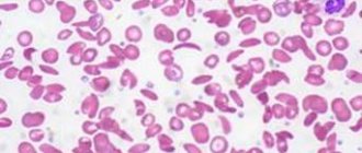
If the disease is present, the patient is diagnosed with a serious decrease in hemoglobin, up to a critical level of 20-30 g/l, and agranulocytosis is observed - a decrease in granular leukocytes and monocytes. The number of lymphocytes may be normal or reduced, platelets are always reduced, sometimes they are not detected at all. Erythrocyte sedimentation rate – increases to 4-60 mm/h. A study of a bone marrow sample reveals an increased content of adipose tissue - 90%, which includes elements of stroma and lymph, but hematogenous cells are present in very small quantities.
General characteristics of the disease
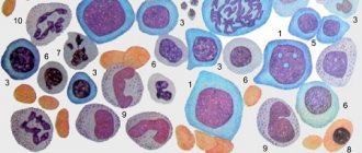
Hypoplastic anemia is characterized by dysfunction of the bone marrow, as a result of which its self-healing processes are disrupted, which leads to damage to hematopoietic germs. A reduced content of platelets, leukocytes, and erythrocytes is accompanied by a violation of their maturation at various stages of formation.
The disease affects young people, mainly women and adolescents.
A characteristic sign of the pathology is complete depletion of the bone marrow in the final stages, as well as a pronounced impairment of its functions.
Symptoms include severe anemia, thrombocytopenia, and leukopenia. If left untreated, the pathology progresses rapidly, which can lead to death.
Treatment of aplastic anemia
Treatment of idiopathic and other types of aplastic anemia is a very complex task that requires a comprehensive individual approach. When developing tactics, CELT specialists take into account the diagnostic results and the patient’s indications. The patient is placed in an isolation room with aseptic conditions, which eliminates the risk of developing infections and their complications. Drug therapy consists of taking:
- Glucocorticoids – in identifying autoimmune mechanisms and the formation of antibodies against one’s own blood cells;
- Cytostatics – in the absence of effect from treatment with glucocorticoids for autoimmune anemia;
- Cyclosporine “A” – to suppress the production of TNF and interferon “y”;
- Anabolic steroids – to stimulate hematopoietic function;
- Androgens – to stimulate the formation of red blood cells.
All patients with aplastic anemia receive transfusions of red blood cells and/or platelets, in volumes determined based on the clinical picture and peripheral blood parameters. In addition, the patient may be prescribed a splenectomy, a surgical procedure aimed at removing the spleen. Bone marrow transplantation can provide the most favorable prognosis. It consists of transplanting donor or own hematopoietic stem cells, previously removed from the iliac bones by puncture. Unfortunately, the procedure is not widely available due to the difficulty of selecting a compatible donor. If this is not possible, the patient is prescribed palliative therapy with cyclosporine A.
The hematology department of our clinic welcomes candidates, doctors and professors of medical sciences with over twenty-five years of practical and scientific experience. You can make an appointment with them online or by contacting our operators. Highly qualified specialists also work in the urology department. You can make an appointment with them for cystoscopy of the bladder.
At CELT you can consult a hematologist.
- Initial consultation – 3,500
- Repeated consultation – 2,300
Make an appointment
By making an appointment with a hematologist, you can get a comprehensive consultation. The doctor is competent to treat various blood diseases, most of which can be identified in the early stages and prescribe timely treatment to cope with the disease quickly and easily.
Complications
In severe cases of aplastic anemia, as well as in the absence of timely treatment, the patient may develop the following complications:
- massive internal bleeding;
- hemorrhages in internal organs, including the brain;
- severe bacterial and viral infections;
- blood cancer;
- chronic bleeding;
- death of the patient.
Only specialized medical care will help reduce the risk of such complications.
Features of the course of pathology during pregnancy
The combination of pregnancy with hypoaplastic anemia is quite rare. If pathology occurs, the prognosis for a pregnant woman is unfavorable, since in most cases it ends in death.
Anemia that develops during pregnancy has a particularly unfavorable course. As a rule, it is detected in the second trimester, accompanied by a rapid deterioration in hematological parameters and the addition of infectious diseases. Terminating pregnancy does not stop the development of pathological processes in the body. Therapeutic measures do not bring results.
If there are adaptation mechanisms in a woman’s body to a violation of homepoiesis, it is possible to maintain pregnancy
Pregnancy, which occurs against the background of hypoplastic anemia, causes its exacerbation. In this case, urgent diagnosis of impaired hematopoiesis is necessary, followed by early termination of pregnancy and removal of the spleen.
Detection of anemia in late pregnancy requires an individual approach in matters of delivery. It can be performed either by caesarean section or by maintaining the pregnancy until spontaneous miscarriage occurs.
Cases of a favorable outcome of delivery have been described, but the child was diagnosed with iron deficiency anemia for several months.
Diagnostic indicators
To confirm the diagnosis of “hypoplastic anemia,” the patient must undergo many studies that can confirm the presence of a disorder in the hematopoietic process. A distinctive feature of this type of anemia is that the spleen and lymph nodes do not enlarge.
Severe hemorrhagic syndrome is accompanied by the development of hypochromic anemia
Laboratory and instrumental diagnostics determine the following changes:
- Clinical blood test (reduced level of red cells, hemoglobin, leukocytopenia).
- Blood biochemistry (increased concentration of serum iron, increased saturation of iron with transferrin).
- Myelogram (reduction of cellular elements of erythrocytes, granulocytes, dysplasia of homepoiesis sprouts).


