Author: Strokina O.A., therapist, functional diagnostics doctor. February, 2021.
One of the most informative methods for studying the heart is scintigraphy (nuclear scanning). To carry out the procedure, a medication containing radioactive isotopes (radionuclides) is used. The drug is injected into the patient's body intravenously and, circulating in the bloodstream, is gradually absorbed by the heart muscle.
Based on the degree of saturation of myocardial tissue with radionuclides, experts evaluate its functionality: active absorption indicates normal heart function, and vice versa, “empty” areas may indicate ischemia (death) of cardiac tissue.
Perfusion scintigraphy is a specific method for diagnosing coronary heart disease (CHD) using radioactive thallium. The procedure is performed with functional tests, and unlike conventional electrocardiography (ECG) and even echocardiography (ultrasound of the heart) with physical activity, it allows you to most accurately determine the location of the ischemic zone.
Indications
Myocardial scintigraphy is prescribed by cardiologists both to patients with already confirmed ischemic heart disease, and to clarify this diagnosis. Based on the results of the study, the doctor plans further treatment tactics or determines the need for surgical intervention on the coronary vessels.
Scintigraphy is prescribed for the following indications:
- preventive examination of persons at risk for coronary artery disease: patients with diabetes and/or hypertension,
- heavy smokers with extensive experience,
- patients with heredity unfavorable for cardiovascular diseases,
- people with high cholesterol,
- women over 55 years of age and men over 45 years of age;
Indications for cardiac scintigraphy
Doctors can recommend cardiac scintigraphy both to patients who have already identified certain heart pathologies, and to patients for whom it is impossible to make a diagnosis due to lack of information. After receiving the results, the doctor will plan further treatment tactics and determine the diagnosis. It is worth noting that this method can act as both primary and additional.
First of all, representatives of the risk group for developing IHD (coronary heart disease) should think about undergoing a heart scintigraphy procedure as a preventive measure:
- long-term heavy smokers;
- people with high cholesterol;
- patients with hypertension and/or diabetes;
- men over 45 years of age and women over 55 years of age.
This diagnosis is prescribed in situations where it is necessary to assess the effectiveness of the prescribed course of treatment as accurately as possible. In addition, the study is recommended for athletes before competitions and patients who are scheduled for surgery or other manipulations on the heart.
Scintigraphy is also prescribed to patients who have had a heart attack, because the technique allows you to assess the performance of the heart muscle. Angina pectoris is also an indication for scintigraphy.
Very often people complain of pain of unknown origin in the chest area. Most people do not pay attention to this, although this may be the first signal of the development of a dangerous heart disease. Doctors recommend that when the first such symptoms appear, immediately contact a specialist who will most likely give you a referral for heart scintigraphy, because this technique is the most informative and reliable. Today, scintigraphy in Moscow is done in almost every large medical center, so no one should have problems finding a suitable institution.
Contraindications
You should not undergo cardiac scintigraphy during pregnancy and breastfeeding (except in emergency situations). There are no more contraindications for conventional scintigraphy.
A much larger list of absolute contraindications is available when performing scintigraphy with stress tests (exercise test or pharmacological test):
- severe general infections with high fever;
- acute stage of myocardial infarction
- unstable angina;
- severe heart failure;
- severe pathologies of the heart valves (aortic stenosis);
- myocarditis (inflammation of the heart muscle);
- the presence of a chronic aneurysm with a thrombus;
- acute cerebrovascular accident (stroke);
- acute thrombophlebitis;
- severe respiratory failure.
Relative contraindications for testing with stress tests include:
- arterial hypertension with blood pressure? 180/100 mm Hg;
- high heart rate, more than 110 per minute;
- serious rhythm and conduction disturbances;
- diabetes mellitus in the stage of decompensation;
- hypothyroidism;
- thyrotoxicosis;
- psychoneurological disorders.
Myocardial scintigraphy
ECG-synchronized myocardial perfusion scintigraphy at rest
Principle of the method:
Physical activity is carried out in the form of a standard veloergometric test (“bicycle”) under the supervision of an ECG and functional diagnostic doctors. When an individual level of physical activity is reached, a small standard amount of radionuclide is injected intravenously and distributed into the heart according to the blood flow in it. After a certain time, the patient is invited to a special device - an emission tomograph.
A resting state study consists of a conventional intravenous injection of a radiopharmaceutical, its distribution in the heart muscle in a resting state, followed by assessment of blood flow in the heart using an emission tomograph.
During the examination itself, the patient lies motionless on his back with his arms raised to his head. The tomograph detectors move around it, recording data on blood circulation and the work of the heart muscle.
The combination of both methods makes it possible to identify circulatory disorders in the heart when a person performs significant physical activity and changes in blood circulation in the heart at rest, as well as establish the exact location of the problem area in the heart muscle, assess the ejection fraction and contractility of the heart.
Duration of the examination:
After intravenous administration of the drug, for its maximum accumulation in the heart, the waiting time is 40-60 minutes.
The direct examination with a tomograph takes about 20 minutes.
Indications:
- determination of the presence, localization, extent and severity of myocardial ischemia (coronary heart disease, angina) or scar lesions (myocardial infarction, myocarditis)
- determination of the functional significance of the lesion of the main coronary bed identified by coronary angiography
- diagnosis of reversible and irreversible myocardial damage (vitality assessment)
- differential diagnosis of angina and cardialgia
- preoperative determination of myocardial viability (ischemic cardiomyopathy, acute myocardial infarction with severe local dysfunction)
- monitoring and evaluation of treatment effectiveness (thrombolysis, restenosis after coronary artery bypass grafting or balloon dilatation)
- diagnosis and stratification of perfusion disorders in non-coronary diseases (diabetes mellitus, arterial hypertension, diffuse connective tissue diseases)
Contraindication: pregnancy
Special instructions:
When breastfeeding, the study is possible, but it is necessary to refrain from feeding for 24 hours after intravenous administration of the isotope.
Preparation:
Early in the morning (around 6 am) - light breakfast.
Avoid drinking coffee and cigarettes for 6 hours.
Stress myocardial scintigraphy requires discontinuation 2 days before the study of medications from the group of beta-blockers (bisoprolol, etc.), calcium channel antagonists (amlodipine, etc.) and sinus node IF-channel inhibitors (Coraxan). All other groups of medications are taken without changes.
Myocardial scintigraphy at rest does not require discontinuation of any medications.
The department performs ECG-synchronized myocardial perfusion scintigraphy with physical activity
Registration for the study:
It is necessary to pre-agreed the day and time of the study by phone:
268−26−31
SPECIALISTS
Plotnikov Konstantin Anatolievich
radiologist of the highest qualification category
Varennikova Anastasia Aleksandrovna
, radiologist of the highest qualification category
Preparing the patient for scintigraphy
Scintigraphy does not require special preparation, but the patient must follow some recommendations.
- The day before the test, you need to exclude products containing caffeine (coffee, tea, chocolate, cola) from your diet;
- Before the procedure (for 12 hours) you should not eat (diabetes patients are allowed to eat light, low-fat food);
- You will have to temporarily interrupt or limit the intake of certain medications, for example, cardiac glycosides, nitrates, beta locators (your doctor will advise you on this matter);
Women who are breastfeeding should express breast milk beforehand to use after the procedure. Radioisotopes are excreted in the secretions of the mammary glands within 2 days, so it is not recommended to feed a child with this milk.
The patient must notify the doctor if:
- he suffers from bronchial asthma;
- he takes drugs to increase erection (during the procedure, a man may develop an attack of angina, which can only be controlled with nitrates, which are incompatible with Viagra, Levitra, etc.).
Before the procedure, women need to make sure that they are not pregnant.
Methodology
Half an hour before the start of the study, a radiopharmaceutical (RP) is administered intravenously to the patient. During the examination, the patient lies on the tomography table with his arms thrown back behind his head to reduce the likelihood of artifacts appearing in the images. The gamma camera then scans the myocardium, capturing radio waves and converting them into a series of images.
Scintigraphy lasts from 2 to 4 hours. During this time, the heart is examined at rest and during physical or pharmacological stress. By comparing the obtained images, the doctor can determine the presence or absence of areas of ischemia in the heart muscle - during exercise, the blood supply to the myocardium increases, so all existing disorders are clearly visualized in the images.
Load scintigraphy
For exercise scintigraphy, an exercise bike or treadmill is used. A functional test is carried out starting with low intensity and gradually increasing its level. At the same time, the dynamics of blood pressure and heart rate are monitored, and an electrocardiogram is taken.
If it is impossible to conduct scintigraphy with natural physical activity for the patient, it is simulated with the help of medications that cause increased heart rate and increased blood pressure.
At the peak of the intensity of physical activity, the patient is again given a radiopharmaceutical (radiopharmaceutical), and 30 minutes later the heart is re-scanned in different projections.
Myocardial scintigraphy (myocardial perfusion scintigraphy) is a study of the blood supply (perfusion) of the left ventricular myocardium using radiopharmaceuticals (RPs), which are distributed in healthy heart tissue. The radiopharmaceutical is administered intravenously and accumulates in the heart muscle, then the radiation from the accumulated drug is captured by the detectors of the recording device.
Myocardial scintigraphy is a unique diagnostic method that makes it possible to objectively assess the blood supply to the heart muscle.
Indications for myocardial perfusion scintigraphy:
- Detection and differential diagnosis of coronary heart disease (CHD): Angina pectoris and stable angina pectoris, episodes of unstable angina pectoris.
- Assessment of the significance of coronary artery lesions in patients diagnosed with coronary artery disease.
- Assessment of the risk of cardiovascular complications (myocardial scintigraphy is performed if the diagnosis of coronary artery disease has already been made).
- Evaluation of the effectiveness of coronary angioplasty, bypass surgery and thrombolysis; assessment of the effectiveness of revascularization.
- Repeated scintigraphy for resumption of angina after interventions on the coronary vessels.
- Confirmation of the diagnosis of acute myocardial infarction (AMI) in the presence of a questionable ECG.
- Study of the blood supply to the myocardium of the left ventricle in its non-coronary lesions (cardiomyopathies, myocarditis, early complications of diabetes mellitus).
- Determination of treatment strategy and quality control of treatment provided.
There are 2 options for performing myocardial perfusion scintigraphy:
- Myocardial scintigraphy at rest - performed within 1 day.
- Myocardial scintigraphy at rest and with load (bicycle ergometry) - carried out over 2 days.
According to experts, scintigraphy of the myocardium at rest, i.e. without stress test, not very informative.
However, in some cases this diagnostic method is indicated for:
- Chest pain of unknown etiology.
- If it is impossible to perform physical activity, permanent cardiac pacing (PAC), left bundle branch block (LBBB).
- Asymptomatic disorders during other stress tests and when they are not informative.
Due to the current impossibility of performing a functional load, myocardial perfusion scintigraphy is performed only at rest.
When studying cardiac perfusion at rest, it is actually possible to confirm the presence of scar changes (infarct zones) of the myocardium, as well as assess blood supply, while studying the myocardium at rest and with stress expands the diagnostic capabilities of the technique.
Contraindications for scintigraphy:
- pregnancy, restriction during breastfeeding (cancellation for 48 hours).
to perform a load test:
- myocardial infarction in the last two days (48 hours);
- unstable angina with a high risk of cardiovascular complications;
- untreatable arrhythmias accompanied by hemodynamic disturbances;
- severe clinical aortic stenosis;
- severe heart failure;
- pulmonary embolism;
- dissecting aortic aneurysm;
- acute myocarditis, pericarditis or infective endocarditis;
- severe non-cardiac diseases that may affect the performance of the test, or may worsen during the test (including infections, renal failure, thyrotoxicosis);
- severe emotional disorders, psychoses.
Preparing for the study
If you plan to do only the study at rest, medications are not canceled.
During exercise tests, after consultation with your doctor, it is recommended to discontinue β-blockers (atenolol, bisoprolol, nebivolol, etc.) 3 days before the exercise test.
Features of the study: it is advisable to conduct a stress test of the heart as part of hospitalization in a hospital for several days to monitor the patient’s condition before and after the stress test and conduct additional diagnostic tests.
The study is carried out strictly on an empty stomach, and have full-fat yogurt (sour cream or cheese) with you.
The procedure is carried out 30–60 minutes after the administration of the radiopharmaceutical and takes up to 30 minutes.
The conclusion is issued on the day of the study.
Patients must have with them an extract from the outpatient card/medical history with an examination by a cardiologist or therapist, an archive of electrocardiograms, the results of heart studies, if available (ECHOCG, Holter monitor, ABPM, VEM, Treadmill test, etc.), which he must provide to the radiologist.
To examine the patient under load, you must bring comfortable shoes and sweatpants for bicycle ergometry.
Cost of radioisotope research:
| Service code | Name of service | Cost, rub. |
| A07.03.001.001 | Whole body bone scintigraphy | 8 700 |
| A07.03.001.002 | Whole body bone scintigraphy combined with single photon emission computed tomography of bones and computed tomography of the thoracic spine | 20 500 |
| А07.03.001.003 | Whole body bone scintigraphy combined with single photon emission computed tomography of bones and computed tomography of the lumbosacral spine | 27 500 |
| A07.03.001.004 | Whole body bone scintigraphy combined with single photon emission computed tomography of bones and computed tomography of the pelvic bones | 27 500 |
| A07.03.003 | Single photon emission computed tomography of bones | 7 400 |
| A07.09.004 | Single photon emission computed tomography of the lungs (perfusion) | 14 000 |
| A07.09.004.001 | Single-photon emission computed tomography of the lungs (perfusion), combined with computed tomographic angiography of the pulmonary artery and its branches | 29 300 |
| A07.10.003 | Single-photon emission computed tomography of the myocardium at rest | 8 800 |
| A07.10.003.002 | Single-photon emission computed tomography of the myocardium, perfusion with functional tests | 15 700 |
| A07.10.005 | Single photon emission computed tomography combined with myocardial computed tomography | 18 800 |
| A07.14.002 | Scintigraphy of the liver and spleen | 9 800 |
| A07.14.002.002 | Liver scintigraphy with labeled red blood cells | 22 200 |
| A07.14.004.001 | Single-photon emission computed tomography of the liver and spleen combined with computed tomography of the hepatobiliary zone | 19 800 |
| A07.22.002 | Thyroid scintigraphy | 6 500 |
| A07.22.010 | Single photon emission computed tomography of the parathyroid glands | 9 600 |
| A07.22.010.001 | Single photon emission computed tomography of the parathyroid glands combined with computed tomography of the parathyroid glands | 29 700 |
| А07.28.002 | Scintigraphy of the kidneys and urinary system | 8 700 |
| A07.28.004 | Angionephroscintigraphy | 8 000 |
| A07.30.031 | Three-phase scintigraphy of soft tissues and bones | 11 400 |
Myocardial scintigraphy results
When reading the scintigraphy results, the doctor can determine:
- intensity of coronary circulation;
- degree of blood supply insufficiency;
- localization of necrotic areas;
- foci of ischemia during exercise;
- scars after a heart attack.
Also, based on the results of the study, the likelihood of developing possible complications is calculated, a program of conservative treatment is planned, or the method of surgical intervention is determined.
Scintigraphy is not informative in assessing the size of the heart and the condition of the coronary vessels. That is, this method will not allow one to determine the location and degree of narrowing of blood vessels.
Diagnosis of cardiac diseases
If there are symptoms indicating cardiovascular disease, it is necessary to conduct a comprehensive instrumental and laboratory examination to confirm the diagnosis, study the extent of the spread of the disease and search for the causes of its occurrence. Cardiology uses a wide range of instrumental and laboratory diagnostics.
— Blood tests (complete blood count, biochemical blood test, hemostasis, serology, immunology, cardiac enzymes)
Changing a number of blood parameters helps assess the degree of heart performance. For example, elevated cholesterol levels (can be a sign of coronary heart disease), abnormal levels of thyroid hormones (can affect heart rhythm), cardiac enzymes (chemicals released from damaged heart muscle cells indicate a myocardial infarction).
— Determination of the level of specific cardiac enzymes in the blood
Blood samples taken over a number of days will help identify the dynamics of changes in the level of enzymes - specific blood proteins that increase against the background of a developed myocardial infarction and decrease with recovery.
— Chest X-ray
The study allows you to evaluate the size and shape of the heart; detect the presence of fluid in the lungs, which may be evidence of ineffective heart function.
— Angiography of the vessels of the neck and brain (cerebral angiography)
An invasive method for diagnosing the location and degree of narrowing of the lumen of the vessel(s), based on the introduction of an iodinated contrast agent into the vascular bed with simultaneous fluoroscopy (radiography) of the neck and brain in patients at high risk of stroke.
— Coronary angiography
Complex invasive diagnostic test. Used to determine indications for surgical intervention in patients diagnosed with coronary heart disease and angina pectoris. This is an x-ray diagnostic method based on studying the passage of a contrast agent through the coronary vessels with subsequent assessment of the degree of their damage (narrowing).
— Echocardiography
Ultrasound scanning allows you to visualize the work of the heart, evaluate the structure (size of the chambers, thickness and structure of the walls), the functional viability of the heart valves and the heart as a whole. A painless examination, especially necessary for patients who have had a myocardial infarction, have heart failure or patients with heart valve diseases.
- Functional diagnostics
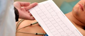
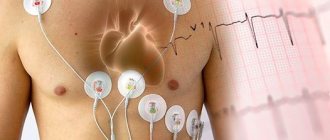
Electrocardiogram (ECG)
Shows the nature of the “electrical activity” of the heart (the rhythm of the heart). Allows you to identify the thickness of the heart muscle (myocardial hypertrophy against the background of high blood pressure), determine the area of myocardial damage after a heart attack, assess the adequacy of blood flow to the heart muscle, etc.
Electrocardiography with physical activity (treadmill test)
Assessment of the state of the heart against the background of physical activity (adequacy of blood supply to the myocardium in conditions of “increased demand”). A good way to diagnose obstruction of the patency of the coronary arteries.
Holter monitoring
The test is also known as a 24-hour ECG. An ECG is recorded within 24 hours to diagnose possible heart rhythm disturbances
— Multislice computed tomography (MSCT)
Computed tomography of the heart or assessment of coronary calcification (CT calcium scoring)
MSCT (multislice computed tomography) of the coronary vessels ( CT coronary angiography, CT cardiac angiography )
CT angiography of cerebral vessels, aorta, large vessels
CT scan of the brain
— MRI
MRI of the heart
MRI of the brain
Vascular MRI
— Radioisotope study of the heart (single-photon emission computed tomography)
One of the most modern and promising diagnostic methods. It is based on the use of radioactive isotopes (thallium 201 and technetium 99 atoms) and recording their distribution in the cardiovascular system using gamma radiation detectors. Allows you to assess myocardial perfusion, visualize the passage of blood through the heart and great vessels, conduct a differential diagnosis of myocardial ischemia, determine the location and size of the infarction, assess the nature of metabolic disorders in the heart muscle (determine the degree of viability of myocardial cells).