Home — For the public
- Map of medical organizations
- Vaccination
- Clinical examination
- Fluorography
- Addresses and opening hours of clinics
- Emergency rooms
- Oncology
- Where to take an HIV test
- Healthy child's office
- Services
- Prevention of CVD
- Disease Prevention
- World Patient Safety Day
- Newspaper "Medical News"
- specialist
- School of Health
— Disease prevention
- HIV infection
- All about vaccination
- All about proper nutrition
- Hepatitis
- Flu
- Dementia
- Schoolchildren's health
- STD
- Tick-borne encephalitis
- Whooping cough
- Measles
- Legionellosis
- Meningococcal infection
- Oncology
- Acute intestinal infection
- Pediculosis
- First aid
- Pneumococcal infection
- Pneumonia
- Prevention of rabies
- Dependency Prevention
- Rotavirus infection
- Diabetes
- Cardiovascular diseases
- Injuries
- Tuberculosis
- Tularemia
- Physical activity
- Obstructive pulmonary disease
- Exotic infections
- Ecology
- Why is swimming in ponds dangerous?
— Cardiovascular diseases — Diagnosis of CVDs
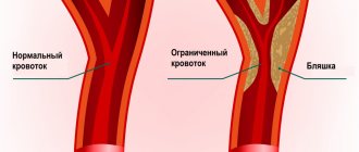
The widespread prevalence of cardiovascular diseases (CVDs) and the continued high mortality rate from them necessitate the introduction into clinical practice of modern effective methods for the early diagnosis of these pathologies.
In recent years, there has been a steady trend of “rejuvenation” of CVD, including myocardial infarction, both among men and women of working age.
Diagnostics in cardiology
Diagnosis of heart disease begins with questioning the patient and examination. The result is a preliminary diagnosis and a plan for further management of the patient. Using laboratory and instrumental methods of further examination, the cardiologist can assess the severity of the disease and decide on further tactics for managing the patient. When a doctor examines a patient, he pays attention to skin color, the presence or absence of shortness of breath, swelling, etc. Quite often, in patients with heart disease and the development of cardiac failure, one can see “blueness” of the lips, tip of the nose, fingers, especially under the nails. And the cardiologist calls this “cyanosis” acrocyanosis (peripheral cyanosis). There are also patients whose skin everywhere on all parts of the body has a “bluish” color - this often happens with very severe heart lesions, heart defects, diseases of the lungs, and body. A cardiologist calls this “cyanosis” central cyanosis. When examining a patient, the cardiologist also pays attention to the pulsation in large vessels, thanks to which a lot of information can be obtained about the state of the cardiovascular system.
24-hour Holter ECG monitoring
Depending on the nature of the pathological process, the clinical picture at the current time may not provide clear criteria for establishing a clinical diagnosis. In this case, the doctor prescribes diagnostic studies carried out over a wider time range, during the patient’s daily activities, allowing not only to monitor the activity of the cardiovascular system, but also to identify those trigger factors that lead to pathological changes. This group of studies used in outpatient practice includes 24-hour Holter ECG monitoring (Holter CMECG) and 24-hour blood pressure monitoring (ABPM).
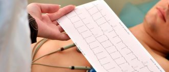
The 24-hour ECG monitoring system consists of an ECG recorder (which the patient usually wears on his belt in the provided case) and a system of electrodes (wires) attached to the patient's body. At the end of the study, the doctor transfers the ECG data into a computer program, and after performing digital analysis, interprets the results and draws up a medical report.
Indications for daily ECG monitoring are:
- suspicion of cardiac arrhythmia and conduction disturbances;
- suspicion of coronary heart disease;
- assessment of the correct operation of the artificial pacemaker (pacemaker);
- history of fainting, attacks of dizziness and sudden weakness.
To conduct the study, it is important to properly prepare the skin for the placement of electrodes: the hair at the places where the wires are connected is shaved off, and the skin is degreased. It is advisable for the patient to wear loose, comfortable clothing during the examination. Water procedures (bathing, showering) are excluded for the duration of the SMEKG.
During the study, the patient leads a normal lifestyle (works, plays sports, walks), recording all complaints that arise during the monitoring process in a special diary. In addition, the diary indicates possible medication intake and changes in types of physical activity.
Electrocardiography
Quite often, a patient comes to a cardiologist with previously taken ECGs. This is not surprising, because the ECG shows the cardiologist the state of the atria, ventricle, and how the heart feels at the moment, whether it receives enough blood, whether there are rhythm disturbances. All this can be read from the ECG, but information from the ECG is information here and now at some fraction of a second, but also if we talk about previously occurring disasters, such as myocardial infarction, then the ECG can tell. To have even more detailed information (this is often to understand what the blood supply to the heart is like when the patient moves, sleeps, laughs, cries. This method is called Hm-ECG (this is an ECG during the day). Such a study can give a cardiologist a lot of information, because that this method monitors the patient’s condition for 24 hours and the likelihood of detecting changes can be much greater, since there is a lot of time involved in monitoring the condition of the heart. Thanks to ECHO-CG, it is possible to uniquely and safely determine the condition of the heart and large vessels for the patient. This method allows you to assess the condition of the heart valves, see how the walls contract, how actively and in what volume blood is ejected from the heart, see with the help of a Doppler study the direction and what intensity the blood moves through the parts of the heart.
Instrumental diagnostic methods
Electrocardiography (ECG)
Electrocardiography (ECG) is one of the basic methods for diagnosing cardiac pathologies, without which not a single examination can be performed.
ECG - a method of studying the electrical activity of the heart has several varieties:
- ECG – standard.
- ECG – mapping.
- Holter monitoring ECG.
- Bicycle ergometry.
Using an ECG, electrical potentials are recorded in all parts of the heart, as well as the characteristics of the passage of impulses through the conduction system of the heart.
The ECG technique reveals:
- rhythm disturbances;
- change in heart rate;
- heart attack;
- ventricular hypertrophy;
- ischemic and cardiodystrophic changes.
To measure the electrical potentials of the heart muscle, a special device is used - a cardiograph. Sensors are placed on the patient's body, and the signals received from them are displayed on paper or film using recorders.
Electric potentials are shown on a graph as different lines. For each line on the ECG tape, strict normal parameters are defined, deviations from which indicate disturbances in the functioning of the heart.
The graph is studied using mathematical methods (advanced models of cardiographs perform this work automatically). Based on the results obtained, the doctor gives a conclusion indicating the parameters of the heart and its conduction system: heart rhythm, heart rate, electrical axis of the heart, conductivity, pacemaker.
ECG mapping
ECG mapping is a modern modified ECG method in which the recorded cardiac impulses are recorded in the form of cartograms. The method is based on recording multiple (from 64 to 224) ECG leads from the entire surface of the chest. When analyzing the data obtained, distribution maps are compiled, consisting of successive phases of the cardiac cycle. This significantly increases the diagnostic capabilities of the ECG, especially if the pathological process is difficult to detect with standard 12 leads.
Bicycle ergometry is a testing technique during which the patient performs dosed physical exercise on an exercise bike or treadmill while recording electrical potentials. The method makes it possible to detect latent heart failure or rhythm disturbances. Symptoms in such cases appear only with increased physical activity.
24-hour Holter blood pressure monitoring
24-hour Holter blood pressure monitoring is an examination in which blood pressure measurement is combined with an electrocardiogram recording.
The device for measuring pressure consists of a cuff, a sensor that captures the pulse waves of the artery, a connecting tube and a recorder that records pressure readings over time. A cuff connected by a tube to a microprocessor is fixed on the patient's shoulder. The device operates automatically: air is pumped into the cuff at specified intervals. The air is then gradually released and pressure levels are recorded on an electronic storage device.
Additionally, electrodes are installed on the patient’s chest, which send data about the electrical impulses of the heart to the device’s memory card.
Holter ECG monitoring for 24 hours or more allows you to detect heart rhythm disturbances and assess their frequency. Holter cardiac monitoring is a method of continuously recording an ECG during a person's normal daily activities. During the examination, the patient keeps a diary in which he records all types of activities (morning exercises, walk, rest, and so on). The doctor, analyzing the ECG readings, compares them with the patient’s records. This allows you to find out quite accurately what actions provoked certain changes in the cardiogram.
Echocardiography
Echocardiography is a study of the heart using ultrasound. Through a special sensor, which is applied to the chest in the area of the projection of the heart, ultrasonic waves propagate deep into the organ. The waves reflected from the tissues return to the sensor and are converted into electrical signals, which, after computer processing, are displayed on the monitor screen in the form of an image.
Echocardiography reveals:
- Congenital, acquired heart defects.
- Intracardiac thrombi.
- Damage to valve flaps.
- Hypertrophy or hypotrophy of the heart chambers.
- Free fluid in the pericardium (the sac around the heart).
- Pathologies of large vessels (aortic aneurysm).
- Changes in the speed and direction of internal blood flow
- Neoplasms in the heart muscle.
In addition, the method makes it possible to assess the anatomy and functional state of the heart: the shape and size of the organ, the thickness of the heart wall, the volume of the cavities of the atria and ventricles, the condition of the valves, pressure in the cavities, blood flow speed and other characteristics.
The procedure does not require special preparation. It is recommended several days before the ultrasound to limit the consumption of strong drinks, tea, coffee, alcohol and other energy drinks that affect the functioning of the heart. To obtain reliable results on the day of the procedure, the patient should avoid psycho-emotional unrest, physical stress, and quit smoking.
The most common in medical practice is the transthoracic method, when the examination is carried out through the anterior wall of the chest. If there are contraindications to this method (obesity, pulmonary emphysema, prosthetic heart valves), when acoustic barriers prevent the free passage of ultrasound waves to the object of study, echocardiography is performed by placing the sensor in the esophagus.
Transesophageal echocardiography provides a clearer image of the heart structures due to the proximity of the transducer and the absence of dense tissue or bone in the ultrasound path.
Doppler examination of the heart and blood vessels
The study is based on the Doppler effect, when ultrasonic waves are reflected from moving blood cells (erythrocytes) with a changed frequency.
The Doppler mode of echocardiography allows you to assess blood flow in the chambers of the heart and great vessels, identify reurgitation, determine the ejection fraction and stroke volume.
Angiocardiography
Angiocardiography is an X-ray diagnostic method that involves introducing a contrast agent into the vascular bed and taking a series of images of the heart and blood vessels. The method combines three studies:
coronary angiography - an image of the coronary arteries supplying the heart;
left-sided ventriculography – obtaining images of the cavity of the left ventricle when it is filled with contrast;
examination of the right heart (atrium and ventricle) and pulmonary artery.
The method is invasive and is performed according to strict indications for the diagnosis of congenital and acquired defects of the heart and great vessels. often before heart surgery to clarify myocardial parameters.
Exercise tests
There is another very interesting method for identifying the degree of decrease in blood circulation (this is the so-called myocardial ischemia) these are tests with physical activity: veloergometry (the patient with the ECG electrodes connected is on a bicycle and pedals). The state of the heart and blood vessels is assessed in response to physical activity. A similar method is the treadmill test (only the patient is on a treadmill). Thus, we see that thanks to the combination of all examination methods, it is possible to collect a detailed picture about this disease. Thus, the cardiologist has a clear picture of the disease and is already moving along the precisely planned path of treatment for this disease.
Expert electrocardiographers
Professional cardiographic systems designed specifically for cardiology clinics and departments. Manufactured using the latest technical advances, electrocardiographs are distinguished by their modern ergonomic design, functionality and ease of use. The high-resolution touch screen displays all 12 leads. The quality of electrocardiogram printing will satisfy even the most demanding cardiologist.
Description of ECG
The ECG graph analyzes:
- An isoelectric line is a horizontal line recorded during a period when no excitation of activity is noted in the heart). It is the starting point for the changes listed below;
- teeth - deviations from the isoelectric line (the upper tooth is called positive, the lower - negative); The teeth in the figure are: P, Q, R, S, T, U
- a segment is an isoelectric line between teeth; the figure shows the segment ST and PQ
- intervals are fragments of the ECG graph that include ……… and the adjacent tooth; The figure shows the QT and PQ
In the ECG display we see (from left to right):
- P wave - characterizes depolarization of the atrial muscle
- segment PQ – reflects the time of depolarization through the atrioventricular node
- PQ interval - reflects the time of depolarization from the sinoatrial node to the atrioventricular node
- QRS complex – corresponds to depolarization of the ventricular myocardium
- QT interval - reflects the time of the action potential of the ventricular myocardium (depolarization + repolarization)
- ST segment – reflects the period of ventricular repolarization
- T wave - corresponds to ventricular repolarization
- U wave - not observed in practice