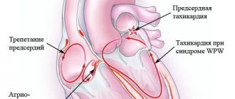
What is sinus bradyarrhythmia in children?
Sinus bradyarrhythmia is a congenital or acquired heart disease in which the beat frequency decreases to a critical level. As a result, the patient’s blood circulation slows down, internal organs and systems receive less oxygen and nutrients, and suffer from hypoxia. Pathology can occur at any age, but the most dangerous consequences are observed in children.
Sinus bradyarrhythmia in a child is diagnosed if the number of beats per minute does not exceed 60 times. The development of the disease is not always accompanied by visible symptoms and disorders.
It occurs more often in premature babies, with some forms of cerebral palsy. The reason lies in the dysfunction of the sinus node, better known as the pacemaker. There are moderate and severe forms of the disease.
Moderate
Moderate bradyarrhythmia in rare cases is a congenital anomaly of the body. Sometimes this feature appears in children who are actively involved in sports. But any form is dangerous to health and can disrupt the formation of organs and brain development. Unpleasant symptoms often appear when air is exhaled from the lungs.
Sinus bradyarrhythmia in a child is when the number of beats per minute is less than normal
Often, a moderate form of the disease appears with constant hypothermia and is formed as a protective reaction to low temperatures. On inspiration, the pulse is restored to normal levels, so diagnosis is difficult and requires a combination of different techniques.
Expressed
When the heart rate drops below 40–45 beats per minute, a diagnosis of severe bradyarrhythmia is made.
The following signs indicate it:
- frequent burning in the heart area;
- the child looks weak, sick, drowsy;
- The baby gets tired quickly with light loads.
The form of the disease is dangerous due to sudden attacks with loss of consciousness and memory. They worsen children’s general well-being, school performance, and limit communication with peers.
Sinus bradycardia: causes, dangers and treatment
Sinus bradycardia is a violation of the heart rate, expressed in its slowing down to less than 60 beats per minute. It can be either a variant of the norm or indicate pathology. The physiological form is often observed in athletes; the norm is a slow heartbeat in sleeping people.
Risk factors, types
Physiological bradycardia indicates that in humans the inhibitory effect of the nervous system on the activity of the heart predominates.
Physiological sinus bradycardia does not cause concern in the following cases:
- heart rate of at least 50 beats per minute;
- no complaints from the patient;
- The rhythm is not disturbed (the same time intervals between beats).
Pathological form
Pathology includes a slowing of the heart rate in the following cases:
- absence of prerequisites (there are no factors that could lead to bradycardia);
- manifestation of bradycardia with sudden attacks;
- arrhythmic pulse;
- There are complaints about poor general health.
8
24/7
Causes
Sinus bradycardia is not a disease in itself. This is a symptom that may accompany the development of other diseases. These include:
- vegetative-vascular dystonia;
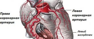
- acute and chronic forms of heart disease (these are myocarditis, heart attacks, cardiomyopathies, weakness of the sinus node);
- brain lesions (TBI, meningitis, tumors, meningoencephalitis);
- excessive drinking, smoking, drug use;
- intoxication with chemicals, work in hazardous industries;
- severe infections (associated with damage to any organ - intestinal infections, pneumonia, abscesses);
- cancerous tumors of the neck and chest area;
- the use of medications that slow down the heart rate (asparkam, verapamil, etc.);
- diseases of the endocrine system (adrenal insufficiency, hypofunction of the thyroid gland, manifested by hypothyroidism);
- severe liver and kidney diseases.
Symptoms
The severity of clinical manifestations is directly related to how many beats the pulse becomes slower.
With a heart rate in the range of 50 - 60 beats, the clinical picture looks like this:
- complaints about the general condition – dizziness, weakness are practically absent;
- consciousness and breathing remain normal;
- blood pressure does not change.
Thus, in this case, only a slow pulse will indicate bradycardia.
With a pulse between 30 and 40, the symptoms are as follows:
- there are complaints of general malaise, dizziness, headache and chest pain, shortness of breath;
- changes in consciousness are observed, which are manifested by drowsiness, lethargy;
- blood pressure decreases;
- the pulse not only slows down, but also becomes weak;
- breathing quickens, shortness of breath appears.
When the pulse is 30-39 beats, severe sinus bradycardia occurs:
- the complaints listed in the paragraph above become pronounced, the patient is forced to remain in a supine position;
- general condition – critical;
- disturbances of consciousness are manifested by coma, i.e. consciousness is absent;
- blood pressure is greatly reduced;
- pulse is not detected;
- shallow breathing (may be absent).
Sinus bradycardia can manifest itself in different ways: be paroxysmal or chronic. The attack can last several minutes, hours, days. The chronic form is more favorable from the point of view of prognosis, especially if the deceleration is small - from 50 to 60 beats. In such cases, the body can readjust and adapt to the slower rhythm.
Sudden attacks are very dangerous, especially if the slowing of the heartbeat was preceded by tachycardia.
8
24/7
Diagnostics
The primary diagnosis can be made based on the patient's complaints and examination. It can be reliably confirmed or excluded by performing an ECG.
Sinus bradycardia on the cardiogram will be reflected by an increase in the intervals between the ventricular complexes and the correct sinus rhythm.
With this diagnosis, the cardiogram is almost identical to normal, with the exception of rarer contractions. If at the time of the cardiogram it was not possible to register an episode of bradycardia, then a Holter study should be performed. This procedure involves the patient wearing a special device during the day, which automatically takes and records all the necessary indicators.
Ultrasound is used to detect lesions of the organic nature of the heart.
Laboratory tests may also be performed. They include analysis of the electrolyte composition of the blood, analysis of hormones, determination of toxins, as well as bacteriological analysis if infectious causes of palpitations are suspected.
Treatment
If physiological sinus bradycardia occurs, no treatment is required.
Indications for treatment are:
- dyspnea;
- fainting;
- general weakness;
- hypotension;
- heart rate drops to 40 beats/min.
Sinus bradycardia requires outpatient treatment, but in severe cases the patient may be hospitalized.
Drug treatment
How to treat sinus bradycardia if it is not physiological?
For bradycardia, it is necessary to take drugs that stimulate contractions and also neutralize the effect of the autonomic nervous system on the conduction pathways. Such medicines include:
- Anticholinergics. Atropine - inhibits the activity of the parasympathetic nervous system, thereby accelerating the heart rate. The drug is intended for injection and can be used subcutaneously or intravenously. It is characterized by a number of side effects, so it is not suitable as a drug for regular use, but it is effective as an emergency aid.
- Adrenergic agonists. Isoprenaline – intended for oral or intravenous use. This is an analogue of adrenaline, simulating the sympathetic department of the autonomic nervous system and activates the functioning of the heart receptors.
- Eufillin. This is a bronchodilator, but it is also indicated when it is necessary to increase the pulse rate. Admission is carried out by intravenous infusion.
It is advisable to take these medications during the period of recovery of the heart rate; when the indicator is brought to normal, they are canceled.
It is also possible to use herbal medicine. Eleutherococcus and ginseng will help increase heart rate and raise blood pressure.
In the chronic form of sinus bradycardia, it is necessary to use drugs aimed at activating metabolic processes. This:
- antioxidants and cardioprotectors (antioxycaps, omega-3);
- drugs that stimulate metabolism in cardiomyocytes (mildronate);
- nootropics (piracetam);
- vitamin complexes.
Complex forms of bradycardia, accompanied by loss of consciousness and developing against the background of lesions of an organic nature, are eliminated through surgical intervention. In such cases, a pacemaker is installed.
Emergency help
Emergency assistance is necessary when the patient experiences a deterioration in their general condition due to a decrease in heart rate of less than 50 beats per minute.
It is necessary to place the patient on his back and provide access to oxygen.
After this, you should check for consciousness and breathing. If they are absent, cardiac massage and artificial respiration are performed.
The following medications can be used:
- Zelenin drops. Used for oral administration in all cases of bradycardia while maintaining consciousness.
- Atropine sulfate. It is administered by injection - subcutaneously or intravenously when the heart rate decreases below 50 beats per minute.
- Dopamine. Taken when heart rate drops below 50 beats per minute with a drop in blood pressure.
- Adrenalin. Administered intravenously or subcutaneously for cardiac arrest (heart rate less than 30 beats per minute).
Forecast
Elimination of symptoms of sinus bradycardia is observed in 95% of cases.
In almost all patients (90-95%), drug treatment is sufficient.
5-10% of cases require implantation of a pacemaker. This procedure restores normal rhythm in all cases immediately after the intervention.
If treatment is not started when a problem is detected, complications may develop. Negative consequences include worsening manifestations of ischemia, myocardial infarction, stroke, heart failure, and cardiac arrest.
Prevention
First of all, preventive measures should include a healthy lifestyle. This includes giving up drinking alcohol, smoking, and controlling your diet. You will need to exclude sweets and fatty foods from the menu. It is necessary to play sports, but it is recommended to discuss the amount of permissible physical activity with your doctor.
Any diseases must be eliminated immediately after detection to prevent them from becoming chronic.
Medicines may only be taken as prescribed by a specialist. You should not try to eliminate the symptom yourself.
If alarming symptoms occur, you should immediately consult a doctor, undergo examinations and begin heart treatment if pathology is detected.
If there are any abnormalities in the functioning of the heart, it is imperative to undergo preventive examinations. And the sooner the patient signs up for a consultation, the better.
8
24/7
Why does the disease develop in children and adolescents?
In newborn babies, the heart beats at a frequency of 120–140 beats per minute. As you grow and mature, your heart rate decreases to a stable rate of 65–75 times. If the level drops below 60 beats, an examination is necessary to exclude dangerous pathologies. They develop as a result of inflammatory or congenital anomalies of the sinus node, increased tone of the vagus nerve.
Among the causes of sinus bradyarrhythmia in preschool age:
- complications after suffering from pneumonia, tonsillitis, otitis media;
- constant hypothermia;
- intracranial pressure;
- predisposition to cardiovascular pathologies;
- metabolic disorders and thyroid dysfunction;
- oncology.
In adolescents, the cause of sinus bradyarrhythmia is often associated with improper formation of hormonal levels. In rare cases, the child’s body reacts to taking energy drinks, illegal drugs, or poor eating habits (bulimia or anorexia in girls 13–15 years old).
Signs of illness
Sinus bradyarrhythmia in a child is sometimes asymptomatic and is detected by chance during a comprehensive diagnosis of other diseases. The main signs depend on the age, developmental characteristics of children, and the stage of the disease. Depending on the cause, the problem may be physiological, organic, neurogenic, drug-related or toxic.
Your child may have the following symptoms:
- increased sweating at rest;
- feeling of lack of air;
- loss or confusion of consciousness;
- heaviness in the chest;
- poor appetite;
- low concentration, deterioration in school performance;
- discharge on the face of the nasolabial triangle.
Tingling or burning in the heart with bradyarrhythmia always occurs when exhaling. If you hold your breath for a few seconds, the discomfort disappears.
Physiological
For natural reasons, a slowdown in heart rate is observed during a night's rest or daytime sleep, after waking up. At rest, the pulse drops to 65–60 beats, the changes are practically not felt by the child. During physical activity or active play, the indicator rises to a normal and comfortable level.
Among the dangerous physiological causes of the disease is the appearance of benign or malignant tumors in the heart area, on the myocardial muscle. The child complains of shortness of breath and tingling in the chest, has pale skin, and may lag behind peers in development in terms of weight and height.
Organic
Bradyarrhythmia can form due to improper development of the sinus node in the heart. The number of beats per minute decreases to 45–50 units, and deformation and necrosis of myocardial tissue begins.
The problem occurs against the background of serious diseases:
- vascular ischemia;
- cardiosclerosis;
- cardiomyopathy.
In a young child, sinus bradyarrhythmia of the organic type develops with inflammation of the pericardium of the heart. This is a dangerous complication after a sore throat or otitis media, in which characteristic symptoms are observed: constant low-grade fever, muscle weakness, low activity in play and study, joint pain.
Neurogenic
Sinus bradyarrhythmia in a child can be a consequence of pathologies of the nervous system: intracranial pressure, childhood neuroses, vegetative-vascular dystonia.
In addition to pain in the heart area, young patients feel:
- dizziness;
- low blood pressure;
- attacks of panic or irritability.
The problem requires an integrated approach to treatment, the help of an endocrinologist, cardiologist and neurologist.
Medicinal
In childhood, bradyarrhythmia of this type develops extremely rarely. It occurs in children with asthma, kidney pathologies or severe lung diseases, who constantly take potent drugs to maintain vital functions. Damage to the sinus node of the heart can occur as a side effect after undergoing chemotherapy for oncology.
Toxic
Increasingly, doctors are observing babies who have sinus bradyarrhythmia after toxic poisoning. The reason is associated with living in a disadvantaged area, constant contact with toxic substances in the house, kindergarten, and school. Damage to the heart is caused by pathogenic bacteria and viruses in typhoid fever, hepatitis A or B.
In everyday practice, a doctor often has to deal with the phenomenon of bradycardia in children. Bradycardia is a rare heartbeat or a slow heart rate below normal for age.
The criteria for bradycardia in children of different age groups have significant differences. So, for example, for an 8-year-old child, bradycardia is considered to be a decrease in heart rate (HR) of less than 66–74 beats per minute, and for a 2-month-old child, a heart rate of less than 119–144 will be considered bradycardia. In an adult (over 18 years of age), bradycardia is a decrease in heart rate below 59 beats per minute. The older the child, the slower the frequency of his rhythm becomes. A baby is born with a heart rate of 140–160 beats per minute, and by the age of 8–11 years, its rhythm slows down to 75–95 beats per minute. This is due to the fact that as the child grows, the volume of blood circulating in the body increases, the size of the heart increases, the ability of the heart muscles to contract decreases, the vessels become less elastic, and the length of the vessels in the body increases; the susceptibility of the cardiovascular system to vasoactive substances circulating in the blood changes. For example, a newborn baby’s heart weighs only 15–25 g; it can push only 3–4 ml of blood. The heart of a healthy adult weighs about 200–250 g. Such a heart can push out from 70 to 90 ml of blood with one contraction. Therefore, normally, the heart rate in children will be higher than in adults, and will depend on age, height, weight, physical activity, sleep or wakefulness. Accordingly, with a physiologically high heart rate in a child, bradycardia values will be in a certain range for each age group.
The prevalence of bradycardia in practically healthy children without obvious heart pathology is quite high. Most often, changes are detected by chance during preventive examinations, clinical examinations, and during examinations after illnesses (mainly of an infectious nature). At the same time, children with bradycardia most often do not make complaints about poor health, tolerate physical activity well, and lead a normal lifestyle. But, unfortunately, bradycardia may not always be asymptomatic and harmless.
The reasons for bradycardia in children can be different. More often, bradycardia in a healthy child is a normal reaction of the body to accelerated growth and rapid/or slow weight gain (this is especially typical for young children - up to 3 years old, as well as during adolescence). If a child regularly attends a sports section, and physical activity is common for him, then such a small athlete will most likely have bradycardia. Also, a slowing of the heart rate without clinical manifestations occurs in children with underweight, with retarded physical development, in children who are often ill, after suffering from colds and infectious diseases, in premature infants.
What happens in the body during bradycardia?
When heart rate decreases, the speed of blood movement through the vessels also slows down. This leads to a decrease in the supply of oxygen and the removal of carbon dioxide in organs and tissues of all systems. The organs most vulnerable to hypoxia are affected first: the brain and heart. Therefore, with bradycardia, a child may experience dizziness, headaches, weakness, increased fatigue (cannot tolerate even moderate physical activity), the child wants to sleep all the time, concentration decreases, appetite decreases, and fainting may occur. If your child faints, you should consult a doctor immediately. For the most part, such loss of consciousness is not dangerous, but there are a number of clinical conditions that still require immediate treatment.
Therefore, a child with bradycardia should be monitored by a cardiologist, undergo timely preventive examinations and, if necessary, receive treatment. The doctor determines the risks and dangers of bradycardia for each small patient individually, based on complaints, life history, physical fitness, clinical manifestations, finding out whether the child is taking any medications that slow down the heartbeat, and whether there are any concomitant diseases that can lead to bradycardia ( kidney disease, heart disease, thyroid gland, pancreas disease, obesity, diabetes, neurosis...). And one of the most important points is assessing the dynamics of heart rate using the ECG (what bradycardia is - moderate/severe, regular/irregular, is it stable). There may be other more serious rhythm disturbances, accompanied by a rare pulse - atrioventricular block of the 2nd and 3rd degree, sinoatrial block of the 2nd and 3rd degree, sick sinus syndrome. In such conditions, it may be necessary to install an artificial pacemaker (pacemaker). Having received all the information about the child’s condition, the doctor, if necessary, will prescribe an additional examination (usually an ECG for rhythm, an ECG with stress (physical, drug), Holter ECG monitoring, echocardiography, blood tests - CBC, determination of blood viscosity, electrolyte composition , study of hormonal status, etc.).
Parents of children observed with bradycardia should properly organize the optimal motor mode for the child (on the recommendation of a cardiologist), monitor the fullness of sleep, and avoid overwork. It is important to maintain normal body weight and eat right. Avoid salty, smoked, spicy and fatty foods, as well as fizzy drinks. Increase the amount of foods containing elements and vitamins “necessary” for the heart - bananas, oranges, persimmons, lettuce, spinach, pumpkin, potatoes, dried apricots, dairy products, fish, porridge (millet, rolled oats, buckwheat).
Results: Most often, bradycardia in children is temporary and as they grow older, it can disappear without a trace. In children, this condition is quite easy to correct if necessary. But the decision about what to do with bradycardia and whether it needs to be treated is made only by a cardiologist.
Is it normal for a child who plays sports to develop the disease?
At the initial stage of bradyarrhythmia, the child leads an active lifestyle. But excessive sports activities in preschool age can provoke disturbances in the development of certain parts of the heart. This causes the appearance of painful symptoms during the transition period.
With regular sports activities in children, in 90% of cases the physiological type of the disease is diagnosed.
With a constant load, the heart gets used to working at an increased pace for several hours a day, so at rest the pulse drops to 60 beats per minute: this is how the body “forces” the myocardial muscles to rest between heavy workouts. This condition does not cause concern among doctors, does not entail dangerous complications, and is detected in 50% of professional athletes, football players, and cyclists.
Some features of the ECG in children involved in sports
S.A. IVYANSKY, L.A. BALYKOVA, A.N. Urzyaeva, L.S. ZAGRYADSKAYA, Yu.O. SOLDATOV, A.V. SAMARIN
Mordovian State University named after. N.P. Ogareva, Saransk
Children's Republican Clinical Hospital of the Ministry of Health of the Republic of Moldova, Saransk
Republican medical and physical education clinic of the Ministry of Health of the Republic of Moldova, Saransk
Ivyansky Stanislav Alexandrovich
Art. Lecturer at the Department of Pediatrics, Medical Institute, Mordovian State University. N.P. Ogareva
430032, Saransk, st. Rosa Luxemburg, 15, tel. (8342) 35-30-02, e-mail
The article presents the results of an ECG examination of 198 children aged 10-15 years playing football and tennis, with a sports experience of 2-5 years (initial training group) and 5-7 years (special sports training group).
It has been established that the level of average heart rate in athletes differs from the corresponding indicator in peers who do not engage in sports after 5 years of regular exercise, while the level of the minimum heart rate of athletes remains at a comparable value. Limits have been established for the duration of the QTc (460-480 ms), requiring additional diagnostic measures.
ECG changes requiring additional examination (bradycardia less than 2%, AV block of the second degree, ST-T changes, heart rhythm disturbances, prolongation of the QTc interval above 460-480 ms) were recorded in 4.9% of cases in the initial training group and in 9% of cases in the special sports training group. Key words:
child athletes ECG, heart rate, QT interval, signs of maladaptation.
SA IVYANSKY, LA BALYKOVA, AN URZYAEVA, LS ZAGRYADSKAYA, Yu.O. SOLDATOV, AV SAMARIN
Mordovian State University named after NP Ogarev, Saransk
Children Republican Clinical Hospital of the Ministry of Health of the Republic of Mordovia, Saransk
Republican medical exercises dispensary of the Ministry of Health of the Republic of Mordovia, Saransk
Some features of electrocardiography in children going in for sports
The article gives the results of electrocardiography of 198 children 10-15 years old, playing football and tennis with sports experience of 2-5 years (group of initial training) and 5-7 years (group of special athletic training).
It was found that the level of the average heart rate of the athletes is different from the corresponding figure of those not involved in sports after 5 years of regular exercise; the level of the minimum heart rate of athletes is at a comparable value. We set the limits of the interval length QTc (460-480 ms), requiring additional diagnostic procedures.
ECG changes that require further investigation (bradycardia less than 2%, AV-block II degree, ST-T changes, arrhythmias, QTc interval prolongation above 460-480 msec) were recorded in 4.9% of cases in the group of initial training and 9 % in the group of special sports training. Key words:
young athletes, ECG, heart rate, QT duration, desadaptation sings.
Currently, the attention of sports specialists continues to be drawn to the state of the cardiovascular (CVS) system as it determines the level of sportsmanship and health status of young athletes. The most accessible and informative method for examining the cardiovascular system in athletes continues to be standard electrocardiography (ECG). Despite its simplicity and accessibility, the economic feasibility of widespread implementation of ECG screening in sports practice continues to be actively discussed in America [1], although European specialists have been using this approach since the late 80s [2-4]. In our country, an ECG performed at rest and during exercise is included (according to Order of the Ministry of Health of the Russian Federation No. 613) in the list of required examination methods for admission to mass and professional sports and is carried out at least 2 times a year during sports activities [5] .
Interpretation of ECG data from examinations of young athletes can cause certain difficulties, since the available regulatory documents are based mainly on the results of examinations of adult athletes [2, 4-6]. An additional number of problems arise due to the fact that even in practically healthy children and adolescents who do not engage in sports, many ECG parameters vary within very wide limits [7, 8]. In addition, gender, age, living conditions, constitutional features, psycho-vegetative status and the nature of training loads have a significant impact on the electrophysiological parameters of the myocardium in young athletes (as well as athletes over 18 years of age).
The only large-scale study in this direction was carried out by V.N. Komolyatova et al. (2013) and concerned “elite” athletes (members of youth Olympic teams) aged 15-18 years [8]. However, in real everyday practice, doctors are much more likely to encounter children involved in sports at the initial preparation stage (1-3 years of classes) and the educational and training stage (4-6 years of classes), and untimely detection of cardiac pathology in them can lead to the most serious consequences .
That is why our study set the goal of identifying the features of a standard ECG in child athletes with no more than 6 years of training experience, in comparison with healthy peers leading a normal lifestyle, and determining the interpretation of the results obtained.
Materials and methods
Using standard 12-channel electrocardiography (Schiller AT-1), during a mandatory examination, 192 children and youth sports school students aged 11-14 years (among them 158 boys and 40 girls) involved in football and tennis were examined. The criteria for inclusion in the study were: availability of access to sports activities based on the results of a previous in-depth medical examination (IME) and the absence of acute and chronic diseases in the acute stage at the time of examination. Athletes with clinically significant somatic pathology (bronchial asthma, Gilbert's disease, atopic dermatitis, etc.), including those taking certain medications for medical reasons, were excluded from the study.
The examination was carried out during the inter-competition period. All child athletes were at the stage of initial or educational training and were divided into groups according to sports affiliation and level of sports skill (Table 1). Thus, 4 study groups were formed, with group No. 1 in each sport including children training for 2-3 years, and group No. 2 - for 4-6 years, with a load intensity of at least 6-6 years. 7 hours a week.
Table 1.
Age and sex characteristics of the study and control groups
| boys | girls | duration of classes | average age, years | |
| Children playing football, No. 1 | 36 | 0 | 2,3+0,68 | 11,3+1,16 |
| Children playing football, No. 2 | 72 | 0 | 5,4+0,71 | 11,4+1,09 |
| Children playing tennis, No. 1 | 24 | 22 | 2,4+0,84 | 12,1+1,32 |
| Children playing tennis, No. 2 | 26 | 18 | 4,7+0,57 | 13,1+1,03 |
| Control group No. 1 | 10 | 0 | 12,4+1,25 | |
| Control group No. 2 | 7 | 7 | 0 | 12,6+1,09 |
Control groups were selected on the “experience-control” principle for young football players from healthy untrained boys of the same age, and for tennis players from children comparable to them in gender and age.
ECG data were analyzed manually at a recording speed of 50 mm/s, measuring the duration of the RR, PQ, QT intervals, the polarity and height of the main ECG waves, assessing the nature of the basic rhythm, the presence of cardiac rhythm and conduction disorders, as well as disorders of the final part of the ventricular complex (ST segment and T waves). The corrected QT interval was calculated using the Bazett formula. The data obtained were interpreted according to the recommendations of L.M. Makarov [7].
The research results were subjected to statistical processing using the statistical software package Statistica7.
Results and discussion
Analysis of the results of a standard resting ECG indicated a lower level of average heart rate in group of football players No. 2, who had been training for more than 3 years (p<0.05), relative to the corresponding control group (Table 2). In the group of tennis players with similar experience in sports, the average heart rate values, although they were significantly lower than the corresponding indicator in control group No. 2, did not reach statistically significant differences (p>0.05). This fact is probably due to the peculiarities of the training process in a sport such as tennis, in the form of a significant proportion of special exercises and technical training and relatively small volumes of speed-strength load, which probably somewhat slows down the processes of cardiovascular remodeling in the initial stages of training.
It is worth noting that the values of the minimum heart rate in the groups of athletes, regardless of the length of experience in sports, did not reach statistically significant differences (p>0.05) with the same indicator in the control groups (Table 2). This fact is in good agreement with the results of observations of highly trained young athletes conducted at the Center for Syncope and Cardiac Arrhythmias in Children of the Federal Medical and Biological Agency of Russia [8] and makes it possible to interpret the changes in heart rate detected in athletes from the perspective of population norms. Thus, we believe that heart rate values that are in the range of 2-5% for a population of children of the same sex who do not engage in sports should be considered unphysiological for child athletes, and pathological - below 2%. It is bradycardia of this level that should be considered as potentially dangerous and requiring additional in-depth examination.
Table 2.
Some ECG indicators in child athletes
| Football No. 1 | Football No. 2 | Tennis No. 1 | Tennis No. 2 | Control No. 1 | Control No. 2 | |
| Wed. Heart rate, beats/min | 74,1+6,42 | 70,8+2,21* | 75,5+4,37 | 72,8+4,92 | 80,9+6,85 | 82,6+7,38 |
| Min heart rate, beats/min | 58,3+5,97 | 56,2+4,89 | 59,3+4,77 | 57,1+5,3 | 60,8+6,45 | 58,2+6,28 |
| QT, ms | 343+17,3 | 354+14,3* | 338+15,9 | 351+12,8* | 340+14,8 | 347+18,6 |
| QTс, ms | 371+8,6 | 399+10,3* | 379+7,2 | 393+9,1 | 368+7,9 | 376+8,2 |
Note: * - differences in the corresponding values of the group of children playing football are significant at p<0.05
When analyzing the duration of the QT interval in young athletes, its increase was revealed, which was adequate to the slowdown in heart rate and at the same time reached significant (p<0.05) differences from the corresponding control indicators in both groups of athletes at the educational and training stage of preparation (training for over 3 years). As for the duration of the QTc interval, its differences from the indicator of untrained peers were recorded only in highly trained football players (group No. 2), which obviously reflects the process of formation of myocardial hypertrophy and confirms our previous statement about the powerful impact of speed-strength “ragged” loads and psycho-emotional stress (in such an aggressive sport as football) on the processes of myocardial remodeling. At the same time, a clear relationship between the duration of QTc and the duration of regular training was revealed both in the group of football players (r = 0.52) and in the group of tennis players (r = 0.58). This makes it advisable to monitor this indicator in young people during sports activities.
We identified 3 football players (1.6%) involved in a special sports training group with a QTc interval duration of more than 460 ms. Moreover, in one athlete, the extension of the electrical systole was combined with a single ventricular extrasystole. According to current recommendations, this required an in-depth examination and, in one case, withdrawal from sports activities. It is worth noting that recently the possibility of increasing the permissible duration of the QTc interval for adult highly trained athletes to 480 ms and even 500 ms (especially for women) has been actively discussed in the literature. However, in our opinion, for children under 18 years of age, a slowdown in the QTc interval over 460 ms should already be considered pathological and require the exclusion of congenital and acquired long QT interval syndrome.
In a large-scale study by V.N. Komolyatova revealed parity indicators of the average duration of the QTc interval and higher values of the average absolute QT in athletes compared to the population [8]. Although, of course, the proportion of people with a longer QTc interval among athletes will be slightly higher than among untrained people. Obviously, this fact is due to an increase in myocardial mass and neurohumoral changes as a result of regular and intense sports activities.
The average duration of the PQ interval in our study did not differ between athletes and practically healthy children leading a normal lifestyle, although the proportion of people with slowed atrioventricular conduction was slightly higher among athletes of group No. 2 (1.8-2.1%), relative to the control (0%). The average duration of the QRS complex was slightly higher among athletes, which is obviously due to the fairly high prevalence of incomplete blockade of the right bundle branch (12.8%) among the examined athletes.
Analysis of the frequency of various ECG syndromes in athletes confirmed the well-known opinion about the high prevalence of bradycardia among people involved in sports. The greatest prevalence of sinus bradyarrhythmia was recorded among athletes of special sports training, especially in the group of football players. In addition, in the same group, sinus node dysfunction was detected significantly more often than the control group (p<0.05): pacemaker migration (MPM) and sinoatrial block, as well as pre-excitation syndromes.
Other researchers demonstrate similar results [8,9]. Conduction disturbances such as atrioventricular (AV) block of the first degree, incomplete blockade of the right bundle branch of His, which are a classic “ECG attribute” of an athletic heart and regarded as physiological, were somewhat more common among athletes (p>0.05), predominantly in groups of special sports training (both among football players and among tennis players).
Second degree AV block and partial block of the left leg of the Gis were recorded only in the group of football players of special sports training and, according to international recommendations, were regarded as potentially dangerous and, of course, required additional examination. A single extrasystole (ventricular and supraventricular) was recorded significantly more often (p<0.05) in the group of football players of special sports training and was not found in the control and groups of athletes of initial training.
Changes in the ST-T segment in the group of football players at the training stage were recorded more often (p<0.05) than in the control group, where they were not recorded in any child. It is known that repolarization disorders in athletes may reflect the formation of working hypertrophy and/or electrolyte and neurohumoral changes. The nature and severity of these changes can vary widely among athletes, reflecting both adaptive changes, which should be regarded as benign, and the formation of potentially dangerous disorders, which are a sign of pronounced myocardial remodeling due to stress and physical overexertion [10].
It is probably not possible within the framework of this work to discuss in more detail the possible causes and mechanisms of the formation of repolarization disorders in athletes, but it should be said that: 1) T wave inversion of more than 1 mm in two or more leads except III, AVR and V1 (excluding V2 and V3 in women under 25 years of age), not disappearing or appearing after FN; 2) depression of the ST segment by 0.5 mm below the isoelectric line between point j and the beginning of the T wave in leads V4, V5, V4, I and AVL or by 1 mm in any leads; 3) a pathological Q wave with a depth of more than 3 mm or a duration of more than 40 ms in any leads, excluding AVR, AVL and V1, should be a reason to exclude organic pathology.
An analysis of the standard ECG findings showed that among athletes at the initial level of training, 4.9% (4 people) of children had pathological ECG results in the form of severe bradycardia in the region of 2-5%) and conduction disturbances such as AV block of the second degree. Among athletes in the sports training group, pathological and borderline ECG results in the form of changes in the ST segment, single extrasystole, conduction disturbances such as AV block of the second degree and pre-excitation phenomena (WPW) were recorded in 7% (8 people) of cases, mainly among football players.
conclusions
Thus, according to the results of the study, the average level of heart rate in athletes differs from the corresponding indicators of their healthy untrained peers (downwards) only after 3 years of regular exercise, while the minimum heart rate in all groups remains comparable. This fact allows us to speak about the possibility of assessing the criticality of bradycardia from the standpoint of ECG standards proposed for healthy peers leading a normal lifestyle.
The average values of the absolute QT interval in trained young athletes were higher than those in the control group, and in terms of the QTc interval, they did not differ for either the average or threshold values. The prevalence of potentially dangerous ECG changes requiring additional examination (bradycardia less than 2%, second degree atrioventricular block, ST-T disturbances, phenomena of ventricular preexcitation, extrasystole, prolongation of the QTc interval above 460-480 ms), among athletes in the special sports training group was 9%, while among athletes in the initial training group it was only 4.9%.
LITERATURE
1. Maron BJ, Doerer JJ, Haas TS et. al. Sudden deaths in young competitive athletes: analysis of 1866 deaths in the United States, 1980-2006. - 2009. - Vol. 119, No. 8. - R. 1085-92.
2. Corrado D., Basso C., Pavei A., Michieli P., Schiavon M., Thiene G. Trends in sudden cardiovascular death in young competitive athletes after implementation of a preparticipation screening program // JAMA. - 2006. - Vol. 296. - R. 1593-601.
3. Hevia AC, Fernández MM, Palacio JM, Martín EH, Castro MG, Reguero JJ ECG as a part of the preparticipation screening program: an old and still present international dilemma // Br J Sports Med. - 2011. - Vol. 45, No. 10. - P. 776-779.
4. Steinvil A., Chundadze T., Zeltser D., Rogowski O., Halkin A., Galily Y., Perluk H., Viskin S. Mandatory electrocardiographic screening of athletes to reduce their risk for sudden death proven fact or wishful thinking ? // J Am Coll Cardiol. - 2011. - Vol. 57, No. 11. - P. 1291-1296.
5. Order of the Ministry of Health and Social Development of Russia No. 613n. — Access mode https://www.rg.ru/2010/10/01/sport-dok.html, free.
6. Corrado D., Pelliccia A., Heidbuchel H. et al. Recommendations for interpretation of 12-lead electrocardiogram in the athlete // European Heart Journal. - 2010. - Vol. 31. - R. 243-259.
7. Makarov L.M. ECG in pediatrics. - M.: Medpraktika-M, 2006. - 544 p.
8. Standard ECG parameters in children and adolescents / ed. M.A. Shkolnikova. - Moscow, 2010. - 232 p.
9. Papadakis M., Sharma S. Electrocardiographic screening in athletes: the time is now for universal screening // Br J Sports Med. - 2009. - Vol. 43. - R. 663-8.
10. Uberoi A., Stein R., Perez MV et al. Interpretation of the Electrocardiogram of Young Athletes Circulation. - 2011. - Vol. 124. - R. 746-757.
REFERENCES
1. Maron BJ, Doerer JJ, Haas TS et. al. Sudden deaths in young competitive athletes: analysis of 1866 deaths in the United States, 1980-2006. - 2009. - Vol. 119, No. 8. - R. 1085-92.
2. Corrado D., Basso C., Pavei A., Michieli P., Schiavon M., Thiene G. Trends in sudden cardiovascular death in young competitive athletes after implementation of a preparticipation screening program // JAMA. - 2006. - Vol. 296. - R. 1593-601.
3. Hevia AC, Fernández MM, Palacio JM, Martín EH, Castro MG, Reguero JJ ECG as a part of the preparticipation screening program: an old and still present international dilemma // Br J Sports Med. - 2011. - Vol. 45, No. 10. - P. 776-779.
4. Steinvil A., Chundadze T., Zeltser D., Rogowski O., Halkin A., Galily Y., Perluk H., Viskin S. Mandatory electrocardiographic screening of athletes to reduce their risk for sudden death proven fact or wishful thinking ? // J Am Coll Cardiol. - 2011. - Vol. 57, No. 11. - P. 1291-1296.
5. Prikaz Minzdravsocrazvitija Rossii No. 613n. — Rezhim dostupa https://www.rg.ru/2010/10/01/sport-dok.html, svobodnyj.
6. Corrado D., Pelliccia A., Heidbuchel H. et al. Recommendations for interpretation of 12-lead electrocardiogram in the athlete // European Heart Journal. - 2010. - Vol. 31. - R. 243-259.
7. Makarov LM JeKG v pediatrii. - M.: Medpraktika-M, 2006. - 544 s.
8. Normativnye parametry JeKG u detej i podrostkov / pod red. MA Shkol'nikovoj. - Moskva, 2010. - 232 s.
9. Papadakis M., Sharma S. Electrocardiographic screening in athletes: the time is now for universal screening // Br J Sports Med. - 2009. - Vol. 43. - R. 663-8.
10. Uberoi A., Stein R., Perez MV et al. Interpretation of the Electrocardiogram of Young Athletes Circulation. - 2011. - Vol. 124. - R. 746-757.
How dangerous is the disease?
More severe complications occur with severe forms of the disease. The child’s body constantly experiences a lack of oxygen, the blood supply to soft tissues deteriorates, and the elimination of toxins and decay products slows down. This leads to the death of heart tissue and nerve fibers, and concomitant diseases of the kidneys, liver, and thyroid gland develop.
Among the dangerous complications:
- fainting;
- brain hypoxia;
- heart failure;
- thrombosis;
- increased risk of ischemic stroke;
- tachycardia.
In the absence of treatment or ignoring the recommendations of doctors, the child develops cardiopathy or angina pectoris, requiring surgical intervention.
Is treatment necessary?
Moderate sinus bradycardia in a child does not always require drug treatment. After diagnosis and identification of the cause, most children continue to play sports, attend school and clubs without restrictions. Doctors recommend annual ultrasound examinations to monitor the condition of the myocardium and sinus node.
In severe cases, treatment with special medications, physiotherapy, and vitamin complexes is required. The task of doctors is to prevent the development of complications that can lead to disability. If the situation worsens as you get older, corrective surgery is performed or a pacemaker is installed.
Which sport is not suitable
Parents should take into account that if a reduction in heart rate in a child is caused by a congenital heart defect, then he should avoid exhausting physical activity.
In general, sports that involve static loads are contraindicated for people with heart disease. They are characterized by prolonged tension of various muscle groups, while no action is performed. As a result, a tense, immobilized muscle compresses the vessels, the heart receives additional stress, and it has to push blood through the narrowed vessels.
In addition to static loads, it is advisable for children with bradycardia to be wary of explosive loads. The fact is that during such exercises, adrenaline is released into the blood. This helps increase blood pressure and blood sugar levels. As a result, the heart requires more oxygen. With bradycardia, when the body is already suffering from a lack of oxygen, this situation can result in not very good consequences.
Currently, there is a classification by J. Mitchell, which includes sports undesirable for people with heart pathologies. According to Mitchell's findings, a high risk of loss of consciousness is possible during activities:
- diving;
- horse riding;
- cycling
- gymnastics;
- karate;
- skiing;
- judo;
- bodybuilding.
But you shouldn’t be too critical of the listed sports. Everything is very individual and largely depends on the training program, the emotional state of the child, as well as the degree of bradycardia. Only a consultation with a cardiologist will help you finally determine what the risks are when playing a particular sport.
Diagnostic measures
Measuring your pulse daily can help detect irregular heart rhythms. If a decrease is observed for more than 7–10 days, it is better to consult a pediatrician or cardiologist for advice.
For a comprehensive examination, the following diagnostic methods are recommended:
- Taking an anamnesis, interviewing parents (doctors are interested in the presence of a genetic predisposition to diseases of the cardiovascular system).
- Daily monitoring of rhythm and blood pressure using a special device (Holter technique).
- Electrocardiogram.
- Ultrasound of the heart or echocardiography.
- MRI of the chest in severe forms of the disease.
- Analysis of thyroid hormones.
- Doppler of the arteries and vessels of the brain in the neurogenic form.
A clinical blood test helps identify hidden inflammation, shows anemia and other disorders. Diagnosis is carried out without hospitalizing the child to the hospital.
Methods for treating bradyarrhythmia
Only after a comprehensive examination does the doctor select a treatment method. For moderate forms of the disease in children and adolescents, a special diet and correction of the daily routine are recommended. A vitamin complex with magnesium, potassium and Omega-3 amino acids strengthens the heart muscle and immunity, helping the child cope with the stress of school and section.
If the heart rate is 40–50 beats, a therapeutic course in a hospital is recommended. The child is given special medications to eliminate hormonal imbalance or infectious inflammation due to pericarditis. In the organic or toxic form, treatment of the underlying cause is required.
Drug therapy
If the heart rate is slow, drugs are administered to normalize the heartbeat.
These are special cholinergic protectors that stimulate myocardial function without sudden surges in blood pressure:
- Metacin. Taken intravenously or subcutaneously 2–3 times a day, 0.5–1 mg. For a child, the daily dose should not exceed 6 mg.
- Atropine. When the heart rate decreases to 20–25 beats, the medicine is administered at a dose of 300 mcg every 6 hours. During an attack of bradyarrhythmia, a one-time dosage of 10 mcg per 1 kg of baby’s weight is used.
- Amiodarone. Improves coronary blood flow, used in the form of tablets or injections. For children, the dose is up to 10 mg per day. Course duration is up to 2 months.
To stimulate signal conduction from the sinus node, it is recommended to take beta-agonists:
- Ephedrine (Norepinephrine, Adrenaline);
- Izadrina;
- Alupenta (Eufillina).
For neurological problems that provoke attacks of bradyarrhythmia, doctors select sedative medications. When treating children, preference is given to herbal remedies with minimal risk of complications: Glycine, Pantogam, Citral.
Surgery
If drug therapy does not help stabilize the heart rhythm, surgery is performed. A common method is radiofrequency cardiac ablation, or RFA.
With its help, the cardiac surgeon cauterizes the area of the muscle that causes the signal to slow down or removes excessive tissue growth on the wall of the sinus node. As a result, the incorrect path of impulses is interrupted, the frequency enters the normal range of 65–75 beats.
Before the operation, the child in the hospital undergoes a comprehensive examination for blood clotting time, viral hepatitis, and receives consultation with an otolaryngologist.
The cauterization procedure is as follows:
- Anesthetic and sedative drugs are injected into the femoral artery for anesthesia.
- Through a vein, the doctor passes a thin probe from sensors directly to the inflamed area of the heart.
- Using a visual intravascular sensor, the surgeon determines the point of impact.
- After cauterization, a change in heart rhythm is observed for 10–15 minutes, after which the probe is removed and the patient is sent for rehabilitation.
The operation has minimal complications: there are no painful incisions, healing and recovery time is reduced. Already on the first day the baby can get up, move and eat. On days 3–5 he is discharged for home treatment.
A more traumatic method is to install a pacemaker. The miniature device becomes an artificial pacemaker, replacing the function lost by the heart. The device is permanently implanted into the child and is adjusted individually.
The procedure for installing a pacemaker is carried out by experienced cardiac surgeons and includes the following steps:
- Electrodes are inserted into the device through a puncture in the vein. They will pass through large vessels directly to the device.
- A subcutaneous pocket no larger than 5 cm in size is formed in the armpit, into which the pacemaker is placed.
- The connection occurs through the lateral vein of the arm.
- The electrodes are additionally attached to the endocardium using reliable hooks.
- Additionally, the device is tested and the results are recorded in a computer program.
The operation to install a pacemaker lasts at least 3 hours. The child receives antibiotics for 3–5 days to prevent rejection of the implanted device. Complete rehabilitation takes no more than 10 days.
Recipes for folk remedies
At the initial stage of the disease, drug treatment and traditional methods can be combined. Some plants contain beneficial substances that stimulate the functioning of the heart muscle and saturate tissues with mineral compounds and phytoncides. The most accessible recipes at home:
| Hawthorn decoction | In a clean container you need to brew 2 tbsp. l. herbs, pouring 0.5 liters of boiling water over it. This tea calms the child after an attack and restores sleep. |
| Calendula tea | Dried flowers are crushed, brewed at the rate of 250 ml of hot water and 2 tsp. plants. The product should be drunk 2 times a day for up to 10–14 days. |
| Honey with dried fruits | In a clean bowl, chop 200 g of dried apricots, prunes and walnuts, mix with 0.5 liters of natural honey. The composition is given to the baby 3 times a day in a spoon after meals. The vitamin mixture saturates with magnesium, potassium, amino acids, supporting heart function. |
With an integrated approach, sinus bradyarrhythmia is treatable. Many children “outgrow” the disease by the age of 16–18 and get rid of shortness of breath and muscle weakness. Modern methods make it possible to completely relieve a child from the consequences of the disease and reduce the risk of dangerous complications, disability or death by 90%.
Article design: Vladimir the Great
Treatment of bradycardia
If your children get tired quickly and at the same time complain of heart pain, it is worth visiting a specialized pediatric cardiologist in Saratov, Elena Nikolaevna Shulgina and, if the diagnosis of bradycardia in children is confirmed, undergo a course of medication, as well as physiotherapy, exercise therapy, and adhere to preventive methods. Among such methods that help in the fight against moderate bradycardia and its other types are:
- increased water consumption (at least 2 liters per day);
- include cottage cheese, lean meat and seafood in your diet;
- eat a lot of vegetables, bananas and oranges.
Even with a diagnosis of congenital heart disease, with timely treatment, it is possible to normalize the child’s life, fill it with all the joys and usual activities. That is why it is extremely important to contact pediatric cardiology in Saratov at the First Children's Medical Center for consultation as early as possible.
With confirmed bradycardia in children, moderate physical activity and even hardening are allowed. A pediatric cardiologist in Saratov will help you determine acceptable loads and create a sports training calendar. With a competent approach, it is possible not only to improve physical fitness, but also to increase the heart rate, and therefore improve overall well-being. Suitable types of sports include swimming, golf, rowing, and race walking, but wrestling, football, and cycling are strictly prohibited.
Make an appointment with a cardiologist
Choose a doctor