Indications for ECG
The referral for the procedure is given by a cardiologist
.
Many groups of people should undergo regular ECGs in order to detect diseases early. Risk factors for heart disease include:
- smoking;
- age over 40 years;
- obesity;
- increased cholesterol levels in the blood;
- poor nutrition;
- passive lifestyle;
- the presence of abnormalities in the structure of the heart;
- increased level of stress;
- diseases of the nervous or endocrine system;
- diagnosed vascular atherosclerosis.
- increased physical activity
An unscheduled ECG is recommended for the following symptoms:
- chest pain;
- palpitations and interruptions;
- shortness of breath;
- high blood pressure;
- chest injury;
- suspicion of acute and chronic heart disease (for example, with symptoms of coronary heart disease).
If you notice the above symptoms, do not worry right away. Many of them - for example, shortness of breath or high blood pressure - occur during stress and physical activity. It is necessary to monitor your own condition and the dynamics of symptoms. If signs of heart disease bother you even in the absence of exercise, or do not improve over a long period of time, you should consult a cardiologist.
for examination.
How to do an ECG for women
ECGs are done for women in the same way as for men. The only peculiarity is that the girls take off their bra, since the impulse does not pass through the fabric of the bra. For the same reason, it is not advisable to wear tights or stockings.
Are there any special features during pregnancy?
There are no contraindications for ECG during pregnancy. This is the same stage of monitoring the health of the expectant mother as an ultrasound. That is why women should not refuse to perform such a study.
During pregnancy, the heart experiences increased stress. During pregnancy, an ECG is prescribed 2 times. In addition, an electrocardiogram is performed not only on the woman, but also on the fetus - this study is called CTG (cardiotocography).
During pregnancy, the following changes appear on the cardiogram:
- displacement of the electrical axis of the heart to the left;
- increased heart rate, single extrasystoles;
- negative T wave in the third and fourth leads;
- shortened PR interval;
- pathological Q wave in the third lead and aVF (right arm lead).
Preparing for an ECG
The procedure does not require hospitalization. Performing an ECG
takes 10-15 minutes and, as a rule, is combined with an appointment with a cardiologist. Proper preparation for an ECG will help avoid false results and the need for repeated examination.
- On the eve of the procedure, the patient is advised to get enough sleep and rest, and try to avoid stress.
- You should not exercise before the procedure.
- It is not recommended to use greasy lotions and creams in the chest area before an ECG.
Types of ECG of the heart
There are several types of ECG of the heart muscle. They allow the patient’s health to be assessed as objectively as possible under different conditions.
The classic electrocardiography procedure described above is carried out over a short period of time and helps to assess the condition of the organ at the time of the procedure. However, a number of heart problems manifest themselves only during certain human conditions - eating, physical activity or sleep, and therefore it is very difficult to record an attack in a clinic at a doctor’s appointment. In order to solve the above problems, it is necessary to do special types of electrocardiography.
Holter ECG monitoring . Today, continuous Holter ECG is common, providing continuous recording of the work of the heart muscle for 24-72 hours. It is also called 24-hour electrocardiography. Now there are even devices that are implanted under the skin (in the area of the heart) and provide continuous recording for 1 year.
Another type of 24-hour electrocardiography is intermittent ECG monitoring. It is used when symptoms of cardiac muscle dysfunction do not appear very often. It does not have to be done in a clinic. The device for such measurements is portable and can, if necessary, be worn on the wrist like a watch. The back panel of the device is equipped with small metal disks that work on the principle of electrodes. When symptoms of cardiac pathologies appear, you need to press a button on the device and data recording will begin.
Stress ECG . In some cases, patients need to have a stress ECG. During it, a person performs physical exercises: running on a treadmill, etc. The results of such electrocardiography are compared with the data obtained after the corresponding procedure at rest. This allows for the most effective assessment of heart function.
It is important to know that in some cases, stress electrocardiography is contraindicated. It is imperative to inform your doctor about high blood pressure, angina pectoris, arrhythmia, heart and lung diseases, suspected heart attack, etc.
Carrying out the procedure
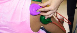
Before the procedure, the patient undresses to the waist and lies down on the couch. The doctor attaches electrodes or special suction cups to the patient's chest, after which the device turns on and the heart impulses are recorded on paper.
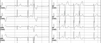
The resulting electrocardiogram includes teeth of different lengths and heights, which display cardiac impulses. The ECG is interpreted by a cardiologist. When deciphering, heart rate, contraction intervals and other indicators of heart function are assessed.
In the branches of the “My Clinic”
On
Gorokhovaya St., 14/26
(metro station Admiralteyskaya, Admiralteysky district) and on
Varshavskaya st., 59
(metro station Moskovskaya, Moskovsky district) there are cardiologists who will conduct a heart examination and draw up a treatment or prevention plan.
You can sign up for an examination by calling 493-03-03 or on our website. Make an appointment
Carrying out an ECG
Electrocardiography can be carried out as part of a comprehensive therapeutic or cardiological examination or be an independent diagnostic procedure. Modern equipment allows you to conduct an affordable ECG in the clinic and at home.
During electrocardiography, the person is in a horizontal position. Special electrodes are attached to the legs, arms and chest. They connect to the ECG machine, the doctor checks all the settings and starts the process. Analysis of cardiac muscle activity is displayed on a paper electrocardiography tape.
During the diagnosis, you should lie still and maintain a natural breathing rhythm. Sometimes during the ECG procedure, the doctor will ask you to hold your breath. The whole process takes about 5-10 minutes and is absolutely painless and safe. There are currently no contraindications for its implementation.
Electrocardiography (ECG) provides valuable information about the condition of the heart.
The essence of this method is to register the electrical potentials that arise during the work of the heart and to display them graphically on a display or paper.
An ECG is a very informative, inexpensive and accessible test that provides a lot of information about cardiac activity. An ECG is a recording of the electrical activity of the heart. Recording is made from the surface of the patient's body (upper and lower limbs and chest). Electrodes (10 pieces) are glued on or special suction cups and cuffs are used. Taking an ECG takes 5-10 minutes.
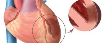
Each myocardial cell is a small electrical generator that is discharged and charged when an excitation wave passes through. An ECG is a reflection of the total operation of these generators and shows the processes of propagation of an electrical impulse in the heart.
The ECG is a valuable diagnostic tool. Using it, you can evaluate the source (the so-called driver) of the rhythm, the regularity of heart contractions, and their frequency. All this is of great importance for the diagnosis of various arrhythmias. The duration of various intervals and ECG waves can be used to judge changes in cardiac conduction. Changes in the terminal part of the ventricular complex (ST interval and T wave) allow the doctor to determine the presence or absence of ischemic changes in the heart (impaired blood supply). An important indicator of the ECG is the amplitude of the waves. Its increase indicates hypertrophy of the corresponding parts of the heart, which is observed in some heart diseases and in hypertension. ECG is, without a doubt, a very powerful and accessible diagnostic tool, but it is worth remembering that this method also has weaknesses. One of them is the short duration of the recording - about 20 seconds. Even if a person suffers, for example, from arrhythmia, it may not be present at the time of recording; in addition, recording is usually done at rest, and not during normal activity. In order to expand the diagnostic capabilities of the ECG, they resort to long-term recording, the so-called Holter ECG monitoring for 24-48 hours. Sometimes it is necessary to evaluate whether a patient has changes on the ECG that are characteristic of coronary heart disease. To do this, an ECG test is performed with physical activity. To assess tolerance (tolerance) and, accordingly, the functional state of the heart, the load is carried out in doses, using a bicycle ergometer or treadmill.
The study is also carried out on children from 3-4 months old using specialized children's sensors.
1. Suspicion of heart disease and high risk for these diseases. The main risk factors are:
- Hypertonic disease
- For men - age after 40 years
- Smoking
- Hypercholesterolemia
- Past infections
- Pregnancy
2. Deterioration of the condition of patients with heart disease, the appearance of pain in the heart area, the development or intensification of shortness of breath, the occurrence of arrhythmia.
3. Before any surgical interventions.
4. Diseases of internal organs, endocrine glands, nervous system, diseases of the ear, nose and throat, skin diseases, etc. if there is suspected involvement of the heart in the pathological process.
5. Expert assessment of drivers, pilots, sailors, etc.
6. Presence of professional risk.
On the recommendation of a therapist (cardiologist), for differential diagnosis of organic and functional changes in the heart, electrocardiography with medicinal tests (with nitroglycerin, with obzidan, with potassium), as well as an ECG with hyperventilation and orthostatic load, is performed.
At the Medical Center, receptions are conducted by:
Shafransky Dmitry Sergeevich - ultrasound doctor, functional diagnostics doctor. Work experience more than 15 years. Clinic specialist No. 1. Doppler ultrasound of the veins and arteries of the upper and lower extremities, vessels of the head and neck. Functional diagnostics: ECG, ECG, blood pressure monitoring, respiratory function.
Lepeshkina Olga Evgenievna - ultrasound diagnostics doctor, functional diagnostics doctor. The highest category. Work experience over 25 years. Ultrasound of the heart with Doppelography, vessels of the neck, ultrasound of the veins, arteries of the extremities.
Preparing for an ECG
Avoid drinking coffee, strong tea, and tonic drinks for 6 hours before the test. If the procedure was preceded by significant physical and emotional stress, you should rest for 30 minutes.
Decoding indicators
ECG is assessed according to several criteria:
- The rhythm is correct and regular. No extraordinary contractions (extrasystoles).
- Heart rate. Normally - 60–80 beats/min.
- Electrical axis - normally R exceeds S in all leads except aVR, V1 - V2, sometimes V3.
- Width of the ventricular QRS complex. Normally no more than 120 ms.
- QRST - complex.
QRST - normal complex
Brief designation of the main elements of the film:
- P wave - shows atrial contraction;
- PQ interval is the time the impulse reaches the atrioventricular node;
- QRS complex - shows ventricular excitation;
- T wave - indicates depolarization (restoration of electrical potential).
Video about ECG norms from the Mass Medika channel.
