A complete blood count means counting the number of cells in a venous blood sample. Capillary blood is not a recommended testing medium for cell counting, but hemogram testing is often performed from a capillary blood sample in intensive care units.
Determination of the leukocyte formula, study of the average size of erythrocytes, platelets, determination of the number of erythrocyte precursors (reticulocytes) and the degree of their maturity, assessment of the erythrocyte sedimentation rate, etc., all this is included in the concept of “clinical blood test”.
A clinical blood test is performed as the first screening test when the patient complains of malaise. An abbreviated clinical blood test, the so-called “troika”, can be performed - counting the number of red blood cells, white blood cells and determining the erythrocyte sedimentation rate (ESR). An abbreviated clinical blood test is not very informative, because can characterize only pronounced pathological processes.
It is more advisable to perform a detailed hemogram from the same volume of blood sample: counting the number of red blood cells with an assessment of their average size (MCV), counting the total number of leukocytes and assessing the leukocyte formula (counting neutrophils, basophils, eosinophils, monocytes, lymphocytes), counting the number of platelets and assessing average platelet size (MPV), reticulocytes and their average size (MRV), reticulocyte maturity (IRF).
Numerous cell characteristics can now be obtained automatically within 3-5 minutes after blood collection. Based on a detailed study of the hemogram, not only a conclusion can be made about the presence of an inflammatory reaction, anemia, but also the nature of other pathological processes, possible previous or ongoing blood loss, deficiency of not only iron, but also vitamin B12 and folic acid.
Research method
The research method depends on the required parameters of the hemogram.
In manual mode, part of a blood sample (3–5 ml) is taken into a capillary to determine ESR, part of the blood sample is used to determine hemoglobin, a drop of blood is used to prepare a smear and further calculate the leukocyte formula. A separate amount of blood is required to prepare a smear and count platelets, and part of the blood sample is needed to study the number of red blood cells and, separately, reticulocytes. In manual mode, if staining and visual assessment of the smear are necessary, the result of a detailed hemogram of a patient in a multi-bed hospital can be obtained at the end of the working day or later.
In automated cell counting and evaluation of different populations, 150 to 300 μl of blood and 100 μl are required for ESR determination. Automatic testing is based on the impedance method of Coulter (1956), which is based on the principle of closing an electrical circuit with each cell successively passing through the sampler aperture. Subsequently, the automated counting method received a number of improvements; in modern analyzers, each cell is assessed according to several parameters: conductivity, light scattering, size, the presence of CD markers on the surface, and, accordingly, membership in different populations. The number of parameters is determined by the device model.
Automatic testing allows you to identify pathological samples that must be visually reviewed by a laboratory diagnostic specialist. Visual control of the hemogram involves preparing a blood smear or slide, which can be made from a drop of blood from an already taken sample, both manually and automatically. Automated blood smear preparation is preferable because There is a uniform distribution of the blood drop and standardized staining. Visual microscopy of the smear is carried out in five fields of view.
An automatic blood test takes 3–5 minutes, unless additional smear preparation and ESR testing are required.
What does blood look like under a microscope?
At high magnification, all three types of blood cells can be seen.
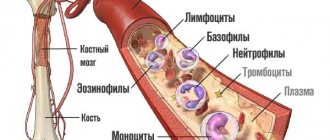
Red blood cells are disc-shaped red cells that transport oxygen throughout the human body. Diameter – 7–10 microns. The color of these cells is due to the content of hemoglobin, a special substance that allows them to carry oxygen molecules. These cells are the most numerous, therefore, when examining human blood under a microscope , you will see them first.
Leukocytes are round-shaped cells ranging in size from 7 to 20 microns. They form the immune system, which protects the body from pathogenic viruses, bacteria and fungi. There are several types of white blood cells: lymphocytes, monocytes, basophils, neutrophils and eosinophils.
Platelets are flat, colorless cells responsible for blood clotting. They have the smallest dimensions - from 2 to 4 microns - so they can only be examined in detail using a professional microscope.
Conditions for sample collection and storage
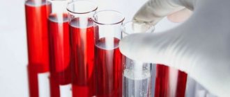
A clinical blood test is performed from venous blood stabilized with potassium salt EDTA, unless otherwise specified in the instructions for the analyzer. Blood collection is performed on an empty stomach. The blood sample should be mixed immediately after collection 9 times by gentle inversion, foam formation and sudden shaking should be avoided. Before testing, the blood sample can be stored at room temperature (23–24 °C) for 24 hours in a rack, in an upright position, away from light.
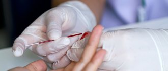
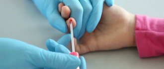
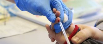
When using a capillary blood sample for clinical analysis, it is necessary to obtain free-flowing drops of capillary blood from the prewarmed puncture area. Collecting capillary blood without squeezing a finger ensures the safety of cells. Applying pressure to the puncture area and collecting a sample from a cooled limb will distort the hemogram results. Capillary blood samples must be stabilized with potassium EDTA, so K3EDTA-treated capillaries should be used for sample collection. Samples can be stored at room temperature (23–24 °C) for 24 hours on a rack, in an upright position, away from light.
Blood smear: execution and research algorithm
N. Puletti
The production of blood smears is technically simple and fast. To obtain maximum information, the assessment of blood cells must be systematized.
In practice, it is not always possible to obtain sufficient objective data when using hematological rapid diagnostics. Examination of blood smears allows one to relatively quickly clarify and supplement the information received. This method allows you to identify some elements that are not apparent during automated clinical blood tests (for example, changes in the shape of red blood cells, a shift in the leukocyte formula to the left [towards immature neutrophils], or the presence of parasites in the blood). In some cases, this method allows a definitive diagnosis to be made.
| Drawing. Different layers of a blood smear. Cells are assessed under a microscope in a thin layer of smear on the pigtails using the shuttle method |
Preparation and staining of blood smears
When collecting material from an ideally calm animal with an average diameter of the vein widening, the blood should quickly flow into a tube containing an anticoagulant. EDTA (ethylenediaminetetraacetate) is most often used because this anticoagulant allows for better preservation of the blood cells being studied. However, to prevent various types of morphological degradation of cells, the time interval between the collection of fresh and well-homogenized blood and the preparation of the drug should be as short as possible (JW Harvey, 2001; D. Walker, 2008).
| Photo 1a. Preparing a blood smear. A small drop of blood is placed on the end of a glass slide using a microcapillary | Photo 1b. Preparing a blood smear. The drop is smeared by placing a second slide at an angle of approximately 45° relative to the first |
Preparation of smears begins by taking a drop of blood (usually one from a capillary tube) onto the edge of a slide. Then the drop is smeared using a second glass, which slides over the first (photos 1a and 1b). A well-prepared blood smear has a so-called “cat tongue” at the end. It indicates that the sample was performed correctly and allows for a high-quality blood examination (figure). Painting is carried out using the standard method (Diff-Quick®, RAL®, Wright-Giemsa®). Before staining, the prepared smear is dried in air by shaking the slide, which avoids the formation of a light unstained zone in the center of the red blood cells and, accordingly, eliminates the erroneous interpretation of hypochromia (photo 2) (D. Walker, 2008). Other coloring artifacts may exist. For example, Wright staining produces a precipitate if the stain is not renewed after a period of time, if the slide was left in the staining solution for a long time, or if it was poorly washed after staining (photo 3) (D. Walker, 2008). As a result, the accumulation of dye can be interpreted as the presence of bacteria or parasites in the blood. In addition, smear staining and cell morphology may be altered when the slide is exposed to formaldehyde fumes. Typically, blood smears are stained using the Romanowsky method (Wright® or Wright-Giemsa®), based on eosin and methylene blue.
| Photo 2. Microscopy of a dog’s blood smear (×1000). In some red blood cells, white light refractions appear in the center (arrows). These are artifacts caused by poor drying of the slide before staining. | Photo 3. Microscopy of a dog’s blood smear (×500). The coloring artifact can be confusing, resembling parasites or bacteria |
Classic coloring differs from fast coloring. Recently, quick dyeing methods such as Diff-Quick® have been advantageous because they are resistant to variations in pH of solutions and the formation of dye deposits. However, they are less effective at detecting polychromatophils and do not stain basophil and mast cell granules well (JW Harvey, 2001; D. Walker, 2008). To obtain a specific visual pattern of reticulocytes, it is necessary to stain with new methylene blue (NBM). In a plastic test tube, a drop of blood is mixed with two drops of NBM. The test tube is left at room temperature for 10 minutes. After mixing, a small drop is placed on a glass slide and smeared in the same way as when performing a blood smear. The slide is then quickly air dried and examined under a high magnification microscope (×50–×100).
Systematic examination of blood smears
When assessing a blood smear, it is very important to be guided by a single research scheme.
A blood smear, made in one thin (monocellular) layer with a roundness at the end, thickens towards the base. Blood cells are assessed on a thin layer, because a thick layer carries little information. At low magnification (×10 or ×20), the edge of the smear, mainly the rounded end, is usually examined for platelet aggregates or broad atypical cells (lymphoblasts or dendritic cells) (Figure 4). Platelet aggregates after their activation are formed in vitro. This phenomenon sometimes occurs as a result of difficult blood sampling, which, for example, is most often noted in cats (E. Duan Lassen, G. Weiser, 2006; SL Stockhman, MA Skkott, 2008; D. Walker, 2008).
| Photo 4. Microscopy of a blood smear of a healthy dog (× 1000). Platelet clots |
The thin layer can have the shape of zigzags or braids, which allows you to very clearly observe different blood cells during a targeted study using the APEL algorithm (A - other [for example, atypical cells, blood parasites]; P - platelets; E - erythrocytes; L - leukocytes).
Blood smear testing is a fairly common method that can quickly diagnose many common disorders in dogs and cats. The main conditions for the effective use of this diagnostic method are strict adherence to the smear preparation technique and systematic research in compliance with the research algorithm.
Basic provisions
— After collecting the material, the blood should be quickly placed in a tube with an anticoagulant in order to preserve the quality of the cells.
— New methylene blue stain allows identification of reticulocytes.
— The assessment is carried out on a thin layer of blood smear with reading under a microscope at the level of its pigtails.
— Systematic blood smear examination refers to the APEL algorithm.
SVM No. 5/2010
Rate material
Like Like Congratulations Sympathy Outrageous Funny Thoughtful No words
2
What do orthophosphoric acid salts indicate?
Phosphoric acid salts
in the blood indicate a pronounced disturbance of phosphorus-calcium metabolism, calcium deficiency in the body, which again is a prerequisite for the development of various diseases. Phosphoric acid is formed during the disposal of proteins, heavy physical activity, consumption of products with thickeners, flavors, etc. Phosphoric acid is a “marker” of calcium loss. It saturates itself with calcium ions, and when the body removes it, it throws out the calcium accumulated in it along with it.
Plasma analysis
The liquid part of blood is plasma
, gives us a fairly large amount of information about the state of the internal environment of the body. The presence of various inclusions in it: cholesterol crystals, uric and orthophosphoric acid salts, bacteria, larvae, fungi and their spores, the rapid appearance of fibrin threads is an indication of a predisposition to the development of a particular disease. Cholesterol crystals form when lipid metabolism is disrupted. Failure of the digestive process, liver and pancreas function leads to hyperlipidemia, which over time leads to a pathological condition - hypercholesterolemia. Cholesterol crystallizes, adsorbs waste products and bacterial waste products on its surface, becomes fixed in the vessels and becomes the basis of atherosclerotic lesions. This can cause the development of cardiovascular pathology.
Hemoscanning of blood: how it is done
The microscope is connected to a video camera, which displays the image on the monitor and also makes it possible to take photographs and videos of interesting objects. It is known that blood performs very important functions in the body. First of all, blood is a transport system that connects the work of all organs and systems. Blood interacts with every cell of the body, delivering them oxygen, bioactive, nutrients, and removes waste products, toxins and waste products that cells release during their functioning. Blood transports immune cells to areas of inflammation or the introduction of foreign agents (infections, allergens), and delivers platelets to areas of bleeding. The condition and composition of the blood is reflected in all pathological processes that occur in the body - inflammatory, infectious, metabolic, immune, allergic, etc. In case of digestive disorders, the function of the excretory organs (kidneys, intestines, skin and lungs), with dysbacteriosis and parasitosis, the blood becomes acidified, overloaded with toxic substances and free radicals.
What can the platelet count tell you?
Platelets
are cells of the blood coagulation system. Changes in the number, shape, and aggregation of platelets are signs of a violation of homeostasis: with blood loss, deregulation of the coagulation system, dehydration, pH shift to an acidic environment, acute infections, anemia, metabolic disorders, intravascular coagulation syndrome. An increase in the number of platelets and their aggregation is caused by provoking and contributing factors: hypokinesia, obesity, excessive physical activity, cooling, intravenous manipulation, taking hormonal contraceptives.
What examinations need to be done for early diagnosis of cancer?
The best results in cancer treatment are achieved when the disease is detected in the early stages of development. This is why regular self-examinations and screenings are so important. Every person who notices any abnormalities in themselves, for example, bumps or retractions on the skin, should consult a doctor as soon as possible. And the oncologist will prescribe a number of necessary tests. For example, the medical list of such examinations includes:
- visualization of internal organs and tissues;
- taking biological material for cytohistological analysis (biopsy);
- a number of laboratory methods, which include, in particular, clinical, biochemical and some other blood tests;
- genetic testing.
Imaging is a reliable and informative way to diagnose malignant neoplasms. Such techniques are used to determine the location of the tumor, assess its size, and interaction with surrounding organs and tissues. Based on the data obtained, it is possible not only to establish a preliminary diagnosis, but also to determine the stage of the disease, as well as monitor the effectiveness of the prescribed treatment.
Imaging tests in oncology may include:
- X-ray examinations;
- computed tomography;
- ultrasound, radioisotope, endoscopic studies;
- magnetic resonance imaging;
- positron emission tomography, which is usually combined with MRI or CT.
A tumor biopsy involves taking a tissue sample from an area of concern for cancer. Often, a biopsy is performed during an endoscopic examination, when a specialist has the opportunity to interact directly with the tumor. The resulting material is sent for study under a microscope in the laboratory. Such a study will make it possible to accurately understand the nature of the tumor (benign or malignant), determine its origin and stage of development, which is extremely important for developing a treatment regimen.
What types of blood tests are used in oncology?
Typically, several different blood tests are used to diagnose cancer:
- general clinical, with determination of the number of red blood cells, leukocytes and a number of other indicators;
- biochemical, showing the concentration in the blood of certain proteins, electrolytes, cholesterol and other biologically active compounds;
- for glucose content;
- on the concentration of certain hormones;
- for the content of specific cancer markers.
In addition, a number of experimental tests are currently undergoing clinical trials, such as CancerSEEK, the Gallery test and PanSeer. These are promising options for a single blood test that may be adopted as a cancer diagnostic method in the future.
Presence of uric acid crystals in the blood
Presence of uric acid crystals
in the blood allows you to assess the level of protein metabolism in the body, the functioning of the kidneys and liver. Uric acid is a byproduct of protein digestion. As uric acid passes through the liver, it is converted into urea, after which the kidneys remove it from the body. Violation of protein metabolism leads to excessive formation of uric acid, its salts (urates), the formation of stones in the urinary tract, deposits in the joint capsules and articular surfaces.