© Author: A. Olesya Valerievna, candidate of medical sciences, practicing physician, teacher at a medical university, especially for SosudInfo.ru (about the authors)
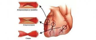
Transposition of the great vessels (GV) is a severe cardiac anomaly when the aorta emerges from the right ventricle (RV) and the pulmonary trunk from the left. TMS accounts for up to 15-20% of all congenital heart defects (CHD); among patients there are three times more boys. TMS is one of the most common forms of congenital heart disease along with tetralogy of Fallot, coarctation of the aorta, ventricular septal defect (VSD), etc.
When the main arteries are transposed, the arterial blood is not enriched with oxygen, since it moves in a vicious circle, bypassing the lungs. The little patient becomes cyanotic immediately after birth, with obvious signs of heart failure. a defect with severe tissue hypoxia, requiring surgical treatment in the first days and weeks of life.
Causes of TMS
The exact reasons for the appearance of pathology in a particular baby are usually impossible to establish, because the mother could have been exposed to a variety of adverse effects during pregnancy. The following may play a role in the occurrence of this anomaly:
- Viral diseases during pregnancy (rubella, chickenpox, herpes, respiratory infections);
- Severe gestosis;
- Ionizing radiation;
- Consumption of alcohol, drugs with teratogenic or mutagenic effects;
- Concomitant pathology in a pregnant woman (diabetes, for example);
- The mother is over 35 years old, especially if this is the first pregnancy.
It has been noted that TMS occurs more often in children with Down syndrome, the causes of which are chromosomal abnormalities caused by the reasons listed above, among others. Children with TMS may also be diagnosed with defects of other organs.
There may be an influence of heredity, although the exact gene responsible for abnormal heart development has not yet been found. In some cases, the cause is a spontaneous mutation, while the mother denies the possibility of external influence in the form of x-rays, drugs or infections.
The formation of organs and systems occurs in the first two months of embryo development, so during this period it is necessary to protect the very sensitive embryo from all kinds of toxic factors. If the heart begins to form incorrectly, then it will not change, and signs of the defect will appear immediately after birth.
| To main |
(Radiation diagnostic news 2002 1-2: 54-55)
A case of corrected transposition of the great vessels.
Bulgak A. G., Vertinsky E. A., Chizh S. A.
BelMAPO.
Corrected transposition of the great vessels (CTGV) is a rare congenital heart defect, the incidence of which ranges from 0.4–1.2% of all congenital heart defects [4]. The anatomical essence of the defect is that as a result of bulbo-ventricular inversion in the normal position of the heart, the morphological right ventricle is located on the left and the aorta departs from it, occupying a left-sided position, and morphologically the left ventricle is on the right, and the pulmonary artery departs from it, occupying a position on the right from the aorta. In Fig.1
the anatomical picture of this anomaly is presented [5].
The defect is corrected by the fact that morphologically the left ventricle communicates through the bicuspid valve with the right atrium, and venous blood enters it, and morphologically, the right ventricle communicates through the tricuspid valve with the left atrium, and arterial blood enters it. Therefore, with isolated CTMS, without concomitant defects, there are no hemodynamic disorders. Characteristic of this defect is the presence of various variants of atrioventricular blockade (in approximately 70% of patients), as a consequence of disruption of the normal topography of the interventricular septum and conduction system [1]. The blockade can be congenital or occurs during life and is first intermittent and then permanent. In this case, the ECG usually shows a deviation of the electrical axis of the heart to the left. In addition to impaired atrioventricular conduction, such patients may experience dysfunction of the tricuspid valve and the anatomical right ventricle, since they are not adapted to the loads placed on the left side of the heart. The prognosis for life in such patients is favorable. In case of development of complete atrioventricular block, implantation of an artificial pacemaker is indicated. [Increase]
| Rice. 1. Corrected transposition of the great vessels. Photo taken from H. Feigenbaum “Cardiac Ultrasound” – London, 1993. | |
Here is our observation.
Patient S., 41 years old, was admitted to the Minsk Emergency Hospital on a referral from the clinic on November 7, 2000.
with complaints of interruptions in heart function, weakness. From the anamnesis it became known that the patient had been experiencing similar complaints for 10-11 years, and therefore she was treated and examined as an inpatient in 1994. Diagnoses of postmyocardial cardiosclerosis and MVP were assumed. Of the diseases he has suffered, he notes colds. Objective examination data: the patient has a correct physique and normal nutrition. The skin is of normal color and clean. Peripheral lymph nodes are not palpable. In the lungs there is vesicular breathing, no wheezing. Heart sounds are muffled, slight systolic murmur at the apex. Ps - 70–76 per minute, arrhythmic. Blood pressure 110/70 mm Hg. The abdomen is soft and painless on palpation. The liver and spleen are not palpable. Stool and diuresis without any peculiarities. General blood test, urine test, biochemical blood test without deviations from the norm. The ECG ( Fig. 2
) shows sinus rhythm, deviation of the electrical axis of the heart to the left, atrioventricular block of the 2nd degree.
Mobitz type 1. During an echo-CG study, the image obtained from the four-chamber position allowed us to suspect ventricular inversion ( Fig. 3
). Using the method of deductive echocardiography [2,3], we identified the morphological right ventricle by the presence of a moderator bundle, the trabecularity of its apex, as well as the corresponding tricuspid valve, which was located closer to the apex than the mitral valve. The pulmonary veins flowed into the left atrium, which communicated through the tricuspid valve with the anatomically right ventricle. The right atrium communicates through the mitral valve with the anatomically left ventricle. In this case, when obtaining a five-chamber position, the first identified main artery was the pulmonary artery, which originated from the anatomical left ventricle. This is due to the fact that the pulmonary artery with this defect is located more posteriorly than usual.
| Rice. 2. The ECG shows sinus rhythm, deviation of the electrical axis of the heart to the left, atrioventricular block of the 2nd degree. Mobitz 1 type. | |
[Increase]
| Rice. 3. During an echocardiography study, the image obtained from the four-chamber position allowed us to suspect ventricular inversion. | |
Thus, the presence of a characteristic ECG pattern and echocardiogram data made it possible to diagnose this rare congenital heart anomaly.
Literature.
- Chazov E.A. “Guide to Cardiology” vol. 3 - M., 1982
- Schiller N., Osipov M.A. “Clinical echocardiography” - M. 1993.
- Feigenbaum H. “Echocardiography” trans. from English - M.1999.
- Braunwald E. “Heart disease” - Philadelphia, 1988.
- Feigenbaum H., “Cardiac ultrasound” - London, 1993.
Blood movement during TMS
I would like to dwell in more detail on how blood moves through the cavities of the heart and vessels during their transposition, because without understanding these mechanisms it is difficult to imagine the essence of the defect and its manifestations.
Features of blood flow during TMS are determined by the presence of two closed, unconnected circulation circles. From a biology course, everyone knows that the heart “pumps” blood in two circles. These streams are separate, but represent a single whole. Venous blood leaves the pancreas into the lungs, returning as arterial blood enriched with oxygen to the left atrium. From the LV, arterial blood with oxygen enters the aorta and goes to the organs and tissues.
With TMS, the aorta begins not in the left, but in the right ventricle, and the pulmonary trunk arises from the left. Thus, we get two circles, one of which “drives” venous blood through the organs, and the second one sends it to the lungs and, in fact, receives it back. In this situation, there can be no talk of adequate exchange, since oxygenated blood does not reach other organs besides the lungs. This type of defect is called complete TMS.
Complete transposition in the fetus is quite difficult to detect. On ultrasound, the heart will appear normal, four chambered, with two vessels branching off from it. The diagnostic criterion for the defect in this case can be the parallel course of the main arteries, which normally intersect, as well as visualization of a large vessel that originates in the left ventricle and is divided into 2 branches - the pulmonary arteries.
It is clear that blood circulation is disrupted to a critical level, and it is impossible to do without at least some opportunity to send arterial blood to the organs. Strange as it may sound, other congenital heart diseases can come to the aid of a sick little heart. In particular, a defect in the septa between the atria or ventricles and an open ductus arteriosus will be beneficial. The presence of such additional communication routes makes it possible to connect both circles and ensure, albeit minimal, delivery of oxygen to the tissues. Additional pathways provide vital activity before surgery and are present in 80% of patients with TMS.
blood flow paths that are pathological for an adult partially compensate for the defect and are present in most patients
Of no small importance in relation to the clinic and prognosis is the state of the pulmonary blood flow, the presence or absence of its overload with blood. From this position, it is customary to distinguish types of TMS:
- With overload or normal pressure in the lungs;
- With reduced pulmonary circulation.
In nine out of ten patients, the small circle is overloaded with “excess” blood. The reasons for this may be defects in the septa, open ductus arteriosus, or the presence of additional communication routes. Depletion of the small circle occurs when the LV outlet is narrowed, which occurs in isolated form or in combination with a ventricular septal defect.
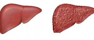
An anatomically more complex defect is corrected transposition of the great vessels. Both chambers and vessels are “confused” in the heart, but this makes it possible to compensate for disturbances in blood flow and bring it to an acceptable level. With corrected TMS, both ventricles with the vessels extending from them change places: the left atrium passes into the right ventricle, followed by the aorta, and from the right atrium the blood moves into the LV and pulmonary trunk. Such “confusion”, however, ensures the movement of fluid in the right direction and the enrichment of tissues with oxygen.
Complete TMS (left) and corrected defect (right), photo: vps-transpl.ru
In the case of a corrected defect, the blood will move in a physiological direction, so the presence of additional communication between the atria or ventricles is not required, and if it is present, it will play a negative role, leading to hemodynamic disorders.
Video: TMS – medical animation (eng)
Publications in the media
Transposition of the great vessels (TMS) is a congenital heart disease characterized by discordance of the ventricular-arterial connection with concordance of the connection of the remaining segments of the heart. In other words, the aorta arises from the morphologically right ventricle, and the pulmonary trunk arises from the morphologically left one. Statistics • 7-15% of all congenital heart diseases • 9.9% of congenital heart diseases diagnosed in infancy • Male to female sex ratio at birth - 3:1 .
Etiology : causes of congenital heart disease (see Tetralogy of Fallot).
Pathogenesis • With corrected TMS, discordance of the ventricular-arterial connection is combined with discordance of the connection of the atria and ventricles (transposition of the aorta and pulmonary artery is accompanied by inversion of the ventricles), and blood circulation does not suffer if there are no associated defects • This article considers only complete TMS • Depending from changes in pulmonary blood flow, complete TMS with increased or normal pulmonary blood flow is distinguished (when combined with a patent ductus arteriosus, atrial and ventricular septal defects, aortopulmonary fistula) and complete TMS with reduced pulmonary blood flow, when accompanying septal defects are combined with stenosis of the left outlet ventricle (complex form of TMS) • If normally the systemic and pulmonary circulations are connected sequentially, then with TMS they function in parallel, being completely separated. Therefore, a prerequisite even for a short life is the presence of communications between the systemic and pulmonary circulation in the form of naturally existing or artificially created defects. In this case, blood is discharged during TMS in both directions • The larger the size of the shunt, the less pronounced the hypoxemia • The most favorable prognosis is noted in cases where a large defect of the interatrial or interventricular septum provides the necessary mixing of blood, and moderate stenosis of the pulmonary artery prevents excessive blood flow in the lungs .
Clinical picture • General cyanosis • Differentiated cyanosis, when the upper half of the body is more cyanotic than the lower, is pathognomonic for TMS in combination with patent ductus arteriosus • Tachycardia • Dyspnea • Increased size of the heart and liver • Edema, ascites • On auscultation - increased both tones, systolic murmur of organic or relative (with atrial septal defect) pulmonary artery stenosis, murmur of VSD or patent ductus arteriosus.
Instrumental diagnostics
• ECG •• Up to 1–1.5 months of life, changes may be absent •• Signs of hypertrophy and overload of the right sections.
• X-ray of the chest organs •• heart takes the shape of an egg lying on its side, with a narrow vascular bundle in the anteroposterior and wide in the lateral projection, and the aortic arch is almost always on the left (highly specific signs) •• Depletion (with pulmonary stenosis) or enrichment (with septal defects) of the vascular pattern of the lungs.
• EchoCG •• Visualization of the abnormal origin of the great vessels •• Assessment of the degree of hypertrophy of the walls and dilatation of the heart chambers •• Diagnosis of associated defects (see Ventricular septal defect, Atrial septal defect, Patent ductus arteriosus) or stenosis of the right ventricular outflow tract.
• Pulse oximetry , study of the gas composition of peripheral blood •• Low oxygen saturation (pathognomonic sign - less than 30%) and pO2 (pathognomonic sign - less than 20 mm Hg) in peripheral blood •• When combining TMS with patent ductus arteriosus - high difference blood parameters of the upper and lower extremities.
• Probing of the cavities of the heart •• Increase in pO2 and blood oxygen saturation in the right sections and decrease in the left •• Tests are performed with aminophylline and oxygen inhalation to determine the prognosis regarding the reversibility of pulmonary hypertension •• See also Ventricular septal defect, Atrial septal defect, The ductus arteriosus is open.
• Right and left atriography and ventriculography , ascending aortography , coronary angiography •• Visualization of the pathological flow of contrast from the right parts into the aorta, and from the left into the pulmonary artery •• Diagnosis of associated defects (see Ventricular septal defect, Atrial septal defect, Ductus arteriosus open) or stenosis of the right ventricular outflow tract •• Both posterior aortic sinuses facing the pulmonary artery become coronary, and there are six normal variants of the origin of the coronary arteries from the aortic sinuses •• In 15% of cases, anomalies of the origin of the coronary arteries are detected.
Drug therapy : in the presence of natural defects between the systemic and pulmonary circulation, in order to prevent their closure, an infusion of PgE1 (alprostadil) 0.05–0.1 mg/kg/min is performed.
Surgery _
• Indications : all patients with complete TMS.
• Contraindications : irreversible pulmonary hypertension.
• Methods of surgical treatment •• Palliative interventions - increasing the size of natural or creating artificial defects between the systemic and pulmonary circulation: ••• Park-Rashkind operation (percutaneous balloon atrioseptostomy), usually carried out within a period of up to 3 months, has no advantages over treatment with Pg ; ••• Blalock-Hanlon operation (open resection of the interatrial septum) is of only historical interest •• Radical correction consists of plastic surgery of the atria with a synthetic patch so that blood from the pulmonary veins enters the right atrium, and blood from the vena cava enters the left ( Mustard and Senning operations) •• Anatomical correction of TMS (Jatenet operation) consists of intersection with subsequent orthotopic replantation of the great vessels, simultaneous ligation of the patent ductus arteriosus, as well as transplantation of the ostia of the coronary arteries into the base of the pulmonary trunk •• When combining TMS with VSD, plastic surgery of the defect is performed septum with simultaneous anatomical correction of TMS according to Zatena •• In case of a complex form of TMS (combination with VSD and pulmonary artery stenosis), the Rastelli operation is indicated - plastic surgery of the septal defect with the formation of the left ventricular outflow tract with a synthetic or autopericardial patch, elimination of patency of the mouth of the pulmonary trunk, implantation of an artificial trunk between the right ventricle and the pulmonary trunk or right pulmonary artery.
Specific postoperative complications • Sick sinus syndrome • Stenosis of the orifices of the vena cava and pulmonary veins • Stenosis of the outflow tract of the right and left ventricles.
Prognosis • Clinically significant circulatory failure usually occurs by 1–1.5 months of life • Half of children die within the first month after birth • Two thirds do not survive to one year of age • The average life expectancy of these patients is 3–19 months • Mortality after Mustard operations and Senning does not exceed 10% • In the long term, good results are achieved in 85-90% of cases • Hospital mortality with anatomical correction of TMS according to Jaten - 2-3%, according to Rastelli - less than 10% • 5-year survival rate with anatomical correction of TMS - 80–90%.
Reduction . TMS - transposition of the great vessels.
ICD-10 • Q25.8 Other congenital anomalies of large arteries.
Manifestations of TMS
During intrauterine development, this heart defect does not manifest itself in any way, because in the fetus the pulmonary circle does not work. After birth, when the baby’s heart begins to pump blood to the lungs on its own, TMS also manifests itself in full. If the transposition is corrected, then the clinical picture is poor; if the defect is complete, its signs will not be long in coming.
The degree of impairment with complete TMS depends on the communication pathways and their size. The more blood mixes in the heart in newborns, the more oxygen the tissues will receive. The optimal option is when there are sufficient holes in the septa, and the pulmonary artery is somewhat narrowed, which prevents volume overload of the pulmonary circle. Complete transposition without additional anomalies is incompatible with life.
Babies with transposition of the main vessels are born at term, with normal weight or even large, and already in the first hours of life signs of congenital heart disease are noticeable:
- Severe cyanosis of the whole body;
- Dyspnea;
- Increased heart rate.
Further, the symptoms of heart failure rapidly increase:
- The heart increases in size;
- Fluid appears in the cavities (ascites, hydrothorax);
- The liver enlarges;
- Swelling occurs.
Other signs of cardiac dysfunction are also noteworthy. The so-called “heart hump” (deformation of the chest) is caused by an enlargement of the heart, the nail phalanges of the fingers thicken, the baby lags behind in development, and does not gain weight well. Certain difficulties arise during feeding, since it is difficult for a child to suckle at the breast with severe shortness of breath. Any movement and even crying can be an impossible task for such a baby.
If an excess amount of blood enters the lungs, there is a tendency to infectious and inflammatory processes and frequent pneumonia.
The corrected form of TMS proceeds much more favorably. In the absence of other cardiac defects, clinical transposition may not occur at all, because the blood moves correctly. The child will grow and develop correctly according to his age, and the defect can be detected accidentally by the presence of tachycardia, heart murmur, or conduction disturbances.
If the corrected transposition is combined with other disorders, then the symptoms will be determined by them. For example, if there is a hole in the interventricular septum, shortness of breath will appear, the pulse will increase, and signs of heart failure will appear in the form of edema and enlarged liver. Such children suffer from pneumonia.
Congenital heart defect. Transposition of the great arteries. Clinical observation
Transposition of the great arteries (TMA) is a group of congenital heart defects related to conotruncal anomalies, the common features of which are atrioventricular concordance and ventricular-arterial discordance. The frequency of this disease ranges from 4.5% to 7% of all congenital heart diseases.
The disease itself is congenital. It causes severe shortness of breath and cyanosis immediately after birth. This problem requires surgical intervention in the first days of life, otherwise the child will live up to 2 years with the best outcome. This is why the clinical case that occurred in the cardiology department of the Children's Clinical Medical Center in the Moscow Region is surprising.
In May 2021, a 16-year-old boy was admitted to the department with complaints of shortness of breath and bluish skin.
From the life history it is known that the child is from a socially disadvantaged family, from 2 term births. Birth weight 3600 g. Height 52 cm Discharged home on the 5th day of life. From 2 weeks on artificial feeding. They refused preventive vaccinations. CHD was diagnosed at the age of 2 months. The parents refused to consult a cardiologist, just as they refused to consult a cardiac surgeon in 2008.
The child was diagnosed with congenital heart disease. Ventricular septal defect. He was observed by a pediatrician at his place of residence. Occasionally consulted a cardiologist.
In May 2021, he was hospitalized at the central district hospital at his place of residence due to a deterioration in his condition - shortness of breath and cyanosis increased.
To continue therapy, the child was transferred to the pediatric department of the Children's Clinical Hospital of the Moscow Region.
On examination, physical development is low and harmonious. Height – 140 cm. Weight – 32.5 kg. Severely lags behind in physical, mental and sexual development. The skin is bluish, with pronounced acrocyanosis. The nails are changed like “watch glasses”, the distal phalanges of the fingers are changed like “drumsticks”. The skin is warm and quite moist. Sat Hb O2 71-79%. Visible mucous membranes are purple, clean, moist. The sclera is injected. The chest is barrel-shaped, asymmetrical, both halves are equally involved in the act of breathing. The act of breathing involves auxiliary muscles (jugular fossa, intercostal spaces).
The area of the heart is visually changed - a left-sided “heart hump”. The apical impulse is diffuse, visualized at 5 m/r along the midclavicular line. “Systolic tremor” is weak, but detectable. Heart sounds are rhythmic, the 2nd tone is increased. Heart rate 89/min. Blood pressure (on the arms) 96/57mmHg. A 3/6 systolic murmur is heard along the left sternal border. The pulse on the radial arteries is symmetrical and of satisfactory quality. The pulsation in the femoral arteries is distinct and symmetrical.
The necessary tests were taken from the child, ECG, ECHO-CG, and other studies were performed. Based on the results of the examination, the following conclusion was made: congenital heart disease, transposition of the great arteries. Subarterial ventricular septal defect 1.9 cm, right-left shunt. Bicuspid pulmonary valve. Valvular pulmonary artery stenosis with max GSD 48 mmHg. The heart chambers are not dilated. Myocardial hypertrophy of the left and right ventricles. Myocardial contractile function is satisfactory. Tricuspid regurgitation stage 1. Mitral regurgitation stage 1. Aortic arch b/o. The pericardium is intact.
With the assistance of the chief freelance pediatric cardiologist of the Ministry of Health of the Moscow Region, head of the department Alfiya Drozdova, the child was examined and operated on by a cardiac surgeon, which significantly improved the boy’s quality of life. For two years, the child continues to be monitored in our cardiology department and receives the necessary treatment.
Methods for correcting TMS
Considering the presence of anatomical changes in the heart, the only possible treatment option for the defect is surgery, and the sooner it is performed, the fewer irreversible consequences the disease will bring.
Emergency intervention is indicated for patients with complete TMS, and before surgery, prostaglandin drugs are prescribed to prevent the closure of the ductus arteriosus, which allows the blood to “mix.”
In the first days of a baby’s life, it is possible to perform operations to ensure the connection of the blood circulation. If there are holes in the partitions, they are expanded; if there are no defects, they are created. The Rashkind operation is performed endovascularly, without penetrating the chest cavity, and consists of inserting a special balloon that expands the oval window. This intervention provides only a temporary effect for several weeks, during which the issue of radical treatment must be decided.
The most correct and effective treatment is considered to be an operation in which the aorta returns to the left ventricle and the pulmonary trunk to the right , as they would be normally. The intervention is carried out openly, under general anesthesia, the duration is from one and a half to two hours or more, depending on the complexity of the defect.
example of surgery for TMS
After the baby is under anesthesia, the surgeon cuts through the chest tissue and reaches the heart. At this point, artificial blood flow is established, when the device plays the role of the heart, and the blood is additionally cooled to prevent complications.
Having opened the way to the main arteries and the heart, the doctor cuts off both vessels slightly above their attachment, approximately in the middle of their length. The coronary arteries are sutured at the mouth of the pulmonary artery, and then the aorta is “returned” here. The pulmonary artery is fixed to the portion of the aorta remaining at the exit from the right ventricle using a fragment of the pericardium.
The result of the operation is the normal arrangement of the vascular tracts, when the aorta leaves the left ventricle, the coronary arteries of the heart also begin from it, and the pulmonary trunk originates in the right half of the organ.
The optimal period for treatment is considered to be the first month of life. Of course, you can live longer waiting for her, but then the intervention itself will become inappropriate. As you know, the left ventricle is thicker than the right and is designed for greater pressure load. With a defect, it atrophies, as the blood is pushed into a small circle. If the operation is performed later than expected, the left ventricle will not be ready for the fact that it will have to pump blood into the systemic circulation.
When time is lost and it is no longer possible to restore the anatomy of the heart, there is another way to correct the blood flow. This is the so-called intra-atrial correction, which has been used for more than 25 years and has proven itself to be an effective method of treating TMS. It is indicated for children who did not undergo the above-described operation on time.
The essence of intra-atrial correction is to dissect the right atrium, remove its septum and sew in a “patch” that directs venous blood from the systemic circle to the left ventricle, from where it goes to the lungs, while the pulmonary veins return oxygenated blood to the “right” heart and then - into an abnormally located aorta. Thus, without changing the location of the main arteries, blood movement in the desired direction is achieved.
Transposition of the great vessels
Transposition of the great vessels (TMS) means the incorrect position of the main vessels leaving the heart, i.e. the aorta and the trunk of the pulmonary artery, relative to each other and relative to the chambers of the heart from which they arise.
There can be many options for transposition: complete, incomplete, corrected, in combination with other defects.
For now we will only touch on the full
transpositions, i.e.
situations when the vessels have completely moved and changed their places. The aorta arises from the right ventricle, and the pulmonary artery arises from the left. The remaining parts of the heart, i.e. the atria with the veins flowing into them, and the ventricles, are normal and do not have any other defects (the term “simple TMS” is sometimes used). It’s not hard to imagine, but this happens to the heart, and not so rarely. It’s as if legs grew where arms should be, and arms grew where legs should be. Fortunately, it doesn't look that scary. Children are born full-term, completely normal, but, unlike tetralogy of Fallot, they are immediately very cyanotic. It becomes obvious that immediately after birth it is very difficult for a child to simply live
.
Venous blood, poor in oxygen, enters the right atrium, the right ventricle, and from here again into the aorta departing from it and into the systemic circulation, without passing through the lungs, without being saturated with oxygen and without giving off carbon dioxide. And blood from the lungs through the pulmonary veins goes to the left atrium, to the left ventricle, and again to the pulmonary artery and to the lungs, which from the point of view of its saturation with oxygen is meaningless, because she is already saturated. Two separate circles of blood circulation are formed.
And, if we previously conventionally depicted the relationship of the circulatory circles in the form of a figure eight lying on its side, then with transposition these are two closed rings not connected to each other.
It is clear that this state of affairs is simply incompatible with life. But nature decides in its own way: it leaves open the oval window (i.e., a natural defect) in the interatrial septum, through which part of the venous blood leaks into the left sections, and then into the lungs. Both circles are connected to each other only by this shunt.
It is clear that the amount of blood that passes through the defect with each cardiac cycle, and on which its saturation depends, is very small and cannot in any way meet the body’s needs. If there are two defects, or a defect of the interventricular septum is added, then this is somewhat better, because the amount of oxidized blood in the arterial system becomes greater. But still it is extremely insufficient.
Children born with complete transposition of the great vessels quickly fall into a critical condition, and if they are not helped in the first days of life, they will die. They will not have attacks of shortness of breath, as with tetralogy of Fallot, but cyanosis appears already in the first hours, and the slightest physical effort - movements, sucking, crying - becomes difficult or completely impossible. What needs to be done? First of all, expand the existing defect, increase its size in order to increase the volume of venous blood passing through it.
This is achieved by the so-called Rashkind procedure, the essence of which boils down to the fact that a catheter with a balloon is inserted into the open oval window, which is inflated, thereby rupturing the interatrial septum and increasing the defect in diameter. The procedure is performed in the X-ray surgery room and preparation for it includes all the points that we described above when we talked about cardiac catheterization or closure of the patent ductus arteriosus.
In case of complete transposition, expansion of the defect must be done urgently. But this expansion - and the increase in the flow of mixed blood - in itself does not solve anything. It only slightly improves the child’s condition and prolongs his life, and something needs to be done further without delay. The effect of the procedure will be very short-lived - only a few weeks, and if you wait longer, the child may die from heart failure and constant oxygen starvation.
It is clear that the ideal method of treating transposition is the complete elimination of this “error of nature” - i.e. surgical relocation of the aorta and connecting it to “its” left ventricle (whereas it now departs from the right), and the pulmonary artery to the right ventricle.
Description of the operation of surgical relocation of the aorta and connecting it to “one’s” left ventricle
After the usual introduction of the child into anesthesia, opening the chest, connecting to the apparatus, artificial circulation is started, with the help of which the blood is simultaneously cooled (this is always done during other operations that may require quite a long time - 1.5-2 hours or more) . The fact is that when cooling, all metabolic processes slow down (like a bear in hibernation), and this protects the body from all sorts of complications in the future. With the help of the device, cooling occurs quickly (as does warming in the final stage of the operation).
The aorta and pulmonary artery are divided in half. The coronary arteries are cut off from the aorta and sutured into the beginning of the pulmonary artery, which will then become the mouth of the new aorta. The cut off “own” aorta is sutured to this area, and then a tube is made from a piece of its own pericardium, which is sewn into the new pulmonary artery, restoring it too.
Thus, true anatomical correction of the defect is achieved. Now everything is fine: the great vessels depart from the ventricles from which they should depart. This operation, as we see, is complex and lengthy. But today it is done quite successfully in several cardiology clinics in the country. However, to count on success, it must be done very early, i.e. in the first weeks of life.
The fact is that at birth both ventricles, right and left, are well developed, have the same muscle mass and wall thickness. With normal development in the right
the load of the ventricle is less than that of the left: it does not need to pump blood into a large circle, i.e.
all over his body, and he gets used to it pretty quickly. A little time will pass - and its wall will become thinner, and the muscle mass will decrease: why work if you don’t have to work? The same thing happens with transposition - but with the left
ventricle.
During transposition, the ventricles of the newborn are also initially identical, and the fact that they work against different resistances of the systemic and pulmonary circulation does not affect them at first. But then the left ventricle becomes thinner and loses mass, but after the operation of moving the vessels, it is he who will have to do the main work. And here it is important not to waste time, because... If you wait several months, then even if the Rashkind procedure has a good effect, radical correction will no longer be possible. The best time for such a correction is the first month of life
.
It must be said that this treatment of complete transposition has been used for 25 years, and in recent years it has become standard. It is clear that to successfully perform this complex operation, not only a well-developed technique is required, but also methods of providing anesthesia, artificial circulation, and management of the postoperative period that are safe for the infant, i.e. everything that leads to ultimate success. This can only be done in large pediatric cardiac surgery centers with extensive experience in treating congenital heart defects.
What to expect after surgery? Overall, the results are very good. Today, hundreds of operated patients lead normal lives, and the life expectancy of some children who were among the first to be operated on is already 20 years or more. Of course, observation by a cardiologist, and sometimes detailed examinations, are mandatory, because As the child and the heart itself grow, problems may arise. However, the main of these problems can be completely eliminated by X-ray surgery, without resorting to repeated surgery.
Unfortunately, radical surgery is not always possible. The main reason is late contact with specialists, when time has already been lost. It is not uncommon to see children with complete transposition at the age of one or two years. They are very blue and are greatly retarded in physical development. Their eyes—intelligent, suffering, and seemingly understanding everything—can never be forgotten. These children can and should also be helped urgently, and such methods exist, they have been used for a long time, and they have proven themselves well. Long before surgical science made possible early and anatomically radical treatment of transposition, another, less radical, but quite effective method was proposed.
The idea is not to correct the anatomy (this was technically impossible then), but to change the paths of blood flow, i.e. the direction of blood from the veins to the lungs, to the pulmonary circle, and the oxidized blood to the left ventricle and the aorta, i.e. in a big circle.
Techniques known as " intra-atrial correction"
“were for a long time the only ones in the treatment of transposition, but they are still used today when, for some reason, it is not possible to perform anatomical correction.
The essence of the operation comes down to dissecting the right atrium, removing the entire interatrial septum and sewing a patch inside the cavity from the child’s own tissues (pericardium, or the wall of the atrium) in such a way that blood from the vena cava is directed to the left ventricle, from which the pulmonary artery arises, and then - into the lungs, and from the pulmonary veins into the right ventricle, into the aorta and into the systemic circulation. These operations, known by the names of their authors - Mustard and Senning, correct hemodynamics, but not the defect itself. Today, the world has accumulated a great deal of experience in such operations, and the fate of the operated patients has been tracked for several decades. Half of them have a normal, fairly active and long life. Over time, the other part may experience complications in the form of rhythm disturbances, insufficiency of atrioventricular valves - after all, the ventricles of the heart remain in place and do not work as intended by nature. People who have had this type of surgery sometimes feel a little blue—especially their lips and fingernails and toenails. There are still restrictions on physical activity. Girls who have reached adulthood may not be recommended to have children, not because the child will have a defect - this is a complete misconception, but because pregnancy and childbirth may be too much physical exertion. But, be that as it may, intra-atrial correction methods saved the lives of hundreds of children. So, even if the moment for anatomical correction is missed, there is a way out, and a good way out.
Moreover, in recent years, operations have begun to move vessels with the removal of old intra-atrial patches. This is not always possible and necessary, and it cannot be called anything other than “aerobatics” in cardiac surgery. But time moves forward, and I just want you to believe: transposition of the great vessels today is a completely curable defect, and not a single child born with it should die. But at the same time, its future depends largely on you and on your faith in the modern possibilities of medicine.
How to get treatment at the Scientific Center named after. A.N. Bakuleva?
Online consultations
Prognosis and treatment results
When a baby is born with transposition of blood vessels, his parents are very concerned about not only the operation, but also what will happen after, how the child will develop and what awaits him in the future. With timely surgical treatment, the prognosis is quite favorable: up to 90% or more of patients live a normal life, periodically visiting a cardiologist and undergoing a minimum of examinations to monitor the functioning of the organ.
With complex defects, the situation may be worse, but most patients still have an acceptable quality of life. After intra-atrial correction surgery, about half of the patients do not experience restrictions in life, and its duration is quite long. The other half may suffer from arrhythmias and heart failure, which is why it is recommended to limit physical activity, and women are warned about the risks during pregnancy and childbirth.
Today, TMS is a completely curable anomaly, and hundreds of children and adults who have successfully undergone surgery are proof of this. Much depends on the parents, their faith in success and desire to help their baby.