Briefly about the treatment method
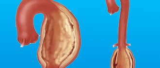
Endovascular treatment of abdominal aortic aneurysm (EVAR) is a minimally invasive alternative to major open surgery for abdominal aortic aneurysm, which reduces the risk of surgery, speeds up recovery time and increases survival rate for this dangerous disease. Endovascular surgery involves only small incisions in the thigh through which an artificial vessel called a stent graft is installed inside the aneurysm. A stent graft is a metal mesh of a special design (stent), which is covered from the inside with a sealed polymer fabric (graft). A metal mesh fixes this device to the walls of the aorta and iliac arteries, and polymer fabric isolates the lumen of the artificial vessel from the aneurysm cavity. Thus, the aneurysmal sac is isolated from the blood flow, and the risk of continued growth and rupture of the aneurysm after installation of the stent graft is significantly reduced. The operation is performed without the use of general anesthesia and is not accompanied by significant blood loss. Installation of a stent graft for an aortic aneurysm is much safer than classical open surgery.
Discussion
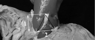
The only effective treatment for aortic aneurysms is surgical correction [14]. Surgery of the aortic arch is associated with high surgical risk [15]. During the formation of the distal anastomosis, it is necessary to use circulatory arrest.
Deep hypothermia with circulatory arrest reduces brain metabolism and its need for oxygen. The advantages of this technique are the absence of trauma to peripheral vessels during cannulation, reducing the risk of embolism, and ensuring a “dry” surgical field [13]. The negative effects of hypothermic circulatory arrest are associated with an increase in the time of cardiopulmonary bypass, which can lead to various blood clotting disorders and severe systemic inflammatory response syndrome. In addition, with circulatory arrest, the time frame for the formation of a distal anastomosis is limited (no more than 35 minutes) [10, 13, 16]. With deep hypothermia and circulatory arrest, neurological disorders in the form of stroke or transient neurological deficit can complicate the course of the postoperative period in 15-20% of patients and remain the main cause of hospital mortality [17]. Bleeding disorders always accompany circulatory arrest and are associated with damage to clotting factors and platelets under the influence of low temperature. At the same time, the frequency of coagulopathic bleeding requiring repeated interventions is 10.9% [18].
In the presented case, a prosthetic operation of the ascending aorta and aortic arch was performed using the hemiarch technique without the use of circulatory arrest. In the postoperative period, there were no neurological disorders, pathology of hemostasis, or complications from visceral organs. An important technical condition for performing this operation is the possibility of applying a clamp to the aorta distal to the left subclavian artery. The proposed methodology requires further study and accumulation of material.
Advantages of treatment at the ISC
The innovative vascular center always strives to introduce the most advanced technologies of vascular surgery into its practice. Considering that endoprosthetics is a safer alternative to open surgery for aneurysms, since 2015 we have practically replaced aneurysm resection operations with this intervention. In our practice, we use the best stent grafts from Western manufacturers, which are selected individually for each patient. Our specialists successfully perform endoprosthesis replacement operations for the most complex forms of abdominal aortic aneurysms.
Indications and contraindications for the treatment method
Indications
- The standard endoprosthetic procedure is suitable for an aneurysm that begins 2-3 centimeters below the orifices of the renal arteries, that is, it has a neck for reliable fastening of the endoprosthesis, preventing blood leakage around the endoprosthesis - “endoleak”.
- Surgery is indicated when the aneurysm reaches a sufficiently large diameter (usually more than 5 cm) that the risk of complications (rupture or thrombosis) exceeds the risk of surgery.
- Installation of a stent graft into an aortic aneurysm is also carried out when the aneurysm is growing rapidly (diameter according to ultrasound increases by more than 0.5 cm per year)
- For embolism (transfer of blood clots from the aneurysmal sac to the arteries in the legs).
- Aortic aneurysms that cause abdominal pain, which may indicate an impending rupture.
When selecting patients for aortic replacement, we proceed from an assessment of the risk of open surgery. Studies that compare EVAR with open surgery have demonstrated fewer early complications with the endovascular approach and lower overall mortality rates. Endovascular treatment requires more careful monitoring and sometimes additional interventions when various leaks are detected. If surgery is planned in young patients in good general health, open surgery is preferable. In elderly patients, endovascular surgery is the method of choice, since the mortality rate after open surgery exceeds 10%. As the technology for manufacturing stent grafts develops, indications for open surgery are becoming less and less common.
Placement of a stent graft for a ruptured abdominal aortic aneurysm provides a significantly better chance of survival compared to emergency open surgery, as well as helping patients with thoracic and abdominal aortic dissection survive if performed in a timely manner.
Contraindications
Anatomically unsuitable structure of the aortic aneurysm for endoprosthetics. Most often this is the absence or a very short section of the normal abdominal aorta below the renal arteries (absence of an aneurysm “neck”). In this case, it is not possible to secure the upper section of the stent graft well to avoid leakage. However, stent grafts with side branches for the renal arteries have now been proposed for use, which allow the stent graft to be placed higher, restoring flow into the renal arteries.
Relative contraindications are small diameter or aneurysms of the iliac arteries, severe calcification and occlusion of the femoral arteries, however, modern stent grafts and the possibilities of hybrid (open and endovascular joint) surgery make it possible to solve these problems.
An order of magnitude better: endoprosthetics of aortic aneurysm
Endoprosthesis replacement of an aortic aneurysm, as vascular surgeons note, is a modern direction in the treatment of patients with this severe pathology. In general, this was the reason for writing this article.
Since 2007, these operations have been carried out with high efficiency at the Interregional Clinical Diagnostic Center. A week ago, a master class was held on the installation of new generation stentgrafts. About this in an interview with the head of the department of vascular surgery, head of the course of cardiovascular surgery at KSMU, Doctor of Medical Sciences, Professor Igor Mikhailovich Ignatiev.
— With the participation of M.I. Generalova (Federal Diagnostic Center, St. Petersburg) and Luchnikov V.M. (Vishnevsky Institute, Moscow) a master class was held, during which we operated on four patients with abdominal aortic aneurysm using new generation stentgrafts.
— What is the essence of the technique?
— There are quite a lot of patients with aneurysms—pathological enlargement of the aorta, which can lead to rupture of the aneurysm with a fatal outcome. This category of patients needs aortic endoprosthesis replacement, that is, from the inside, through small accesses, an endoprosthesis is “inserted” into the aorta with little trauma, which “opens up” and blocks the aneurysm. The operation is minimally invasive and is usually well tolerated by patients.
— Is there an analogue to endoprosthetics to eliminate an aneurysm?
— Open operations. But there are some nuances here. Most of our patients often have severe concomitant pathology - pathology of the heart, kidneys, lungs and other organs. Therefore, such patients usually do not tolerate open, surgical intervention, which poses a high risk.
— What about the new prosthetics that were demonstrated at the master class?
— These are new generation prostheses that are modeled quite well and positioned in the lumen of the aorta. We installed prostheses in the abdominal aorta made in England. They have been significantly modernized and, thanks to this, make it possible to perform more complex options for endoprosthetics. The endoprosthesis is selected individually for each patient based on its size and characteristics.
— During the master class, four patients were operated on. Please tell us about the results.
— All four patients were admitted with a fairly severe form of abdominal aortic aneurysm. One operation was technically very complex - a hybrid, during which, in addition to aortic replacement, a vascular stage was performed - cross-bypass.
Such surgical interventions are performed by two teams - vascular surgeons and x-ray surgeons.
— That is, we can say that you work in symbiosis...
- Yes. Endovascular operations are quite technically complex and require certain skills and experience.
— What was the purpose of the master class?
“Our surgeons are proficient in this technique, and, in principle, they have already accumulated quite a lot of experience, but since new generation endoprostheses are being used, we had to get acquainted with certain of their characteristics and features. M.I. Generalov (Federal Diagnostic Center in St. Petersburg) has good experience in installing new prostheses and was ready to share it with us.
— How many similar operations are planned to be done this year?
- About ten.
— Do your colleagues from the RCH perform similar operations, or do you have a prerogative in this regard?
— They do it too, but, as far as I know, to a lesser extent than at the Interregional Clinical Diagnostic Center. Such surgical interventions remain unique to this day in Russian clinics, because their number, unfortunately, is not large. Every year the pace of this surgical intervention is increasing. If we compare a Russian clinic and a European or US clinic, several hundred operations are performed at this level per year. That is why this direction should be developed here too.
— Are there many patients with this pathology?
— Quite a lot and it is difficult to diagnose. In some cases, the patient's death occurs before the diagnosis of aortic aneurysm is made.
— How can patients be diagnosed in order to prevent disaster and perform endovascular intervention on time?
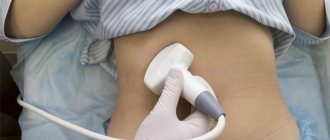
-We work in the regions of the republic, we go out and diagnose. The most accessible diagnostic method is ultrasound. With its help, you can clearly see the expansion of the aorta.
— Are there any symptoms? What can a GP do?
“Unfortunately, at the very beginning the disease is asymptomatic. This makes diagnosis even more difficult. Let me give you an example. One of our patients had an aneurysm with a diameter of about 15 cm, was shown to general practitioners, but no one thought to palpate the abdomen. In a phonendoscope, the aneurysm “makes noise”, so it can be felt, seen and heard, the main thing is to suspect it in time, detect it and carry out surgical intervention.
— What possible complications occur during the operation and in the postoperative course? What is their probability?
— Complications can occur with any, even the most minor intervention, but if technically everything is done correctly, the risk of complications is minimal. The experience of the vascular surgeon himself is important; the higher it is, the lower the risk of any troubles. No fatal things are observed during endovascular intervention, but some nuances in the form of, for example, repeated operations, sometimes occur. But all these situations can be corrected.
— Speaking about experience, could you tell us where the specialists of your department were trained?
— In leading clinics in the USA and Germany.
— What are the advantages of endovascular intervention over open surgical interventions that were previously performed for this pathology?
— Endovascular intervention is less traumatic, easier to tolerate by patients, the period of postoperative rehabilitation and recovery is shorter and, most importantly, the mortality rate is significantly lower than with open intervention.
— What drug therapy is given after this surgery?
— Patients are prescribed aspirin-containing drugs that work to improve the rheological properties of the blood.
— How can you prevent aortic aneurysm?
— The nature of the occurrence of aortic aneurysm is still a controversial issue. On the one hand, there is an opinion that this is a congenital pathology of the matrix of the vascular wall, on the other hand, atherosclerosis also plays an important role. Therefore, when talking about prevention to your patients, it is necessary to mention preventive measures aimed at preventing the development of atherosclerosis.
Ekaterina Lobanova
Preparing for treatment
To determine the indication for endovascular treatment of an aortic aneurysm, patients must be properly evaluated.
- General clinical blood and urine tests
- Biochemical blood test (urea and creatinine)
- Blood tests for infections (hepatitis, HIV, syphilis)
- Ultrasound of the abdominal cavity and aorta
- Ultrasound of the carotid arteries
- ECHO cardiography
- Examination of the stomach (gastroscopy) to prevent bleeding from an ulcer.
- Multislice computed tomography (MSCT) of the aorta and arteries of the lower extremities. This study should show the thoracic and abdominal aorta with branches and arteries of the lower extremities. Based on MSCT, the operation is planned and the size and type of stent graft is selected.
Diagnostics
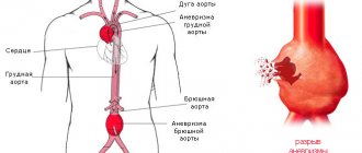
Very often patients do not make any complaints. But if symptoms appear, they are nonspecific. The most common symptoms include pain in the abdomen, back, or chest area. Patients describe this pain in different ways: some feel moderate to severe pain or tenderness in the middle or upper abdomen or lower back, while others feel the aneurysm itself as a beating and pulsating mass in the abdominal cavity. But, as noted, many may not experience any symptoms. Sometimes abdominal aortic aneurysm
may be detected during a routine physical examination. When palpating the abdomen, the doctor may detect a bulge or pulsation in the abdominal cavity. But most often, an aneurysm is detected during medical tests such as an abdominal ultrasound (ultrasound) or computed tomography (CT) scan. The size and location of the abdominal aortic aneurysm, as well as the general condition of the patient, help to choose the right treatment method. If the aneurysm is small, your doctor may recommend surveillance (regular examinations to monitor its size). A large aneurysm or a rapidly enlarging aneurysm has a high chance of rupture and requires treatment.
Pain relief during treatment
To access the aorta, the femoral arteries are usually used, which are exposed openly through small incisions. For pain relief, epidural anesthesia is used (the insertion of a catheter into the spine through which an anesthetic solution is supplied). Full patient monitoring is mandatory. Respiratory equipment for artificial ventilation of the lungs is at the ready. For intravenous infusions, a subclavian venous catheter is used.
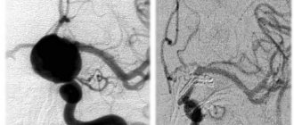
How the treatment method works
The operation is performed under sterile conditions, under the control of an X-ray angiographic unit, by a joint team consisting of a vascular and endovascular surgeon.
The patient's femoral arteries are accessed by a vascular surgeon using 4-5 centimeter incisions. The femoral arteries are isolated and placed on supports that allow them to be manipulated.
A diagnostic catheter is passed through the brachial or radial artery in the arm into the abdominal aorta for angiographic evaluation of the aneurysm and the condition and origin of the renal arteries. This is very important, since it is impossible to close the renal arteries with a stent graft, as renal failure will develop.
The endoprosthesis reliably closes the aneurysm cavity from the inside, conducting blood without contact with the aneurysmal sac, which over time leads to thrombosis of the aneurysm and a decrease in its size.
After opening the femoral arteries, special tubes (intraducers) are installed in them, through which special conductors are passed into the aorta above the aneurysm. The main body of the stent graft is then passed through a guidewire through a small incision in the right femoral artery. This is the main prosthesis placed in the aorta and the right leg, which should be located in the right iliac artery. Next, the main body of the endoprosthesis is carefully positioned in the area of the “neck” of the aneurysm, so that when opening in the area of the renal arteries, only the special crown of the endoprosthesis is placed, but not the closed section of the stent-graft. Monitoring is carried out using a diagnostic catheter placed above the aneurysm.
After opening the main branch and the right leg, a guide is inserted into the hole intended for the second branch and taken out beyond the stent graft. The left leg of the stent graft is passed along this guidewire for placement in the left iliac artery. After opening this leg, the structure is assembled.
To ensure a tight fit of the stentgraft to the walls of the aorta, it is additionally straightened with special balloons. After this, control angiography is performed, which should show uniform filling of the endoprosthesis, no leakage of contrast in addition to the prosthesis, and normal patency of the arteries below the prostheses.
The stent graft delivery devices are then removed from the arteries. The holes in them are sutured with a vascular suture. Control drainage is inserted into the wound. Blood flow in the lower extremities and kidneys is monitored using angiography from the upper catheter.
results
There was no intraoperative, in-hospital, or 30-day mortality after the first and second stages of retrograde prosthetics. In 1 patient after TAA replacement, retrograde dissection developed on the 7th postoperative day. The patient underwent emergency reconstruction of the SOA and its arch. Intraoperative data and the range of postoperative complications are presented in Table. 3.
Table 3. Intraoperative data and postoperative complications
In-hospital mortality after the second stage of antegrade aortic replacement was 5% ( n
=1). In this patient, the postoperative period was complicated by multiple organ failure due to hypocoagulable bleeding. Repeated thoracotomy was performed in 3 patients, revision of the retroperitoneal space due to bleeding was performed in 3. Respiratory failure requiring prolonged ventilation (within 72 hours) developed in 5 patients. Tracheostomy was required in 1 patient. Acute renal failure requiring hemodialysis developed in 1 patient. There were no cases of stroke or paraplegia.
The interval between operations was 7±2 months, the duration of hospitalization was 14±9 days.
During the observation period there were no deaths among discharged patients. MSCT of the aorta 3 and 6 months after surgery revealed no leaks of contrast material in any of the patients; the prosthetic segments were without kinks or deformations and were passable along their entire length. In one case, stenosis of the left renal artery without signs of nephrosclerosis was detected. After stenting, the patency of the artery was restored.
Possible complications during treatment
Complications of the intervention can be divided into those that relate to the course of the operation itself or to the design and location of the endoprosthesis. Thus, myocardial infarction that occurs immediately after the intervention is a complication of the operation, not the prosthesis. On the contrary, the development of leaks along the stent graft is a complication associated with the endoprosthesis.
The longevity of the results of endovascular treatment of aortic aneurysm depends on careful monitoring of the condition of the prosthetic section of the aorta and taking action when complications are identified.
One of the main reasons for complications with EVAR is that the contact between the proximal neck of the aneurysm and the stent graft is loose, due to the complex anatomy of the neck and the discrepancy between the diameter of the stent graft and the size of the neck. Most often, this is due to the anatomical features of the neck and one can expect that with the advent of special fenestrated or branched stent grafts, this complication will become a thing of the past. Currently, the use of such stent grafts is possible, but their installation is more complex, and the devices themselves are much more expensive than standard ones. If the contact is not tight, the aneurysm does not turn off completely and can continue to grow and even rupture. Therefore, leaks (endoleaks) must be eliminated immediately after they appear.
Complications associated with surgery
- Aortic dissection
- Contrast-related renal failure
- Thromboembolism is the transfer of blood clots through the bloodstream into the underlying arteries of the legs.
- Acute obstruction of the intestinal arteries - mesenteric thrombosis
- Bleeding in the access area in the groin area
- Access area infection
- Myocardial infarction, heart rhythm disturbances, respiratory failure.
Complications associated with stent graft
- Migration of the endoprosthesis
- Aneurysm rupture
- Stent graft thrombosis
- Leaks (endoleaks) into the aneurysm cavity
There are 5 types of leaks:
Type I - Leakage in the upper and lower places of stent graft attachment (in the area of the renal and iliac arteries). The latter lies almost freely in the aorta and aneurysm without performing its protective function for the aneurysm. Type II - Retrograde blood flow into the cavity of the aneurysmal sac from branches of the aorta such as the lumbar and inferior mesenteric artery. This type of leak occurs most often and carries the least risk. Immediate treatment is not required, since the cavity may become thrombosed and the endoleak will disappear spontaneously. Type III - leakage between overlapping parts of the stent (for example, between the main body of the stent graft and the left leg) or rupture of the endoprosthesis covering. Type IV - leakage through the stent graft wall due to the porosity of the coating material. With the development of technology for manufacturing stent grafts, this type of endoleaks is disappearing. Type V—expansion of the aneurysmal sac without detectable leakage. Spontaneous growth of an aneurysm, the causes of which are not clear.
Prognosis after treatment method
If the endoprosthetics operation is performed correctly and with a good immediate result, then the risk of aneurysm rupture is reduced many times and a year after this procedure is compared with the results of an open aneurysm resection operation. The patient can be discharged home 2-3 days after surgery. Taking into account the low postoperative mortality after endovascular treatment, the risk of endoprosthetics is significantly less than the risk of open surgery. Therefore, in Western countries, more than 80% of patients with abdominal aortic aneurysm are operated on using the endovascular method. However, for a lasting positive result, the patient must be under dynamic observation.
After the procedure
First, the operated patient is transferred to the intensive care ward, where he spends a day under the supervision of anesthesiologists and resuscitators. There, the patient recovers from anesthesia with continuous monitoring of blood counts, diuresis, respiration, heart function and blood pressure.
The next day, the patient is sent to a regular ward, where he is given infusions to replenish blood loss and correct the electrolyte-water balance. Particular emphasis is placed on pain relief and antibacterial therapy.
The patient is prohibited from drinking for several hours after surgery. This helps avoid vomiting and nausea due to water entering the digestive tract. In general, the patient spends up to 4-7 days in the hospital. The full rehabilitation period covers up to 2-3 months.
Observation program after treatment method
The next day after the operation, an examination by a vascular surgeon is carried out, wounds in the groin areas are bandaged, and drains are removed. To assess the effectiveness of the operation, an ultrasound of the abdominal aorta and arteries of the lower extremities is performed.
Every 3 months after surgery, it is necessary to perform MSCT of the aorta to identify possible leaks. Such studies are carried out during the first year. Then they need to be repeated annually. If MSCT is unavailable, ultrasound of the abdominal aorta may be performed.
If leaks are detected, the patient is hospitalized in the clinic for correction, since once these complications arise, they do not go away on their own, but require intervention to correct and prevent aneurysm rupture.
Why is the procedure necessary?
This intervention helps prevent such a dangerous phenomenon as aneurysm rupture. If you ignore timely treatment, this can lead to death. According to statistics, in 40 out of 100 patients, rupture occurs during the first year of illness.
Consequently, resection saves lives and protects against various complications. In particular:
- prevents digestive tract disorders;
- relieves portal hypertension syndrome;
- prevents organic changes in the digestive system;
- does not allow intestinal ischemia to occur, etc.