- August 7, 2018
- Orthopedics and traumatology
- Elena Kerra
Few people have never had bruises on their body in their life. They could be large or small, painful and completely imperceptible, appearing after a blow or for unknown reasons.
If you had to go to the hospital as a result of an injury, doctors always use the word “hematoma” in the medical history. Because of this, some patients worry that something dangerous has happened to their health, and ask what is the difference between a hematoma and a bruise? In essence, nothing, because both formations have an identical etiology, that is, they arise for the same reasons. Sometimes you can hear the opinion that a bruise is a small and very mild injury, and a hematoma is large and serious. This statement is not entirely true, since there is a classification of hematomas according to severity, which includes a mild degree. Still, there is a difference between a bruise and a hematoma. Our article is about this and various types of injuries with vascular damage.
What is a bruise
Almost every one of us first becomes acquainted with these formations on the body in childhood, because there is not a single child in the world who has never fallen or hit something. Therefore, everyone knows what a bruise is. This is a bluish (hence the name) spot that appears at the site of the bruise. If bruises form accidentally in children, then in older people, especially among representatives of the stronger half of humanity, they can “bloom” on the face or other parts of the body as a result of a fight. Among the peaceful part of the adult population, bruises appear from accidental bruises, after injections, intravenous injections, squeezing the skin with great force, pinches or bites, for example, from a pet. In all these cases, small blood vessels rupture in the subcutaneous layer, and blood flows out of the wounds into the resulting cavity. That's what a bruise is.
In the cavity, the blood can be in a liquid state, thickened or already completely coagulated. If an injury causes a rupture of not only blood vessels, but also the skin, the blood flowing from the wound can be seen visually. If the top layer of skin remains intact, it will appear as a bluish or purple bruise.
Reasons why bruises often appear on the legs
The appearance of bruises on the legs without a blow or injury is an alarming sign indicating health problems. Most often they occur suddenly and may be accompanied by bruising. In this case, you should not delay a visit to the doctor, since if the blood vessels are in normal condition without injury, hemorrhage does not occur in the upper layers of the skin. In case of injuries, they can remain on the legs for quite a long time and this is a type of norm that does not require special treatment.
Among them may be:
- Vitamin deficiency, in which due to a lack of vitamins C, K, P, the elasticity of the walls of blood vessels decreases and their fragility increases. If you adjust the menu and replenish it with rose hips, black currants, chokeberries, then the lack of vitamins can be compensated. Most often, such bruises appear in the spring, when the manifestations of vitamin deficiency are most severe.
- Reduced strength of the connective tissue that protects the capillaries. In this case, the appearance of bruises on the legs without a contusion becomes possible with the slightest pressure. This needs to be diagnosed (which is only possible in a specialized center). If the diagnosis is established, sometimes the fragility of blood vessels can be eliminated by taking special medications.
- Lack of platelets, in other words, a blood disease in which there are problems with blood clotting. The localization of bruises is not limited to the legs alone; they also appear on other parts of the body.
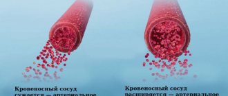
- Varicose veins, which not only cause bruising, but also spider veins. The appearance of bruises in this case may be accompanied by a feeling of heaviness in the legs, swelling, and bloating of the veins. As a treatment, the doctor will prescribe you: taking special medications that increase the tone of the veins; sclerotherapy (bonding of blood vessels through subcutaneous administration of drugs); wearing special knitwear that maintains muscle and vein tone).
- Hemorrhagic vasculitis. The reason for the appearance of this disease lies in the fact that the immune system fails and the cells of the blood vessels are perceived as foreign. The IS begins to produce antibodies that destroy the walls of blood vessels. This leads to inflammation, and as a result, the appearance of hematomas.
- Yellow bruises may indicate ruptured blood vessels. This is typical for people actively involved in sports. Under heavy load, capillaries under the skin of the extremities inevitably burst. As a result, bruises appear. Therefore, if after a fitness class your legs hurt a lot and bruises appear, we recommend reducing the level of stress during training.
- The appearance of bruises on the body may be a consequence of developing diabetes, because contrary to popular belief, this disease does not only affect blood sugar levels. Diabetes causes a metabolic disorder that affects blood circulation. Bruises appear, seemingly for no apparent reason, because the vessels become more fragile and brittle, and blood clotting worsens.
- Another reason for the appearance of bruises under the knee at the back, most often is a tendon sprain, varicose veins or internal arterial thrombosis.
- Also, if bruises appear on their own, seemingly for no apparent reason, this may be a reaction to the use of analgesics, antidepressants and anti-inflammatory drugs. The fact is that the components of these drugs reduce blood viscosity, which in turn leads to the appearance of hematomas.
You should start by diagnosing the causes, for which you should visit a phlebologist to rule out problems with the blood. If many small bruises appear on the body, we are talking about a blood disease. By taking tests, the number of platelets in the blood is determined and if a deficiency is detected, appropriate treatment is prescribed. If everything is normal according to hematology, you cannot do without examining the blood vessels to find out why bruises often appear on the body and legs: on the foot, on the inside of the thigh, on the knees, on the calves.
As preventive measures, it is advisable (even in the absence of problems with blood vessels) to wear comfortable shoes, dose physical activity, avoiding stagnation on the one hand, but also without straining it excessively, on the other, adjusting the diet. At the first signs of varicose veins, it would be a good idea to take special medications that strengthen the walls of blood vessels, but only a specialist can say about the advisability of taking them, as well as recommend a specific drug and its dosage. And then the problem that women often develop bruises on their legs for no reason will not bother you.
At the Antireflux phlebological center, our specialists will help you identify the true causes of bruises on your legs and body. After conducting the necessary tests and diagnostic studies, the necessary medications and treatment regimen will be selected according to the cause of the bruises.
Watch our video about why bruises can form for no reason:
What is a hematoma
If a person goes to the hospital after an injury or criminologists become interested in his situation, the medical term “hematoma” will appear in official papers describing the victim’s state of health. This means that a person has bluish-colored spots on his body that appeared after closed injuries that caused ruptures of blood vessels. The location and size of hematomas may be different, but this does not change the essence of the matter. In all cases, they are formed due to the fact that from ruptured vessels, under the pressure created by cardiac activity, blood flows into the space formed under the skin. The larger the damaged vessel or the more extensive the damage, the larger the hematoma and the more significant the body’s reaction to it. But pain depends not only on the degree of vascular damage, but also on the individual sensitivity threshold of each person.
Why it is not recommended to treat a hematoma on the leg at home
If there is large accumulation of blood under the skin, it must be removed. There are enough videos and articles on the Internet describing this procedure at home, but you absolutely cannot follow such advice. Firstly, a person risks damaging his leg even more and disrupting its normal functioning, even to the point of disability. Secondly, and more likely, you can introduce an infection into the body and provoke even greater inflammation. Thirdly, only an experienced doctor using special equipment will do this carefully, but independent attempts can permanently disfigure the skin and leave unaesthetic scars on it.
Difference
What is the difference between a hematoma and a bruise? From the above it is clear that both are accumulations in the formed cavities of a large or small amount of blood that has flowed out from ruptured blood vessels. There are several differences between these two concepts.
- The word “bruise” is used colloquially, and “hematoma” in official medicine, criminology, jurisprudence, and scientific literature.
- The medical term hematoma comes from the fusion of two Greek words, ema, meaning blood, and oma, meaning tumor. People called bruises the spots on the skin that appear after bruises or other injuries, because of their characteristic blue with various shades of color.
- A bruise is always visible to the naked eye because it forms in the subcutaneous fat layer. A hematoma can appear not only in the subcutaneous tissue, but also in the internal organs, so sometimes instrumental examination is required to detect it.
- Bruises are commonly called spots that go away without treatment. Hematomas can be so complex that they require mandatory medical intervention.
Classification of hematomas
You will be surprised to know how many types of hematomas exist in official medicine. There are several criteria that determine the differences:
1. According to the state of the exudate and blood that make up the hematoma:
- fresh (the blood in it is liquid);
- organized (blood already coagulated or heavily thickened);
- infected (pathogenic microorganisms have been added to the blood);
- festering.
2. By size:
- small, dotted;
- large (over 3-5 mm in diameter).
Such bruises on the body can be located singly or in groups.
3. By location:
- subcutaneous hematoma;
- subserous (effusion of blood into internal cavities - abdominal, pulmonary);
- intracranial (subdural, epidural);
- intramuscular.
4. According to clinical signs:
- pulsating;
- encysted;
- not pulsating.
We can say that a bruise is a subcutaneous hematoma, not pulsating, large, fresh or organized.
Sometimes hematomas are divided according to the type of damaged vessels:
- arterial;
- venous;
- mixed.
A bruise can appear when the integrity of both small venous vessels (venules) and arterial vessels (capillaries) is damaged.
Etiology of bruises
To the above explanations of how a hematoma differs from a bruise, we can add the reasons why a person develops such formations. First, let's look at where bruises come from. Since they always form in the subcutaneous layer, their appearance can be caused by any injury that disrupts the integrity of small and medium-sized blood vessels located in the subcutaneous fat. These are:
- injury;
- hit;
- compression, squeezing;
- pinch;
- injection (bruises are observed with intramuscular injections, because the syringe needle touched and injured small vessels);
- intravenous injections (bruises are observed because blood has leaked from the punctured vein into the resulting space).
Bruises can appear on the legs due to varicose veins and phlebitis, and on the face after medical procedures, such as tooth extraction.
The weaker the walls of a person’s blood vessels, the more often and in greater numbers bruises “bloom” on his body.
Bruise, bruise, hematoma, lump. How to say it correctly? Let's learn proper terminology.
In practice, we often encounter cases where a wide variety of terms are used to describe the same injury, which often leads to significant confusion. And the greatest confusion arises when, as a result of some impact (usually traumatic) on the body a person develops intradermal and subcutaneous hemorrhage - bruise.
They just don’t call this kind of damage – a bruise, a hematoma, or just a “bump.” Are these all synonyms?
Let's figure it out.
What is the difference, if any, between the terms hematoma and bruise? Both hematoma and bruise can form either as a result of external mechanical impact or as a result of disease.
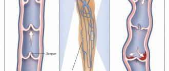
A hematoma is a hemorrhage with a violation of tissue integrity and the formation of a cavity filled with blood. Literally translated, hematoma is a blood tumor (from ancient Greek αἷμα - blood + ōma - tumor).
A hematoma is formed due to the rupture of a vessel, followed by hemorrhage, which leads to tissue separation with the formation of a cavity in them.
A hematoma can be located superficially (under the skin or external mucous membranes), or it can also form deep in soft tissues, such as muscles. Rice. Hematoma of the leg
Rice. Subcutaneous hematoma
Rice. Neck hematoma
Formation of hematomas in the walls of organs is possible.
Rice. Nasal septum hematoma
If a hematoma has formed in the cranial cavity, then in relation to where it is located, the following are distinguished: - epidural (extradural) hematoma (located between the bones of the skull and the dura mater) - subdural hematoma (under the dura mater) - subarachnoid hematoma (under the soft mater) membrane) - intracerebral (parenchymal) hematoma (in the substance of the brain).
Rice. Variants of intracranial hematomas.
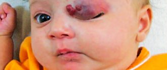
In newborns, there may often be a birth tumor on the head - a subcutaneous hematoma, which forms during childbirth in the presenting part of the head. This is fine. In adults, such a hemorrhage under the scalp is formed as a result of a blow, and is popularly called . ”
Newborns may also have a cephalohematoma (cephalohematoma) on their head. This is a hematoma, formed during childbirth and located between the periosteum and the outer surface of the cranial bones. The tumor is limited to the edges of one or another bone of the skull.
Rice. Variants of birth trauma to the head of a newborn. A. Cephalohematoma (subperiosteal hematoma). B. Birth tumor (subcutaneous hematoma)
So, an important, significant sign of a hematoma is the formation of a cavity filled with blood. If there is no cavity filled with blood, it is not a hematoma. Bruising is bleeding into soft tissue (for example, skin) while maintaining the tissue structure. A cavity in the tissue does not form during a bruise; this is not a cavity formation.
Rice. Bruise
In unprofessional, everyday speech, a bruise is called a bruise. Bruise is not a medical term, but a colloquial one. Not a single doctor, including a forensic expert, uses the word “bruise.” It is not recommended for lawyers to use this word in their professional activities.
Sometimes, wanting to show knowledge of medical terminology, a bruise is called a hematoma. This is fundamentally wrong. If someone wants to call a bruise in Latin, call it hemorrhagic permeation of the skin.
As a synonym for the word bruise, doctors often use the terms “ecchymoses” and “petechiae.” Ecchymosis is hemorrhage in the skin or mucous membrane, the diameter of which exceeds 3 mm. Petechiae are pinpoint hemorrhages in the skin, mucous membranes, serous membranes and internal organs, up to 3 mm in diameter.
Rice. Multiple petechial hemorrhages (point hemorrhages) in the skin of the arm.
Bruises in the skin change color over time and “bloom,” which allows forensic medicine to determine how long ago they formed.
Bruises in an array of soft tissues are called hemorrhages, for example, hemorrhages in the muscles of the thigh, forearm, etc.
Rice. Hemorrhage into the muscles of the forearm.
So, in this brief review, we tried to find out what the difference is between a bruise and a hematoma.
And knowing this difference is very important, since a bruise and a hematoma are different formations, they have different consequences for the body, the tactics of their treatment are different, and the severity of bodily injuries is different.
Etiology of hematomas
If we talk about cavities with blood in the subcutaneous layer, then a bruise and a hematoma are essentially identical formations. The difference between them is observed when we talk about blood-filled cavities in the internal organs. Such formations can also appear as a result of a blow, such as a traumatic brain injury. In addition, hematomas inside the body appear for the following reasons:
- bone fracture;
- ligament rupture;
- dislocation;
- muscle strain, for example, due to high physical activity;
- vitamin deficiency, which caused fragility of blood vessels;
- hemorrhagic vasculitis;
- liver diseases;
- low blood clotting;
- varicose veins;
- phlebitis;
- thrombophlebitis;
- long-term use of Cavinton, Aspirin, and other blood thinning drugs;
- age-related changes.
Hematoma or bruise
A hematoma or bruise is an accumulation of partially coagulated blood under the skin, outside the vessel. It is formed when the walls of a vessel are damaged. The blood goes beyond it and a bruise appears. The size of the bruise depends proportionally on the severity and severity of the injury. Blood entering the tissue causes swelling, pain, and redness.
The cause of a hematoma is tissue trauma. In the main cases, this is a direct, intense, sharp impact on tissue (impact, bruise, fracture, dislocation, rupture). In other cases, hematomas may appear as a result of diseases associated with increased fragility of blood vessels and bleeding disorders. Over time, the hematoma resolves and scars.
Kinds:
- subcutaneous;
- fascial;
- intramuscular;
- pulsating - damage to a large vessel, without thrombus formation;
- non-pulsating - damage to small and medium-sized vessels with the formation of a blood clot;
- not coagulated - appear in the first days after injury;
- curled up - appear within 24 hours after the injury;
- lysed - lack of blood clotting ability;
Severity:
- Easy. Appears within 24 hours, minor pain appears, but there is no impairment of limb mobility. Does not require medical intervention.
- Medium-heavy. Forms within six hours. Accompanied by slight pain, swelling, and redness. Limb mobility is limited. Conservative treatment is mainly used.
- Heavy. Forms within an hour. Severe pain, large swelling, impaired mobility. When infection occurs, surgical treatment is required.
In the affected area, the skin becomes red, then purple-bluish, after a few days - yellow, then after five days - greenish.
Complications:
- suppuration;
- abscess;
- sepsis;
- phlegmon.
Diagnostics.
Examination by a specialist doctor. In the absence of other lesions, additional examination is not required. The diagnosis is made taking into account examination, medical history, complaints and laboratory tests. For deep lesions, ultrasound is used. If a fracture is suspected, take an x-ray.
Treatment.
Under good conditions, the bruise resolves on its own. If for some reason resorption does not occur, the blood-filled cavity disrupts the proper functioning of nearby organs and causes pain.
Conservative treatment uses: rest, cold compress, pressure bandage, ointments, painkillers, physiotherapy, warming.
During surgical treatment, an autopsy is performed, the lesion is drained, and antibiotics are prescribed. Treatment of intracranial hematoma is carried out surgically, but some patients do not require surgical intervention, but only observation.
In general, the prognosis for treatment is positive. Most bruises resolve on their own, and those that were operated on recover within 2.5 weeks.
Prevention is the prevention of injuries and elimination of diseases that lead to the formation of hematoma and early contact with specialists.
Color change
Probably everyone has noticed that bruises on the body tend to change their color over time.
Basically, in the first hours after its occurrence, the hematoma visible to the eye has a red-purple color, which is caused by the content of oxyhemoglobin in the spilled blood. It soon turns into hemoglobin, causing the hematoma to turn blue-violet. In about 5-6 days, hemoglobin breaks down into methemoglobin and verdochromogen, the color of which is green. Therefore, the hematoma acquires greenish tints. The transformation of blood particles continues, and after about a week, verdochromogen breaks down into bilirubin and biliverdin, which color the hematoma yellow. After this, it gradually becomes discolored and is compared with the overall skin tone.
The rate at which the hematoma changes color depends on its size, thickness, location and individual characteristics of the patient’s body. In hematomas of large area and thickness, several colors can be observed simultaneously.
Symptoms of bruises
When talking about the difference between a hematoma and a bruise, we cannot ignore the symptoms of these formations. Bruises can appear almost immediately after vascular injury. In this case, they are classified as early. Such formations rarely cause health problems. Only when they appear on the face do they cause psychological discomfort in some people. Characteristic features of bruises:
- painless unless touched with great force;
- do not lead to swelling of the injured area;
- do not cause pain when moving.
Sometimes it happens that bruises appear a little later after the injury (one or several days). In this case, they are classified as late or very late. Such bruises are practically no different from early ones, but before their visual appearance, a person may experience pain at the site of injury.
Bruises do not require special treatment, but to prevent them from appearing, you can immediately apply ice to the site of the bruise. An already “blooming” bruise can be lubricated with “Bruise-Off”, “Express-bruise” cream or “Thrombocide” gel so that it resolves faster.
Symptoms of hematomas
A person can develop a simple superficial hematoma anywhere on the body. Her ICD 10 code is T 14.0. The symptoms of superficial hematomas are the same as those of bruises.
Intramuscular hematomas are almost always very painful. A person has:
- swelling at the site of injury;
- stiffness of movements;
- difficulty or inability to perform certain movements due to pain;
- Sometimes the victim develops a fever.
If a hematoma occurs in the internal organs, pain in this organ, and sometimes in neighboring areas, always comes to the fore. The rest of the symptoms depend on the location of the injury. So, if a hematoma appears in the gastrointestinal tract, nausea and vomiting may occur. If in the lung area, a cough appears (sometimes with blood), difficulty breathing. A hematoma in the brain is accompanied by headache, nausea, sometimes clouding of consciousness and temporary loss of vision.
Treatment
External hematomas are usually diagnosed based on clinical indications. Often, at the site of the injury, the patient has bruises on the skin, which are an accumulation of blood that has leaked from a ruptured vessel, but without the formation of a cavity. People also call bruises bruises.
External hematomas do not require specific treatment. If the victim complains of pain in the area of the injury, he is recommended to use an ointment with healing and analgesic effects. The following ointments have proven themselves well: “Heparin”, “Lioton”, “Indovazin”, “Sinyak-Off” and others. Traditional healers recommend applying cabbage leaves or compresses with onion and wormwood decoctions to the bruised area.
To identify and diagnose internal hematomas, instrumental studies are performed - MRI, CT, ultrasound. Treatment of these formations is always difficult, requiring the use of specific medications. Hematomas in the brain are especially difficult to treat. In most cases, they require the installation of drainage or urgent surgery to drain the blood. The prognosis for the outcome of such hematomas is often unfavorable.
Treatment of hematoma on the leg
Typically, a hematoma on the leg after a bruise is treated by two methods: conservative and surgical. The first category includes: cold and warm compresses, pressure bandages, taking analgesics, applying ointment, physiotherapeutic manipulations.
The second group involves performing a puncture - surgical removal of accumulated blood from the site of a bruise with the application of a suture and an aseptic bandage.
Treatment of a serious hematoma on the leg must be carried out under the guidance of a doctor; this will speed up the healing process and prevent unpleasant consequences.