The cardiac muscle (myocardium) in the structure of the human heart is located in the middle layer between the endocardium and epicardium. It is this that ensures the uninterrupted operation of “distilling” oxygenated blood into all organs and systems of the body.
Any weakness affects blood flow and requires compensatory restructuring and coordinated functioning of the blood supply system. Insufficient ability to adapt causes a critical decrease in the performance of the heart muscle and its diseases. The endurance of the myocardium is ensured by its anatomical structure and endowed capabilities.
Features of muscle tissue are:
- striated striations formed by myofibrils of cardiomyocyte cells;
- the presence of two types of fibers: thin (actin) and thick (myosin), connected by cross bridges;
- the connection of myofibrils into bundles of different lengths and directions, which makes it possible to distinguish three layers (superficial, internal and middle).
The structure of the heart muscle is different from skeletal and smooth muscle muscles, which provide movement and protection of internal organs
The morphological features of the structure provide a complex mechanism of heart contraction.
Structure and functions
The cellular elements of muscle tissue are elongated, which is why they are called “fibers.” The cytoplasm of cells contains thin protein filaments of myofibril, which can lengthen and shorten (Table 1) . Special organelles and energy production by mitochondria provide contraction and elongation of fibers.
Table 1.
Structure and functions of muscle tissue
| Types of muscle tissue | Structure | Functions | Location in the body |
| Cross-striped | Consists of long and thick fibers (Fig. 1) . They are formed by the fusion of individual cells. There are a lot of nuclei. Striations are caused by alternating light and dark discs. The fibers are combined into bundles. | Voluntary body movements, breathing, facial expressions and a number of other actions. | The basis of skeletal muscles, tongue, pharynx, initial part of the esophagus. |
| Smooth | Individual spindle-shaped cells are small in size, united in bundles (5–10 pieces each). Each cell has one nucleus (Fig. 1) . Thin myofibrils stretch between the ends of the cell. The fabric is devoid of transverse stripes. | Involuntary contractions of the walls of internal organs under the influence of nerve impulses. | Muscular layers of the skin and internal organs (digestive system, bladder, blood and lymphatic vessels, uterus). |
| Striated heart | The cells are elongated, branched, with a small number of nuclei, forming a single network (Fig. 1) . Transverse striation occurs due to shiny stripes at the junctions between cells. | "Engine" of blood circulation. Involuntary contractions of the heart muscle can occur under the control of the autonomic nervous system. | The bulk of the heart. |
Rice.
1. Structure and location of muscle tissue Muscle tissue ensures the movement of the body in space. Muscle contractions are necessary to change the position of individual parts of the body. Muscles, in addition to motor function, perform protective and heat exchange functions.
How does the heart contract?
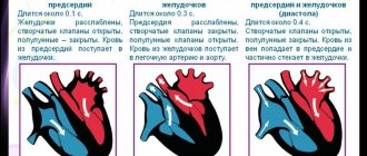
Contractility is one of the properties of the myocardium, which consists in creating rhythmic movements of the atria and ventricles, allowing blood to be pumped into the vessels. The chambers of the heart constantly pass through 2 phases:
- Systole - is caused by the combination of actin and myosin under the influence of ATP energy and the release of potassium ions from the cells, while thin fibers slide over thick ones and the bundles decrease in length. The possibility of wave-like movements has been proven.
- Diastole - relaxation and separation of actin and myosin occurs, restoration of expended energy due to the synthesis of enzymes, hormones, and vitamins obtained through the “bridges”.
It has been established that the force of contraction is provided by calcium entering the myocytes.
The entire cycle of heart contraction, including systole, diastole and the general pause after them, with a normal rhythm fits into 0.8 seconds. Begins with atrial systole, the ventricles are filled with blood. Then the atria “rest”, moving into the diastole phase, and the ventricles contract (systole). Calculating the time of “work” and “rest” of the heart muscle showed that per day the state of contraction accounts for 9 hours 24 minutes, and the state of relaxation accounts for 14 hours 36 minutes.
The sequence of contractions, ensuring the physiological characteristics and needs of the body during stress and anxiety depend on the connection of the myocardium with the nervous and endocrine systems, the ability to receive and “decipher” signals, and actively adapt to human living conditions.
The spread of excitation from the sinus node can be traced by intervals and ECG waves
Heart wall, myocardium
As I have said more than once, the wall of the heart consists of three layers. Each layer has its own cellular composition and performs a specific job or function.
Each layer of the heart wall is important and indispensable for the normal functioning of this organ. And yet none of the layers can compare in importance with the myocardium.
The myocardium consists of many muscle cells. It is thanks to these cells that the heart has the ability to contract and relax, either filling its cavities with blood or pushing out the blood and moving it further along the bloodstream.
The middle layer of the heart wall, the myocardium, is an entire muscular system. Because this thick layer of muscle cells is represented by several layers of multidirectional muscle fibers.
Some muscle fibers are directed strictly horizontally or transversely in relation to the heart. The second part has a vertical or longitudinal direction. And the third part of the muscle fibers goes at a steep oblique slope down to the apex of the heart.
This arrangement of muscle fibers makes the heart muscle especially strong. In addition, this also allows for the most rational distribution of the load between individual parts of the myocardium.
It is also important for the functioning of the heart that the atrial myocardium and ventricular myocardium are completely independent of each other. In other words, the atria have the ability to contract separately, regardless of the contractions of the ventricles and vice versa.
If you have read articles about the work of the heart and the cardiac cycle, then you understand how important this autonomy is.
Human circulatory system
The structure of the human heart
What does a human heart look like?
Where is the human heart?
What's Below the Heart?
Human right atrium
Right ventricle of the heart
Left atrium
Left ventricle of the heart
Atrioventricular orifices
How many valves are there in the heart?
Valve apparatus of the heart
Mitral valve
Tricuspid valve
Semilunar valves
Wall of the heart: pericardial sac
Endocardium: the inner lining of the heart
Cardiac mechanisms that provide contraction
The properties of the heart muscle have the following purposes:
- support myofibril contraction;
- ensure the correct rhythm for optimal filling of the cavities of the heart;
- maintain the ability to push blood through any extreme conditions for the body.
For this, the myocardium has the following abilities.
Excitability - the ability of myocytes to respond to any incoming pathogens. Cells protect themselves from above-threshold stimulation by a state of refractoriness (loss of the ability to excite). In a normal contraction cycle, a distinction is made between absolute and relative refractoriness.
- During the period of absolute refractoriness, for 200 to 300 msec, the myocardium does not respond even to extremely strong stimuli.
- When relative, it is able to respond only to sufficiently strong signals.
This property prevents the heart muscle from “distracting” the contraction mechanism during the systole phase
Conductivity is the property of receiving and transmitting impulses to different parts of the heart. It is provided by a special type of myocytes that have processes very similar to neurons in the brain.
Automaticity - the ability to create its own action potential inside the myocardium and cause contractions even when isolated from the body. This property allows resuscitation in emergency cases and maintains blood supply to the brain. The importance of the located network of cells and their accumulation in nodes during transplantation of a donor heart.
Pacemaker cells (pacemakers) become the main ones if the processes of repolarization and depolarization in the main nodes are weakened. They suppress “other people’s” excitability and impulses, and try to take on a leadership role. Localized in all parts of the heart. The possibilities are restrained by the sufficient strength of the sinus node.
Cross-striped fabric
The cells have a thickness from 10 to 100 microns, a length from 10 to 40 cm. The cytoplasm contains a large number of nuclei and myofibrils, occupying a central position (Fig. 2) . Mature cells have hundreds of myofibrils and more than 100 nuclei. Actin and myosin filaments inside myofibrils are linked to each other (Fig. 3) . The ability for rapid contraction of this tissue is higher than that of others.
Rice. 3. Structure of skeletal muscle
Muscle fibers are covered with a membrane called sarcolemma. There are alternating plates of proteins of different densities that have unequal light refractive indices. Under an optical microscope, such muscles appear to be striated across. The contractile elements are united into muscle bundles covered with a connective tissue membrane. Skeletal muscles are well supplied with blood vessels and nerves.
The importance of biochemical processes in the myocardium
The viability of cardiomyocytes is ensured by the supply of nutrients, oxygen and the synthesis of energy in the form of adenosine triphosphoric acid.
All biochemical reactions occur maximally during systole. The processes are called aerobic because they are possible only with a sufficient amount of oxygen. The left ventricle consumes 2 ml of oxygen per minute per 100 g of mass.
To produce energy, the following are used in the blood:
- glucose,
- lactic acid,
- ketone bodies,
- fatty acid,
- pyruvic and amino acids,
- enzymes,
- B vitamins,
- hormones.
If the heart rate increases (physical activity, anxiety), the need for oxygen increases 40–50 times, and the consumption of biochemical components also increases significantly.
Sports adaptology
Viktor Nikolaevich Seluyanov, MIPT, laboratory “Information Technologies in Sports”
The development of science leads to the emergence of models of the object of research, with the help of which new properties are learned or innovative technologies are developed, and a theory is created. To build a DFT, it is necessary to build a model of an ideal cell, muscle fiber, muscle, neuromuscular system, cardiovascular system, respiratory system, endocrine and immune, and digestive.
The perfect cage
All animal cells are arranged, to a first approximation, the same way. A cell, for example, a muscle fiber, has a membrane - the sarcolemma. The sarcoplasm contains all the usual organelles and numerous nuclei (muscle fiber is a multinucleated cell). Specific organelles are myofibrils.
The structural components of the cell are:
- - plasma, a transparent liquid containing proteins in the form of enzymes for the metabolism of carbohydrates, amino acids, fats (lipids) and other substances, as well as tRNA. In plasma, the construction of new organelles occurs with the help of ribosomes and polyribosomes.
— cell membranes consist of fat (40%) and protein (60%). Protein inclusions perform the functions of: carrier proteins, enzyme proteins, receptors, and structural basis.
- Mitochondria are the energy stations of the cell, engaged in the resynthesis of ATP molecules using oxidative phosphorylation. They consume oxygen, carbohydrates, fats and release carbon dioxide, water, and resynthesized ATP molecules. Metabolic products can also penetrate the mitochondrial membranes into the cytoplasm.
- endoplasmic reticulum - a set of membranes, tubes, vacuoles. There are granular and smooth endoplasmic reticulum. In the granular ER, the synthesis of membrane proteins and other cell components occurs. Smooth ER is involved in lipid synthesis and is well developed in the cells of the endocrine system. A connection with glycogen synthesis is also possible.
- Golgi complex - a network of membranes that perform a secretory function.
— lysosomes are spherical structures containing hydrolytic enzymes (proteinases, glucosidases, phosphatases, nucleases, lipases). Lysosomes participate in intracellular digestion processes. Lysosomes become especially active when the cell becomes acidic and the concentration of hydrogen ions increases.
— ribosomes are elementary apparatuses for protein synthesis.
— microtubules are fibrillar formations that act as framework structures.
- glycogen globules - the supply of carbohydrates in the cell.
- droplets of fat - fat reserves in the cell.
- nucleus - a system for genetically determining protein synthesis. Includes chromatin, nucleoli, karyoplasm and nuclear envelope. Chromatin contains DNA, mRNA is formed here, and ribosomal rRNA is formed in the nucleoli.
After clarifying the structure of the cell, we can consider the physiological processes in the cell. From the point of view of the theory of physical training, the processes of catabolism and anabolism are of interest.
Anabolism is provided by DNA and polyribosomes; anabolism is activated by steroid hormones. Somatotropin (growth hormone) and testosterone are especially important for physical development. Steroid hormones penetrate only into active cells.
Catabolism in the cell is provided by lysosomes. They become especially active when the cell is acidified—the appearance of hydrogen ions in them. In this case, the pores in the membranes increase, and both the processes of diffusion and active transport are accelerated.
Thus, the physical development of active cells is ensured by increasing the concentration of steroid hormones in the blood, while minimizing catabolism (blood acidification). For the coach, the first principles of constructing the training process appear:
1. Control of the activity of the central nervous system and muscles is ensured by the control of the endocrine system (the concentration of steroid hormones - somatotropin and testosterone in the body of athletes).
2. Controlling the concentration of hormones in the blood leads to adaptive changes in muscle fibers (growth of myofibrils and mitochondria).
Endocrine system
The endocrine system includes several glands: pituitary gland, pineal gland, adrenal glands, gonads, pancreas, etc. When performing physical exercises, mental tension (stress) arises in the cerebral cortex, which causes activation of the hypothalamus and activation of the pituitary gland. The anterior lobe of the pituitary gland releases somatotropin, thyrotropin, ACTH, follicle-stimulating (FSH) and luteinizing (LH) hormones into the blood.
Somatotropin (growth hormone) - penetrating into muscle fibers stimulates the synthesis of myofibrils, and synthesis in tendons and bone tissue is activated.
FSH, LH - activate the gonads, which leads to the release of testosterone into the blood, which activates the synthesis of myofibrils in muscle fibers.
It is well known that the concentration of somatotropin and testosterone increases when performing strength, speed-strength and speed exercises, as well as the mass of active muscles. Therefore, the development of muscle fibers occurs most intensively when performing extreme and near-limit exercises in terms of mental stress while minimizing the degree of acidification (catabolism) of MF.
This leads to the following pedagogical principle of sports training:
3. The most effective (stressful) are physical exercises performed with extreme or near-limit mental stress (intensity).
The immune system
The immune system includes bone marrow, thymus, lymph nodes, etc. Bone marrow is responsible for the construction of blood cells. The most important factors in normalizing the functioning of the bone marrow are testosterone and vitamin B12. Therefore, stress loads stimulate the activity and development of the bone marrow, and therefore the immune system.
Muscle
A muscle is made up of muscle fibers. Muscle fibers are usually classified into fast and slow. Muscle composition can be determined using a biopsy. A biopsy is taken from the lateral head of the quadriceps femoris muscle. A piece of muscle tissue is quickly frozen, then thin sections are made and chemically processed using a certain technology. The activity of myosin ATPase, an enzyme that destroys the ATP molecule, is usually determined. Then they look at cross sections of muscle fibers and see the color - black, gray and white CF. The proportion on a certain surface or out of 200 units of MV of the same color is calculated. This muscle composition is inherited. It is practically impossible to significantly change the ATPase activity of MV. In experiments with electromyostimulation, it is possible to temporarily change ATPase activity, but these experiments do not yet have practical significance.
It is important to note that each muscle has its own inherited muscle composition, so taking a biopsy from a single muscle may not provide a complete picture of an athlete's endowment. Pedagogical observation and testing can provide more complete information about an athlete’s talent than a laboratory examination. For example, a set of tests for track and field athletes—a standing jump on two legs, multi-jumps from one foot to another, throwing a shot forward and backward, throwing a grenade—will allow one to evaluate the talent of various muscle groups in a given athlete in comparison with the norms. If most boys 11–12 years old jump 200 cm from a standing position, and one of them jumped 250 cm, then there is no doubt that this boy has a high percentage of fast MVs in the extensor muscles of his leg joints.
There is a way to classify CF according to other enzymes. Of particular interest is the classification of CF according to the activity of mitochondrial enzymes. In this case, they talk about oxidative, intermediate and glycolytic MVs. This muscle composition is not inherited because oxidative muscle fibers are easily converted to glycolytic fibers when training stops. Mitochondria are destroyed, age, and after 20 days only 50% remains of 100%, etc. Sports form is lost very quickly without training.
Muscle fiber has specific organelles - myofibrils. Myofibrils in all animals are the same in structure and differ only in length (the number of sarcomeres). The cross section of all myofibrils is the same. Therefore, the force of contraction of a muscle fiber depends on the number of myofibrils in it.
The sarcomere is a sequential component of the myofibril, consisting of actin and myosin filaments. Branches with heads emerge from myosin. The myosin head is also an enzyme for the destruction of ATP and CrP molecules. When an ATP molecule breaks down, ADP, P, H and energy are formed. To resynthesize an ATP molecule, energy is needed; it is taken from the KrP molecule, which, upon destruction, is converted into free Kr, inorganic phosphate (P) and energy.
Contraction of the sarcomere and myofibril occurs when calcium leaves the cisterns. It attaches to actin active sites and releases them to create a bridge between actin and myosin. The myosin head, when attached to actin, rotates 45 degrees, which allows the filaments to slide relative to each other. The separation of the myosin head from actin requires the expenditure of energy, which is taken from the process of destruction of the ATP molecule by the enzyme - myosin ATPase. Following this, creatine phosphokinase destroys CrP and the energy of this molecule is used for the resynthesis of ATP. Free creatine and inorganic phosphate penetrate through the myofibril to mitochondria or glycolytic enzymes and lead to the initiation of glycolysis and oxidative phosphorylation.
The release of calcium from the tanks occurs upon activation of the CF. After the cessation of electrical stimulation of the CF, the pores in the cisterns close, and calcium pumps continue to pump calcium atoms into the cisterns. After 50–100 ms, most of the calcium ions are pumped back into the tanks. This process is called muscle relaxation.
ATP molecules are large, so they move very slowly through the MV. The intermediary between myofibrils and mitochondria for energy delivery are KrF molecules. These molecules are small and easily move around the CF. Russian scientists (Sachs et al., 1977) called this mechanism the creatine phosphate shuttle.
Therefore, taking creatine with food allows you to increase its concentration in CF. As a result, metabolic processes in the CF are significantly accelerated.
Model of bioenergetic processes in muscle fibers of different types
In glycolytic muscle fibers there is a supply of ATP molecules in the myofibrils, a supply of ATP molecules near the mitochondria, and a supply of ATP molecules in the sarcoplasm. There is a supply of CR molecules, glycogen globules and fat droplets. The mass of mitochondria in glycolytic MVs (GMVs) is small, since it is necessary only for the life of these cells at rest.
Activation of biochemical processes begins from the moment electrical impulses pass through the MV membranes. Pores in the cisterns open, calcium is released into the sarcoplasm, calcium attaches to actin, actin-myosin bridges are formed, ATP and CrP are consumed. Free creatine and inorganic phosphate leave the myofibrils and use the energy of sarcoplasmic ATP molecules to resynthesize CrP. ATP molecules are resynthesized during anaerobic glycolysis. Glycolysis begins with the destruction of the glucose or glycogen molecule and ends with the formation of pyruvate. Pyruvate, due to the absence of mitochondria, is converted to lactate. The combination of the lactate anion with a hydrogen proton leads to the formation of lactic acid, which can be released into the blood in this form. In the blood, the lactic acid molecule dissociates, therefore there is a high correlation between the concentration of hydrogen and lactate (R = 0.99).
Hydrogen ions are formed during the breakdown of sarcoplasmic and other ATP molecules.
The activity of HMB leads to the accumulation of metabolic products H, Kr, F, La, Pyr, etc. in the sarcoplasm.
The reserves of myofibrillar ATP last for 1–2 s, CrF for 5–20 s (depending on the mode of contraction and relaxation of the MV). Then glycolysis intensifies, but its power is no more than 50% of the maximum, and due to the accumulation of hydrogen ions, the process of formation of actin-myosin bridges is disrupted and after 30 s they almost completely cease to form. This phenomenon is usually defined as local muscle fatigue. WMGs are defined as fatigable muscle fibers.
Oxidative muscle fibers are constructed in exactly the same way as glycolytic muscle fibers. The main difference relates to mitochondrial mass. In OMV, the mass of mitochondria is in the maximum ratio with myofibrils, which ensures a maximum oxygen consumption of about 0.3 l/min per kilogram of OMV.
Activation of OMB leads to the formation of actin-myosin bridges and energy consumption of ATP molecules. The concentration of myofibrillar ATP molecules is maintained by CrF. Maintaining the concentration of CrF is ensured in two ways:
- - ATP molecules resynthesized in mitochondria,
- ATP molecules resynthesized in aerobic glycolysis.
This process develops within 45–60 s. By this time, both glycolysis and fat oxidation can occur simultaneously. But as mitochondria function, citrate accumulates in the sarcoplasm, so inhibition of glycolytic enzymes begins and OMB completely switches to lipolysis.
Lipolysis uses fat reserves in droplets; the reserve of this fat in normal people lasts for 30–50 minutes. Fatty acids in the blood slowly enter the musculoskeletal system, so they cannot fully support high-intensity muscle activity.
Mitochondria absorb ADP, phosphorus, oxygen, pyruvate, fatty acids, glycerol, hydrogen ions and release resynthesized ATP molecules, carbon dioxide and water. Therefore, the OMVs do not become acidic and do not become tired.
The oxidation of fats in OMV can stop if lactate ions appear in the sarcoplasm. In this case, fat oxidation is inhibited, and lactate becomes a substrate for oxidation. Lactate is converted into pyruvate by cardiac lactate dehydrogenase, which enters the mitochondria through acetyl coenzyme. Pyruvate also begins to form during glycolysis from glucose and glycogen.
Lactate can enter the OMV only with the simultaneous functioning of the HMV and OMV.
The biomechanical properties of muscle fibers are associated with empirical laws:
- - “strength - length”,
- “strength - speed”,
- “strength - activation time”,
- “strength is the time of relaxation”,
— “force is the energy of elastic deformation.”
These laws must be taken into account when analyzing competitive activity.
Neuromuscular system
Heart and circulation
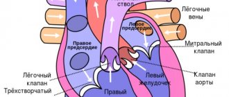
The activity of the heart and blood vessels ensures blood circulation - the continuous movement of blood in the body. In its movement, blood passes through the systemic and pulmonary circulation. The great circle starts from the left ventricle of the heart, includes the aorta, arteries, arterioles, capillaries, and veins extending from it and ends with the vena cava flowing into the right atrium. The pulmonary circulation begins from the right ventricle, then the pulmonary artery, pulmonary arterioles, capillaries, veins, the pulmonary vein flowing into the left atrium.
The function of the heart is to rhythmically pump blood into the arteries. The contraction of muscle fibers (myocardiocytes) of the walls of the atria and ventricles is called systole, and relaxation is called diastole.
The amount of blood ejected by the left ventricle of the heart per minute is called minute volume of blood flow (MVF). At rest, it is normally 4–5 l/min. By dividing the IVC by the heart rate per minute (HR), one can obtain the stroke volume of the blood flow or heart (SV). At rest, it is 60–70 ml of blood per beat.
The frequency and strength of contractions depends on the nervous, humoral (adrenaline) regulation and biomechanical conditions of the ventricles.
When the body is in an upright position, there is a mechanical factor - blood gravity, which complicates the work of the heart and the flow of venous blood to the right atrium. Up to 300–800 ml of blood accumulates in the lower extremities.
During muscular work, the minute volume of blood flow increases due to an increase in heart rate and stroke rate. Note that SV reaches its maximum at a heart rate of 120–150 beats/min, and the maximum heart rate occurs at 180–200 or more beats/min. IOC reaches 18–25 l/min in untrained individuals when maximum heart rate is reached (Physiology of Muscular Activity, 1982). At this moment, the heart delivers maximum oxygen to the body:
VO2 = MOK×Hv×0.00134 = 20×160×0.00134 = 4.288 l/min
Here Hb is the hemoglobin content in the blood, g/l of blood; 0.00134 is the oxygen capacity of hemoglobin in arterial blood.
If the muscles of an untrained person could fully utilize all the incoming oxygen, then this person could become a master of sports in long-distance running (world-class runners consume oxygen at the anaerobic threshold level of 4.0–4.5 l/min). However, there are few mitochondria in muscles, so the maximum oxygen consumption (MOC) for an untrained man is 3–3.5 l/min (45–50 ml/kg/min), for an untrained woman it is 2–2.2 l/min ( 40–45 ml/kg/min). At the level of the anaerobic threshold, oxygen consumption averages 60–70% of MOC, which is 2 times less than that of masters of sports (Aulik I.V., 1990; Sports Physiology, 1986).
Blood vessels
During contraction (systole), the heart pushes blood into the aorta and pulmonary artery, stretching them and creating blood pressure (P). Blood movement is impeded by vascular (peripheral) resistance. The maximum pressure is called systolic blood pressure (SBP), the minimum is called diastolic blood pressure (DBP). Under resting conditions, normal SBP = 120 mmHg. art., DBP = 80 mm Hg. Art. There is an inverse relationship between the distensibility (elasticity) of arteries and blood pressure in the vessels. The more distensible the arteries, the more blood can be pumped without increasing blood pressure (BP). With arteriosclerosis, the aortic wall is less elastic, so it is necessary to pump more blood (the same volume of blood as in a healthy person) so that it passes further through the vessels. Resistance to blood flow depends on the viscosity of the blood and, mainly, on the lumen of the blood vessels. An increase in muscle tension causes vascular closure - an increase in vascular resistance. The accumulation of products of anaerobic processes in the muscle blood (pH, pCO2, decrease in pO2, etc.) leads to working hyperemia - dilation of blood vessels, i.e., a decrease in blood pressure (Physiology of muscular activity, 1981).
Nervous and humoral control are most important in controlling the functions of the vascular system. Sympathetic nerve fibers innervate smooth muscles in the walls of arterial and venous vessels, especially small ones. Blood flow through the capillaries is determined by local factors.
The vasoconstrictor effect is associated with the release of norepinephrine from the endings of adrenergic sympathetic fibers, which causes the effect of contraction of smooth muscle vascular cells that have alpha receptors on the membrane (kidneys, liver, gastrointestinal tract, lungs, skin). The vasodilatory effect (vasodilation) is caused by the action of norepinephrine and adrenaline on smooth muscle cells with beta receptors (vessels of skeletal muscles, heart, adrenal glands) (Human Physiology, 1998).
The reaction of the athlete's body to exercise of varying intensity
Each athlete can test himself by participating in competitions at various distances. Knowing your running speed and time, you can build a graph of personal records. If the time axis is represented as a logarithm of time, then a graph of two straight lines is obtained. The first straight line characterizes maximum speed-strength abilities, the second, an inclined straight line, characterizes the aerobic capabilities of the athlete.
Thus, there are no 4 or 5 power zones for individual athletes, so the classic idea of power zones on the world record curve is erroneous. On the semi-logarithmic graph of world records in athletics, you can see four straight lines corresponding to the 4 best athletes in the world, i.e., each straight line segment represents an individual record curve. The first is sprinters, the second is middle-distance runners, the third is long-distance runners and the fourth is marathon runners.
What compensatory mechanisms does the heart muscle have?
A person does not develop pathology as long as the compensation mechanisms work well. Regulation is carried out by the neuroendocrine system.
The sympathetic nerve delivers signals to the myocardium about the need for increased contractions. This is achieved by more intense metabolism and increased ATP synthesis.
A similar effect occurs with increased synthesis of catecholamines (adrenaline, norepinephrine). In such cases, increased work of the myocardium requires an increased supply of oxygen.
If atherosclerotic narrowing of the coronary vessels does not allow the heart muscle to be supplied in the required volume, then the mediator acetylcholine is released. It protects the myocardium and helps maintain contractile activity in conditions of oxygen deficiency.
The vagus nerve helps reduce the frequency of contractions during sleep, during rest periods, and preserve oxygen reserves.
It is important to consider reflex adaptation mechanisms.
Tachycardia is caused by congestive stretching of the mouths of the vena cava.
Reflex slowing of the rhythm is possible with aortic stenosis. In this case, increased pressure in the cavity of the left ventricle irritates the endings of the vagus nerve, contributing to bradycardia and hypotension.
The duration of diastole increases. Favorable conditions are created for the functioning of the heart. Therefore, aortic stenosis is considered a well-compensated defect. It allows patients to live to an old age.
How to treat hypertrophy?
Typically, prolonged increased load causes hypertrophy. The thickness of the left ventricular wall increases by more than 15 mm. An important point in the formation mechanism is the lag in the growth of capillaries deep into the muscle. In a healthy heart, the number of capillaries per mm2 of cardiac muscle tissue is about 4000, and with hypertrophy the figure decreases to 2400.
Therefore, the condition is considered compensatory up to a certain point, but with significant thickening of the wall it leads to pathology. It usually develops in the part of the heart that must work hard to push blood through a narrowed hole or overcome a vascular obstruction.
Hypertrophied muscle is able to maintain blood flow for a long time in case of heart defects.
The muscle of the right ventricle is less developed; it works against a pressure of 15–25 mm Hg. Art. Therefore, compensation for mitral stenosis and cor pulmonale does not last long. But right ventricular hypertrophy is of great importance in acute myocardial infarction, cardiac aneurysm in the left ventricular area, and relieves overload. The significant capabilities of the right sections in training during physical exercises have been proven.
Thickening of the left ventricle compensates for aortic valve defects and mitral insufficiency
Tips for Maintaining Healthy Heart Muscle Tissue
Regular aerobic exercise can strengthen your heart muscle tissue and keep your heart and lungs healthy. Aerobic activity involves moving large skeletal muscles, which causes a person to breathe faster and increase their heart rate. Doing these activities allows you to exercise your heart. Some examples of aerobic exercise include:
- jogging;
- walking;
- Biking;
- swimming;
- jumping rope;
- dancing;
- climbing stairs.
Doctors give the following recommendations for physical activity:
- Children ages 6 to 17 should get 60 minutes of moderate-to-vigorous intensity physical activity daily.
- Adults over 18 years of age should do 150 minutes of moderate-intensity aerobic exercise or 75 minutes of vigorous-intensity exercise each week.
- Pregnant women should do at least 150 minutes of moderate-intensity aerobic exercise per week.
- Adults with chronic illnesses or disabilities can replace aerobic exercise with two workouts per week to strengthen muscles.
- Regular aerobic exercise can strengthen heart muscle tissue and reduce the risk of heart attack, stroke and other cardiovascular diseases.
Related article: What is Takotsubo cardiomyopathy?
Can the heart adapt to work in hypoxic conditions?
An important property of adaptation to work without sufficient oxygen supply is the anaerobic (oxygen-free) process of energy synthesis. A very rare occurrence for human organs. Turns on only in emergency situations. Allows the heart muscle to continue contracting. Negative consequences are the accumulation of breakdown products and overwork of muscle fibrils. One cardiac cycle is not enough for energy resynthesis.
However, another mechanism is involved: tissue hypoxia reflexively causes the adrenal glands to produce more aldosterone. This hormone:
- increases the amount of circulating blood;
- stimulates an increase in the content of red blood cells and hemoglobin;
- increases venous flow to the right atrium.
This means that it allows the body and myocardium to adapt to the lack of oxygen.
How myocardial pathology occurs, mechanisms of clinical manifestations
Myocardial diseases develop under the influence of various causes, but appear only when adaptation mechanisms fail.
Long-term loss of muscle energy, the impossibility of independent synthesis in the absence of components (especially oxygen, vitamins, glucose, amino acids) lead to a thinning of the actomyosin layer, breaking the connections between myofibrils, replacing them with fibrous tissue.
This disease is called dystrophy. It accompanies:
- anemia,
- avitaminosis,
- endocrine disorders,
- intoxications.
Arises as a consequence:
- hypertension,
- coronary atherosclerosis,
- myocarditis.
Patients experience the following symptoms:
- weakness,
- arrhythmia,
- shortness of breath during physical exertion,
- heartbeat.
At a young age, the most common cause may be thyrotoxicosis and diabetes mellitus. In this case, there are no obvious symptoms of an enlarged thyroid gland.
The inflammation of the heart muscle is called myocarditis. It accompanies both infectious diseases of children and adults, as well as those unrelated to infection (allergic, idiopathic).
It develops in focal and diffuse forms. Proliferation of inflammatory elements affects myofibrils, interrupts pathways, and changes the activity of nodes and individual cells.
We advise you to learn more about inflammatory diseases of the myocardium from this article
As a result, the patient develops heart failure (usually right ventricular failure). Clinical manifestations consist of:
- pain in the heart area;
- rhythm interruptions;
- shortness of breath;
- dilation and pulsation of the neck veins.
Atrioventricular blocks of varying degrees are recorded on the ECG.
The most well-known disease caused by impaired blood flow to the heart muscle is myocardial ischemia. It proceeds in the form:
- angina attacks,
- acute heart attack,
- chronic coronary insufficiency,
- sudden death.
The main morphological substrate for this pathology are areas of the heart muscle depleted of nutrients and oxygen. Depending on the degree of damage, cardiomyocytes change and undergo necrosis.
All forms of ischemia are accompanied by paroxysmal pain. They are figuratively called “the cry of the starving myocardium.” The course and outcome of the disease depends on:
- speed of assistance;
- restoration of blood circulation due to collaterals;
- the ability of muscle cells to adapt to hypoxia;
- formation of a strong scar.
A controversial drug included in the doping list for providing additional energy to the heart muscle
Cardiomyocyte
The word "cardiomyocyte" consists of three ancient Greek words:
- "cardio" - heart
- "myos" - muscle
- "citus" - cell
Therefore, the word “cardiomyocyte” is simply translated: “heart muscle cell.”
Similarities with other muscle cells
The cardiac muscle cell is a middle ground between a striated muscle cell and a smooth muscle cell.
The cardiomyocyte, like the muscle striated cell, has transverse striations. But, just like a smooth muscle cell, a cardiomyocyte has only one nucleus and is not subject to human will.
But there are some other features of the cardiomyocyte. These features distinguish this muscle cell from all other muscle cells.
Features of the cardiomyocyte
First feature
The cardiomyocyte has a nucleus that contains a very large number of chromosomes. But a chromosome is the energy reserve of a cell, it is a kind of battery in which energy is stored.
It is this feature of the cardiomyocyte that makes it so tireless. It is this feature that allows the heart muscle to perform a load that is unbearable for all other muscles of the human body.
Second feature
Cardiomyocytes have processes. With these processes, like hands, the muscle cells of the heart firmly hold onto neighboring cells. And they, in turn, cling to their neighbors.
Therefore, the muscle cells of the heart are closely connected to each other. And the layer of the heart wall, the myocardium, forms a single system, the cells of which intertwine and pass into one another.
The third feature of cardiomycytes
In addition, the places of contact of one cell with another are equipped with some kind of intercalary disks. These disks have a large number of special slits through which an excitation impulse from one cell easily and quickly passes to another cell.
This leads to the fact that the excitation potential born in one cell instantly covers all other myocardial cells. This causes a friendly and powerful contraction of all cardiomyocytes. For this, the myocardium needs on average only 0.3-0.4 seconds.