Extrasystole and parasystole
Extrasystole is an extraordinary and premature excitation of the heart that occurs in various parts of the myocardium and is the most common cardiac arrhythmia.
Extrasystoles are of functional (idiopathic) and organic origin. Clinical picture. In most cases, patients do not notice extrasystoles occurring in them, but some of them note periodic jerky beats in the heart area and a feeling of tightness in the chest, a feeling of “turning over” the heart. With atrial extrasystole on the ECG, along with premature registration of the P wave and the QRS complex, a change in the shape and polarity of the P wave is observed. The P wave of the extrasystole always precedes the QRS complex. The PQ(R) interval may be normal, shortened, or prolonged. The QRS complex is not changed; only with a functional blockade of one of the bundle branches, it is widened and deformed. When atrial extrasystoles are blocked, only the P wave is recorded, the QRS complex of the extrasystoles is absent.
Nodal atrioventricular extrasystole is characterized by the fact that the P wave is negative, recorded after the QRS complex, and is often absent due to the fact that it merges with the latter. The QRS complex is not changed, sometimes widened and deformed, which reflects the presence of a functional blockade of one of the bundle branches.
With supraventricular extrasystole, an incomplete compensatory pause is usually noted.
With ventricular extrasystole, which can occur in any part of the conduction system of the ventricles, the P wave is absent before the QRS complex, which is widened and deformed, the ST segment is displaced discordantly with respect to the main wave of the QRS complex, a complete compensatory pause is recorded (the distance between the ventricular complexes before and after extrasystole is equal to twice the RR interval). If the shape of the QRS complexes of the extrasystoles is the same, then the extrasystoles are considered monotopic; if the QRS complexes differ in different shapes, they are considered polytopic.
Parasystole is a heart rhythm disturbance in which two centers of automaticity operate simultaneously, independent of each other. The main rhythm is most often sinus. The parasystolic (ectopic) center can be located in the atria, atrioventricular junction or ventricles. In this case, the main rhythm is interrupted from time to time by the appearance of ectopic contractions that externally resemble extrasystoles.
In contrast to extrasystole, during parasystole, pre-ectopic intervals vary significantly, indicating a lack of connection with the previous contraction. The appearance of confluent (combined) contractions of the ventricles, the shape of which is something between the shape of sinus and ectopic complexes, is also characteristic.
The prognosis for extrasystole and parasystole depends on the presence or absence of organic heart damage and the severity of ectopia.
Treatment. In most cases, extrasystole in children has no clinical or prognostic significance. Extrasystole not associated with organic heart damage (idiopathic form) in most cases does not require the use of antiarrhythmic drugs, except in cases where the patient has complaints of sensations of interruptions (deterioration in quality of life) or in the presence of cardiac decompensation that appeared or worsened against the background of extrasystole . Chronic foci of infection are sanitized and physical treatment methods are used: electrosleep, alternating magnetic field, sinusoidal modulated currents, etc.
For extrasystole, you can use any of the listed antiarrhythmic drugs
In this case, their side effects should be taken into account, especially negative inotropic and conduction slowing. For these reasons, in children with heart failure, cordarone has an advantage due to the fact that it does not have a pronounced effect on myocardial contractility
{SOURCE}
Recommendations for self-help with extrasystole
- To prevent rhythm disturbances: It is necessary to take the medications prescribed by your doctor, know their names and dosages.
- You should report any side effects from the medications you are taking to your doctor.
- You should accurately and regularly follow all recommendations prescribed by your doctor.
- You need to calm down, sit comfortably, and if you feel dizzy and severely weak, lie down.
Prepared based on materials from the book “Heart rhythm disturbances” Treshkur T.V., Parmon E.V., Ovechkina M.A. and etc.
Diagnostic methods
Supraventricular extrasystoles, what are they?
Diagnosis of parasystole is carried out using the following methods:
Collection and analysis of medical history. By interviewing the patient, the specialist collects information about the presence of complaints (when there is a feeling of interruptions in heart rate, dizziness, loss of strength, pain in the heart and attacks of rapid heartbeat); analyzes family history (information about the presence of close relatives suffering from cardiovascular diseases) and the patient’s life history (does he have chronic diseases, have he had any surgeries, is he taking medications). Physical examination
The doctor pays attention to the appearance and color of the skin, hair and nail plates, breathing rate, heart murmurs and the presence of wheezing in the lungs. Laboratory tests
The patient's blood is taken for general and biochemical analysis, and a general urinalysis is performed. Establishing a hormonal profile
Information about the amount of thyroid hormones makes it possible to exclude diseases of this organ that can cause parasystole. Electrocardiography. 24-hour Holter ECG monitoring. An ECG recording is performed over 1-3 days. If parasystole is detected on an ECG, this recording is used to establish its nature and the location of the source of additional impulses. Echocardiography, which allows you to detect pathological changes in the septa, valves and walls of the heart muscle. Treadmill test or bicycle ergometry test: tests carried out using a special treadmill or exercise bike. Applying gradually increasing physical activity, the presence of ischemia of the heart muscle is detected, provoking the development of parasystole. At the same time, they find out the nature of the change in parasystole as the load changes. Magnetic resonance imaging of the heart muscle. The indication for this procedure, which allows obtaining a three-dimensional image of the heart muscle, is ventricular parasystole. An electrophysiological study consisting of inserting a thin probe into the heart muscle (through the femoral vein). Using this highly informative method, it is possible to accurately determine the nature of the heart rhythm disturbance and the localization of the paracentre.
- Analysis of medical history and complaints.
- Determination of the presence of chronic pathologies, previous surgical interventions, and medications taken.
- Identification of genetic predisposition to pathologies of the cardiovascular system.
- Physical examination. The doctor assesses the condition of the patient’s skin, hair, nails, calculates the frequency of respiratory movements, listens to the heart for murmurs and wheezing in the lungs.
- Taking a general blood and urine test.
- Biochemical blood test. During the procedure, they determine how much bad cholesterol, sugar, and potassium are in the blood. This is necessary to determine the cause of the arrhythmia.
- Analysis for the presence of thyroid hormone levels. The examination excludes the presence of thyroid pathologies that could cause parasystole.
- Electrocardiography. This is the main type of examination if any type of arrhythmia is suspected. Not only parasystole is detected on the ECG, but also heart pathologies that could cause it.
- Daily Holter monitoring. An electrocardiogram is recorded over one or several days. To do this, the patient is connected to a portable device. The procedure allows you to detect the location of the source of arrhythmia.
- Echocardiography is performed to detect structural abnormalities in the heart.
- Carry out load tests. The patient must exercise on an exercise bike or treadmill, after which he is given an ECG. The study allows us to determine how the heart tolerates loads, and also reveals the presence of ischemic lesions as one of the causes of parasystole. Signs of parasystole on the ECG during physical activity are also determined.
- To obtain information about the patient's condition, it is recommended to undergo magnetic resonance imaging.
- For ventricular parasystole, electrophysiological diagnostics are also performed. This is an effective technique that allows not only to confirm parasystole, but also to accurately identify the pathological focus.
Pacing system coding
What is ventricular extrasystole and methods of its treatment
— The first letter represents the chamber or chambers of the heart
that are stimulated: A - atria, V - ventricles, D (dual - double) - for two-chamber systems, if both atria and ventricles can be stimulated.
— Second letter
indicates the camera or cameras whose electrical activity is being analyzed by the device (sensitivity function). In addition to the letters A, V, and D, the letter O indicates that the pacemaker does not have a sensing function.
— Third letter
denotes the nature of the response of the implanted device to perceived information about the spontaneous activity of the heart chamber. The letter I (inhibition) indicates that the pacemaker impulse generation is inhibited by the sensed event, T (trigger) indicates that the pacemaker impulse generation is triggered by the sensed event, D indicates that the sensed ventricular activity inhibits the impulse stimulator, and atrial activity triggers the generation of a ventricular stimulus. The letter O indicates the absence of a response to perceived events (asynchronous stimulation with a fixed frequency.).
— Fourth letter
- R (rate response - frequency response, frequency adaptation.) - is used if the stimulator has the function of adapting the stimulation frequency to the physiological needs of the body. To achieve this, some devices have sensors that record physiological parameters such as physical activity or breathing.
— Fifth letter
is relevant only to multifocal cardiac pacing: O indicates the absence of such function, while A, V and D indicate the presence of a second atrial lead, a second ventricular lead, additional leads in both the atria and ventricles, respectively.
a — Asynchronous stimulation of the ventricles with a fixed frequency (leads I, II, III). High amplitude stimuli precede each ventricular complex. P waves can be seen dissociating with the ventricular complexes. b — Asynchronous ventricular stimulation with a fixed frequency in a patient with first-degree AV block. The first 3 stimuli fall into the refractory period and are ineffective, the 4th stimulus causes premature depolarization.
Examples of sensory dysfunction
: a - sensory dysfunction in a pacemaker operating in the “on demand” mode.
The 1st, 3rd, 5th and 7th stimuli “capture” the ventricles, while the 2nd, 4th, 6th and 8th stimuli fall on the T waves of spontaneous ventricular complexes; b - dysfunction of sensitivity, leading to the fact that in the 6th ventricular complex the stimulus coincides in time with the T wave and causes VF. On-demand ventricular stimulation. Stimulation is inhibited by the sinus complexes (2nd and 4th complexes), the 6th complex is confluent. Immediately before the stimulus, you can see the P wave. It turned out that the sinus node was triggered at the moment when the stimulator had already received the command to generate its own stimulus, and the ventricles were activated by both impulses - from the sinus node and from the stimulator. Drain complexes should not be confused with signs of dysfunctional stimulation.
— Return to the table of contents of the section “
Cardiology."
- Causes of sudden death from cardiac arrest
- Electrical cardioversion (cardiac defibrillation) - technique
- Indications for cardioversion - cardiac defibrillation
- Anticoagulant therapy for cardioversion (cardiac defibrillation)
- Indications for Holter ECG monitoring (continuous electrocardiography)
- Intermittent ECG registration - indications, features
- Indications for cardiac pacing (pacing) for AV block
- Indications for cardiac pacing (PAC) for bundle branch block
- Indications for cardiac pacing (PAC) for sick sinus syndrome (SSNS)
- Electrocardiostimulation (PAC) modes and their coding
Symptoms
Net present value: what is it, what does this indicator represent?
In some cases, parasystole is not felt at all by the patient and is detected accidentally during an ECG. The main electrocardiographic signs of this condition are:
- a multiplicity law is observed for all interectopic intervals;
- instability of adhesion intervals;
- the frequency of parasystoles is about 25-65 beats per minute;
- the presence of confluent ventricular complexes when the parasystolic and sinus complexes coincide;
- parasystoles disrupt the regularity of sinus rhythm.
Other patients with parasystole may complain of:
- increased weakness;
- decreased performance;
- sensations of “jokes”, “revolutions”, “fading” of the heart;
- heartbeat;
- pain in the heart area, accompanied by a feeling of fear;
- pre-fainting states.
If the patient has diseases of the cardiovascular system, parasystole may be complicated by:
- ventricular fibrillation leading to sudden coronary death;
- heart failure.
Parasystole occurs in different ways. The main signs of manifestation are:
- constant fatigue;
- increased heart rate for no apparent reason;
- decreased performance;
- pain in the chest area;
- feeling of anxiety and restlessness;
- heavy heartbeats;
- the appearance of a feeling of “turning over” of the motor;
- heart sinking;
- a sharp drop in pressure, as a result of a pre-fainting state;
- dizziness;
- general weakness.
In some cases, the disease is recognized on an ECG by chance. This indicates an asymptomatic course.
Like most diseases, parasystole has a number of symptoms, except when it is asymptomatic. In the second case, violations are detected by chance, during medical examinations.
Typically, the signs and symptoms of parasystole appear as follows:
- decreased performance, high fatigue, drowsiness, weakness and dizziness, in rare cases, symptoms of shortness of breath appear. Feelings of fear and anxiety may occur;
- rapid heartbeat, from time to time a feeling of strong “jokes” in the heartbeat. The patient also feels as if the heart is about to stop;
- chest pain, lightheadedness.
Diagnostics: signs on ECG and Holter
A complete examination of a patient with ventricular parasystole includes the following methods:
- History: family, life, illness.
- Physical examination.
- Blood tests: general, biochemical, hormonal profile.
- Urine tests: general.
- ECG studies: simple ECG, under load, CMEG, EchoCG.
- MRI.
- EFI.
In simple cases, it is enough to conduct an electrocardiographic study, which will show all three specific signs of pathology.
The ECG speed is 25 mm/second. The distances from the normal wave to the pathological wave are not the same, and the difference will be more than 1/10 of a second, which indicates that the parasystole is not associated with sinus contractions. Confluent complexes occur with the simultaneous arrival of sinus and ectopic impulses.
In the image, the upper arrows point to the parasystoles, and the lower ones point to the places where they would be if not for the refractoriness (decreased excitability) acquired by the myocardium after sinus contraction.
The middle line contains three parasystoles in a row, the last of which is a confluent complex.
The second ECG is slightly different.
In this case, sinus rhythm is much more frequent than parasystole; ECG signs of arrhythmia are therefore different. In particular, two pathological complexes in a row are not recorded.
Separately, I would like to point out the presence or absence of the third electrocardiographic sign of ventricular parasystole, the law of “multiplicity”. It is not always detected, or rather, only in 29% of cases. Several circumstances prevent its manifestation:
- the sinus node imposes its rhythm on the paracenter (PC);
- blockade of entrance to or exit from the PC;
- modulation of the PC operating cycle;
- temporary elimination of PC;
- PC arrhythmia;
And in 8% of cases the reason for the absence of the third sign remains unclear. Because of this unreliability of the “multiplicity law”, it can be neglected when diagnosing ventricular parasystole.
Consequences of parasystole
The most dangerous consequences of uncontrolled parasystole can be:
- ventricular fibrillation, leading to death;
- the formation of chronic heart failure with a gradual decline in the ability of the heart muscle to contract and push blood.
Parasystole is a type of ectopic rhythm, which is characterized by the presence of an active heterotopic focus that functions independently of the main causative agent of the rhythm. Simply put, a double rhythm is formed in the human heart, in which the main rhythm of the sinus node and an additional rhythm of any part of the heart take part.
https://youtube.com/watch?v=OmS-uAfbJNI
This anomaly manifests itself as an extraordinary contraction of the heart and rapid heartbeat. It has been noted that the additional source of cardiac impulse generation produces impulses with a frequency of 20-60 per minute.
What it is
Ventricular parasystole is a type of arrhythmia in which, in addition to a normally functioning rhythm regulation system, electrical impulses are also produced by an additional pathological focus. This is the cause of double rhythm formation, manifested in two forms of arrhythmia:
- extrasystole (extraordinary heart contractions, controlled from a pathological focus);
- episodes of tachycardia (increased heart rate).
An additional center of rhythm formation is called a paracentre. Electrical impulses constantly emanate from it, the frequency of which can vary from 20 to 60 per minute. Parasystole occurs not only in patients with cardiac pathology, but, occasionally, in practically healthy people, and even in active professional athletes.
One study involved 200 patients with ventricular parasystole, ranging in age from 17 to 77 years. Of these, coronary heart disease was detected in 54% of patients; hypertension - 32%; mitral valve prolapse - 8%; without organic cardiac pathologies - 6%.
Treatment
Non-drug therapy
Non-drug treatment consists of following the principles of a healthy lifestyle:
- Doing physical exercises
- Proper nutrition,
- Prevention of psycho-emotional stress,
- Normalization of weight,
- Full sleep
- Fighting bad habits
- Optimal work and rest regime.
Drug treatment
Patients with parasystole are advised to make adjustments to their lifestyle:
- stop drinking alcohol and smoking;
- maintain a sleep schedule;
- stop overeating and drinking hot and spicy foods, coffee and strong tea;
- include more fiber-rich foods in your diet;
- eliminate intense psycho-emotional stress and fatigue;
- control body weight.
Prescription of drug therapy can only be carried out by a doctor after determining the cause of the development of parasystole. In addition to medications aimed at treating the underlying disease, the patient may be recommended to take the following medications:
- antiarrhythmic drugs: Amiodarone, Ritmonorm, Allapinin, Sotalex;
- beta blockers: Bisoprolol, Carvedilol, Concor, Celmprolol;
- Omega-3 drugs: Norvesol, Vitrum Cardio Omega-3, Doppelherz Active Omega-3;
- metabolic drugs: Karinten, Elkar, Kudesan, lipoic acid, magnesium preparations.
After identifying the disease that caused parasystole, you should carefully follow the cardiologist’s recommendations on regimen and treatment. In each specific case, different medications are prescribed, so you should not adopt the experience of neighbors or acquaintances.
In your daily routine, it is imperative to allocate enough time for rest, relaxation, exercise, and sleep.
Treatment will not be successful if you abuse alcoholic beverages (including beer) or smoke. You will have to get rid of these habits.
There is no special diet in nutrition, but nutritionists recommend eliminating strong irritants from food:
- fried and smoked meat dishes;
- high-fat dairy products;
- butter and animal fats;
- strong tea and coffee;
- spicy seasonings and sauces.
Very hot or cold foods are not recommended.
Performing physical exercises, Proper nutrition, Prevention of psycho-emotional stress, Normalization of weight, Good sleep, Fighting bad habits, Optimal work and rest regime.
fried and smoked meat dishes; high-fat dairy products; butter and animal fats; strong tea and coffee; hot seasonings and sauces.
drug therapy; surgery; non-drug therapy.
intolerance to medications; refractory to drug therapy; cases when long-term drug treatment is impossible (pregnancy). Phases of the heart
heart failure; ventricular fibrillation.
healthy lifestyle; balanced fortified diet; giving up bad habits (smoking, alcohol, drugs).
Reasons for appearance
All causes of the disease are divided into two groups:
- cardiac;
- extracardiac.
If the examination fails to detect any reasons for the development of such arrhythmia, then we are talking about idiopathic parasystole.
Cardiac causes include:
- cardiac failure;
- myocardial inflammation;
- heart defects;
- heart attack, etc.
Extracardiac:
- hormonal disorders (hypo- and hyperthyroidism, pathologies of the adrenal glands);
- overdose of certain medications;
- imbalance of electrolytes in the blood;
- disorders of the autonomic nervous system;
- hyperglycemia;
- anemia.
Thus, this type of arrhythmia can be caused by various reasons, including those not directly related to the heart. I would like to advise a person who experiences symptoms characteristic of parasystole to immediately consult a therapist or cardiologist.
Ventricular extrasystole
Reverse
links
This is the most common arrhythmia; it also occurs in organic heart diseases. and without them.
According to 24-hour Holter ECG monitoring, ventricular extrasystole is present in more than 60% of men. In the absence of heart disease, it does not affect the prognosis. After myocardial infarction, the prevalence of ventricular extrasystoles reaches 80%; in these cases, frequent (more than 10 per hour) and paired ventricular extrasystoles are associated with increased mortality. It has been shown, however, that frequent and paired ventricular extrasystole, although an independent risk factor, is not as significant as a low left ventricular ejection fraction. Even the fact that the vast majority of cases of sudden death are caused by ventricular tachycardia and ventricular fibrillation. does not yet speak of a direct connection between ventricular extrasystole and sudden death.
Previously, early ventricular extrasystoles (such as “R to T”) were considered especially dangerous. Such extrasystoles are indeed often observed in acute myocardial ischemia and prolongation of the QT interval. but much more often, ventricular tachycardia and ventricular fibrillation are triggered by ordinary extrasystoles following the T wave of the previous cardiac cycle.
On the ECG, ventricular extrasystoles are manifested by extraordinary wide (usually more than 0.14 s) deformed QRS complexes without preceding P waves (Fig. 231.3. A). The coupling interval to preceding QRS complexes may be constant. If it varies, and the intervals between extrasystoles have a common divisor, then they speak of ventricular parasystole (Fig. 231.4). Extrasystoles with it come from an ectopic focus into which sinus impulses are not conducted. The focus does not discharge under the influence of the excitation that comes into it, but independently generates impulses at a constant interval (on average, deviations from the average RR interval between extrasystoles should not exceed 0.12 s). Parasystole resembles the operation of a pacemaker in V00 mode. Some of the impulses sent are not imposed, but the stimuli are sent with a frequency independent of the main rhythm.
Extrasystoles can be single, or they can follow every (bigeminy), every second (trigeminy) or third (quadrigeminy) normal QRS complex. Two ventricular extrasystoles in a row are called paired extrasystoles, and three or more (with a frequency of more than 100 per minute) are called ventricular tachycardia or unstable ventricular tachycardia.
Ventricular extrasystoles can be of the same or different shapes (monomorphic extrasystole or polymorphic extrasystole, Fig. 231.3. B-C).
In most cases, the extraordinary impulse is not conducted to the atria and does not discharge the sinus node. As a result, the next impulse from the sinus node is not able to excite the ventricles, as it finds them in a state of refractoriness. For this reason, with ventricular extrasystole, a complete compensatory pause is most often observed - the interval between the pre- and post-extrasystolic R waves is equal to twice the normal RR interval. If an extraordinary impulse is conducted to the atria (which is manifested by negative retrograde P. waves in leads II, III and aVF), then it can discharge the sinus node. and the compensatory pause becomes incomplete.
Sometimes retrograde conduction of the impulse to the atria is blocked in the AV node. and the subsequent sinus impulse finds it in a state of refractoriness. This causes a slowdown in AV conduction (prolongation of the PQ interval or loss of the next QRS complex). Such post-extrasystolic prolongation of the PQ interval is a manifestation of hidden retrograde conduction to the AV node.
If after ventricular extrasystoles there is no compensatory pause at all (the extrasystoles are not carried out retrogradely and do not affect the order of the next sinus impulses), then they are called interpolated or intercalated ventricular extrasystoles.
Ventricular extrasystoles can cause interruptions in the functioning of the heart, increased pulsation of the jugular veins (contraction of the right atrium with the tricuspid valve closed) and increased post-extrasystolic contractions of the heart. Very frequent ventricular premature beats (eg, bigeminy) can cause decreased cardiac output with dizziness and fainting.
Case from practice
37-year-old patient S. was under outpatient observation for several years with a diagnosis of ventricular parasystole. The disease was difficult to tolerate, and treatment did not bring any visible effect. It was not possible to identify pathologies that could provoke parasystole.
S. was anxious and suspicious. She was asked to consult a psychotherapist and undergo endocardial EPS. The patient refused both. The ECG detected single and paired gastric parasystoles.
In a calm state, arrhythmia did not manifest itself. The patient was prescribed B-blockers, which were effective, but after stopping their use, the parasystoles returned.
An interesting situation was revealed after daily monitoring. While the woman was quietly driving in the car, sinus rhythm was observed. And then she gets into an accident, and parasystolic trigeminy immediately appeared, the heart rate rose to 150 with paroxysmal constant-recurrent ventricular tachycardia.
After the situation returned to normal, the rhythm changed to sinus tachycardia with single ectopic complexes, with various intervals before them.
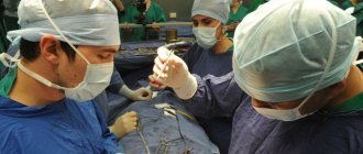
After analyzing these data, the prescriptions were changed: the dose of B-blockers was reduced and Grandaxin, a daytime tranquilizer, was added. The effect was not long in coming. The patient was recommended to consult a psychotherapist and possible radiofrequency ablation.
Causes and development of the disease
There is a difference between atrial and ventricular parasystole (80% of cases). It is also found in the atrioventricular connections (a group of anatomical structures that control the parts of the heart). Cases of multiple lesions, which are called parasystole in a combined form, are not uncommon.
Idiopathic parasystole occurs if the doctor notices the following disorders during diagnosis:
- heart failure;
- chronic myocarditis;
- mitral valve prolapse;
- cardiac ischemia;
- myocardial dystrophy;
- myocardial infarction.
The causes of extracardiac parasystole are:
- pathology of the autonomic nervous system;
- diabetes;
- hormonal disbalance;
- disturbances in the electrolyte composition of the blood;
- thyroid diseases;
- adrenal gland diseases;
- anemia.
Only a cardiologist can determine the exact cause of the disease based on characteristic signs and determine the group, who will conduct a series of studies and prescribe treatment. First, an anamnesis is collected. Next, an examination is carried out, tapping the body (percussion) and listening to sounds using a stethoscope (auscultation).
A thorough study of thyroid hormones and hormonal levels is important. Parasystole is mainly diagnosed using an ECG. With its help, the cause of the disease is established and extrasystole is detected
With its help, the cause of the disease is established and extrasystole is detected.
Echocardiography is used to see structural changes in the heart. Sometimes the patient is continuously examined using an ECG for 1-2 days. Thus, it is possible to diagnose atrial parasystole.
A load test is applied. The patient is given a gradually increasing load (on a treadmill), and the work of the heart is recorded during the procedure. Diagnosis is also possible using MRI. Due to the electromagnetic field, a 3D drawing of the chest is created.
To identify cardiac causes, a complete examination is necessary, including differential diagnosis of defects, physiological abnormalities, and the consequences of vascular atherosclerosis.
Conducting stress tests is recommended to identify the latent form (asymptomatic), the connection with physical activity and the influence of nervous regulation. Bicycle ergometry tests, treadmill walking, and stair tests are used.
If parasystoles appear rarely, then the Holter monitoring method helps: the patient is fitted with electrodes for a day, from which information is recorded even during night sleep. Decoding allows you to determine the cause of the extrasystole.
Dopplerography is a very informative method for identifying heart defects, the degree of mitral valve prolapse, and myocardial reserve reserves. The image on the screen visualizes the contraction process and its phases.
At the same time, a quantitative analysis of the indicators is carried out. Magnetic resonance imaging (MRI) is the method of choice for diagnosing the correct functioning of all parts of the heart muscle and identifying replacement with scar tissue.
An ECG recording records different distances between the ventricular complexes, a violation of their shape, and the direction of the teeth
Biochemical blood tests that determine the level of total cholesterol, sugar, potassium; Determination of the hormonal background of the body, paying special attention to thyroid hormones; ECG is one of the main diagnostic methods that allows you to accurately determine the presence of a disease, and sometimes its cause;
Echocardiography - determines structural changes in the heart; 24-hour Holter ECG monitoring - conducting an ECG for 24-48 hours, which allows you to determine the nature of the parasystole and the location of the focus.
Load test - performing a stepwise increasing load under constant ECG monitoring. MRI is a study of the heart that creates a three-dimensional image under the influence of a magnetic field and electromagnetic waves.
Electrophysiological study, a diagnostic method using a probe inserted into the heart through the femoral vein. The indication for this diagnostic method is ventricular parasystole.
registration of two independent rhythms; ventricular extrasystole is indicated by measuring the coupling interval from the beginning of the q wave; with atrial extrasystoles - from the P wave of the previous complex to the extrasystole;
For an accurate diagnosis, a long-term ECG may be necessary, during which the distance between individual ectopic complexes is recorded. Parasystole is accurately indicated by the multiplicity between two interectopic intervals and the shortest distance between two complexes.
Why do parasystole centers appear?
There are cardiac and extracardiac causes. In some cases, it is impossible to establish a connection with any cause, then the extrasystole is called idiopathic.
Cardiac ones include:
- ischemia or necrosis in the sinus node area and other places with coronary heart disease, which forces different areas to become active and “survive” on their own;
- inflammation of a focal or diffuse nature in acute and chronic myocarditis (rheumatic carditis, acute infectious diseases, sepsis);
- metabolic changes during dystrophy;
- replacement of myocytes with connective tissue cells with disruption of their functions (cardiomyopathies, cardiosclerosis);
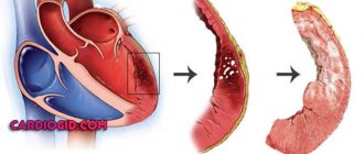
- loss of the ability to restore the required level of energy (circulatory failure);
- hypertrophy of myocardial tissue (hypertension, decompensation in heart failure, cardiomyopathies);
- disruption of the valves (congenital malformations, acquired changes in the valves due to inflammatory processes, injuries).
Non-cardiac causes include concomitant diseases leading secondary to myocardial dysfunction. Most often, these changes are “guided” by endocrine organs when:
- diseases of the thyroid gland (hypothyroidism or hyperthyroidism associated with deficiency or excess synthesis of thyroid hormones);
- diseases of the adrenal glands;
- diabetes mellitus
Activation of parasystolic foci is detected on the ECG when:
- vegetative-vascular dystonia, neuroses;
- anemia (anemia) of various origins;
- overdose of drugs (cardiac glycosides);
- violation of the necessary balance in the electrolyte composition of the blood between potassium, sodium, magnesium and calcium, they are necessary for the implementation of the normal process of excitation and contraction of myocardial cells.
Development mechanism
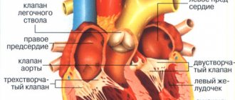
Normally, the excitation that causes the heart to contract into systole occurs in the sinoatrial node (SA node). It is a cluster of special cardiomyocytes in the right atrium appendage, capable of spontaneously generating an electrical impulse at a certain frequency. This property is called automaticity.
However, with various congenital pathologies or heart diseases leading to changes in the structure of the myocardium, it is possible either the formation of areas with slow conduction, which then conduct excitation in the opposite direction, or accumulations of impulse-inducing cardiomyocytes in a place other than the SA node. In the first case, they talk about extrasystole of the re-entry type.
Many scientists agree that parasystole is one of the types of extrasystole. Its peculiarity is the “automatism” of the ectopic focus. Those. complexes are generated with a certain frequency. In this case, 2 pacemakers appear in the heart, each of which generates its own line of impulses.
Some authors believe that, unlike the SA node, the parasystolic center is not influenced by nervous impulses (such as slowing of impulses in the SA node during sleep), the effects of hormones and catecholamines (increased excitation in the SA node under stress) . This is the basis for scientific studies of identifying “true parasystole” against the background of creating “vagal cardiac arrest,” when, with complete shutdown of the SA node, the rhythm is recorded only from the ectopic focus of excitation.
Structurally, the ectopic focus is usually “protected” from the path of propagation of the normal impulse from the SA node. Usually this is a section of altered tissue (connective instead of cardiomyocytes), which delimits the ectopic focus either at the entrance (a frequent substrate for the formation of extrasystoles of the re-entry type) or at the exit (more typical for parasystole).
Types of parasystoles
Depending on the location of the source of the second rhythm, the following types are distinguished:
- ventricular parasystole;
- atrial;
- from the atrioventricular node;
- polytopic (from different places).
In addition, in relation to the normal contraction, extrasystole can be:
- early and late;
- single, group and allorhythmic (constant rhythmic alternation).
According to the frequency of the ectopic rhythm:
- rare (up to 10 per minute);
- medium (10–30);
- frequent (more than 30).
There are temporary and permanent forms. The type of parasystole can be clarified using the ECG picture.
A serious type of parasystole is atrial fibrillation in the form of atrial flutter or fibrillation. With this pathology, there are either many foci of excitation, or the process forms a vicious circle.
Parasystole: definition and features
This is one of the cardiovascular diseases, characterized by the appearance of additional wire impulses that are independent of the main pacemaker. In other words, this is a pathology in which an extraordinary cardiac contraction is recorded. In the international classification of pathologies, such deviations are assigned a separate group - other heart rhythm disorders (ICD 10 code - I49).
The sinus node, located in the right atrium, is responsible for the main rhythm. The frequency and rhythm of the heartbeat is regulated depending on the needs of the body. In case of pathological rhythm disturbance, normalization of its functioning is possible with the help of medications.
Parasystole develops against the background of the appearance in the heart muscle of another source of electrical impulses, which operates independently of brain signals, the action of hormones or medications. Often an additional node occurs in the ventricles, so patients need to know what ventricular parasystole is and how it is formed.
The development mechanism is as follows: the heart muscle receives signals to contract from the main pacemaker, as well as from the additional ventricular node. This phenomenon is called double rhythm formation. It is often accompanied by tachycardia (rapid heartbeat). During daily monitoring of patients with a similar pathology, up to 30 thousand unplanned contractions were recorded. However, it must be remembered that the parasystolic node can also be located in other parts of the organ.
Where should parasystole be classified in the classification of arrhythmias?
Classifications of arrhythmias are based on different characteristics, each with its own disadvantages:
- according to the anatomical location of the ectopic focus - the mechanism of development of disorders is not taken into account;
- according to the mechanism of violation of automaticity, conductivity or excitability - in most cases there is a violation of all functions at once;
- by rhythm frequency - the algorithm for determining the type begins with the diagnosis of normo-, tachy- and bradyarrhythmia, but it requires further clarification using electrocardiographic (ECG) studies;
- depending on the mechanism of impulse occurrence (in a normal and ectopic focus) - separate identification of conduction disorders and combined disorders.
Parasystole is closest to the last option. Let us clarify that we understand the term as additional contractions of the heart muscle in response to impulses coming from the “paracenter” located in any part of the heart.