General information
Mitral stenosis is an acquired monovalvular defect that impairs the activity of the heart muscle as a result of morphological and/or functional changes in the structure of the mitral valve.
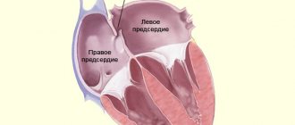
Normally, it has a bicuspid (bicuspid) structure and is located on the border of the left atrium and the left ventricle, preventing the reverse flow (regurgitation) of blood into the chamber of the left atrium in systole, that is, during ventricular contraction. However, infectious lesions, inflammation, autoimmune reactions, overload, and enlargement of the heart chambers can cause pathological changes in the valve system in the form of narrowing of the atrioventricular orifice, which leads to disruption of intracardiac hemodynamics and systemic blood flow. The disease is characterized by a slow course - the first symptoms may appear after 40-50 years, although the formation of pathological narrowing usually begins at a young age. A downward change in the diameter of the atrioventricular orifice occurs in approximately 0.05-0.08% of the population, more often in females.
Congenital mitral valve defects
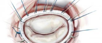
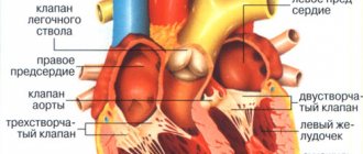
The mitral valve is located between the left atrium and the left ventricle. It is a complex natural mechanism and consists of two valves - thin plates, which, like the sails of a schooner, are controlled by a network of ropes and cords starting from the heads of small papillary muscles of the wall of the left ventricle. These cords are called chords. The papillary muscles, chords and valves are a single apparatus, thanks to the precise and coordinated work of which the “gateway” of the mitral orifice opens and closes with each contraction of the heart.
Isolated congenital defects
Isolated congenital
Mitral valve defects that affect only it are a significant rarity. Valve changes are often combined with other congenital heart defects, which relate either to valve rings in general (AVR) or to underdevelopment of the entire left half of the heart (LVH, i.e. hypoplastic left ventricular syndrome, aortic atresia). However, congenital valve defects with a normally developed left ventricle have been described: valve stenosis and valve insufficiency. Congenital mitral valve stenoses are extremely rare. They may be associated with improper formation of the valves, with a membrane located above the valves, with improper formation of the valves, creating two small holes instead of one large one. In all cases, the cause of heart failure in the form of an enlarged heart, stagnation of blood in the lungs, and enlarged liver can be suspected and identified using non-invasive diagnostic methods. Surgical treatment is possible, and its choice depends on the type of damage to the valve structures: from dissection of the narrowed sections of the valves to valve replacement.
Much more often, the mitral valve is affected in cases of endocarditis or rheumatism, and these, no longer congenital causes, should be excluded first.
Mitral valve prolapse
Congenital mitral valve insufficiency, which would necessitate early referral to cardiac surgery, is extremely rare. But one of the most common
Cardiological diagnoses have recently affected him directly.
This is mitral valve prolapse . Typically, the diagnosis is made in a completely asymptomatic child during a random examination on the basis of a slight murmur, which was previously called a “functional heart murmur” and was not paid much attention to. With the advent and implementation of echocardiography, the causes of this noise have become clearer. Most often, it is caused by inaccurate closure of the valve flaps with each other, as a result of which a stream of blood at the moment of contraction of the ventricle flows back and leaks into the atrium. This may be caused by the fact that one of the sections of the valve bends back under pressure, “prolapses” towards the atrium and allows this stream to pass through. This is prolapse. The degree of mitral valve prolapse can vary - from minor to severe regurgitation (backflow). This degree is clearly visible on echocardiograms. If your child is diagnosed with this, you should neither be afraid nor automatically consider him a “heart patient.” So far this is only an “echocardiographic” diagnosis and, if there are no signs of heart failure, then your child is practically healthy and can do everything that children of his age can do. You just need to be especially careful about colds, sore throats, and carious teeth so that they do not cause infection of the mitral valve, which is more likely to be affected in a child with prolapse than in other children.
A cardiologist will tell you about all this. How to get treatment at the Scientific Center named after. A.N. Bakuleva?
Online consultations
Pathogenesis
Stenosis is a persistent narrowing of any hollow structure in the body of humans and other living beings. In the case of a mitral valve consisting of two connective tissue leaflets, their thickening, fibrosis and subsequent calcification , fusion of commissures or shortening of the chords occur. This leads to a decrease in the size of the atrioventricular junction from normal - 4-6 cm square to 2.5 cm square or more, which causes pathological manifestations of functional class heart failure. As a result, blood does not have time to be completely pumped out of the left atrium during diastole and therefore systole is prolonged.
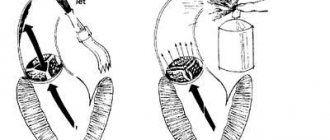
Mitral valve of a healthy person and in case of stenosis
Narrowing of the left atrioventricular orifice impairs the ability of the mitral valve to regulate blood flow from the ventricle to the atrium, and the diastolic flow of arterial blood from the left atrium into the left ventricular chamber is disrupted, which leads to circulatory disorders and causes blood stagnation.
The compensatory mechanism to ensure normal blood filling of the left ventricle is to increase the pressure in the atrium to 25 mmHg, creating a greater pressure gradient to facilitate blood flow through the mitral valve.
An increase in pressure in the cavity of the left atrium also has negative aspects, because it causes an increase in pressure not only in the right ventricle, but also in the pulmonary arteries and throughout the pulmonary circulation. In addition, high pressure in the left atrium leads to hypertrophy of its myocardium. Under conditions of increased work of the atria and the progression of hypertrophy, the volume and mass of the wall of the right ventricle increase, which creates a risk of increased pressure in the pulmonary arteries and in the lungs themselves. Pathogenesis is usually divided into several stages:
- compensation – observed with a slight decrease in the size of the atrioventricular orifice and is asymptomatic;
- subcompensation - emerging circulatory disorders caused by valve dysfunction lead to increased pressure in the pulmonary artery and the first symptoms appear in the form of shortness of breath , increased fatigue, heart pain , etc.;
- depletion of the body's compensatory forces is a condition in which pronounced signs of blood stagnation in the pulmonary circulation, heart failure and atrial fibrillation ;
- sclerotic or dystrophic changes - accompanied by a persistent decrease in myocardial function, severe pulmonary hypertension and arrhythmia .
Pathological specimen of a stenotic mitral valve
Mitral valve stenosis
Every year, medical advances in the field of cardiac surgery are becoming more and more improved, which makes it possible to cure many diseases of cardiovascular pathologies. Today, heart defects pose a great threat to human life and require close monitoring by cardiologists. One of these conditions is mitral valve stenosis, which is accompanied by characteristic symptoms, and with rapid progression, it is complicated by decompensated disorders of myocardial functioning, resulting in death.
Mitral (bicuspid) valve stenosis is a pathological process of acquired etiology, manifested in the form of a decrease in the lumen of the left atrioventricular (atrioventricular) opening. With an anomaly in size, there is a deterioration in the diastolic flow of blood from the left atrium to the left ventricle.
Narrowing of the bicuspid valve forms as an independent disease or against the background of other heart valve defects in the form of stenosis, insufficiency, or a combination of both.
Risk factors
A narrowing of the physiological dimensions of the atrioventricular orifice develops throughout life, most often as a result of rheumatism. These pathological processes are diagnosed several times more often in females over 45 years of age.
8
24/7
Negative factors affecting the cardiovascular system that can provoke a narrowing of the mitral orifice include treatment with ionizing radiation used in the chest area, as well as long-term use of certain medications, in particular those based on wormwood.
Classification of the pathological process
Mitral valve stenosis has varying degrees of severity, depending on which the symptomatic complex develops and therapy is prescribed. There are 4 degrees of the disease, which are determined by the size of the pathological narrowing. Degrees of mitral valve stenosis:
- 1st degree - pathological changes are insignificant, the size of the orifice is about 3 square meters. cm;
- 2 – the clearance is from 2.3 to 2.9 square meters. cm, such changes are considered moderate;
- 3 – pronounced narrowing of the orifice of the mitral valve, dimensions range from 1.7 – 2.2 sq. cm;
- 4 – critical narrowing of the bicuspid valve, the area of which is in the range of 1.0-1.6 square meters. cm.
There is also a classification of clinical manifestations, the indicators depend on the severity of the disturbance in blood flow with mitral valve stenosis:
- Stage 1 – complete compensation of blood circulation by the left atrium; the patient is not bothered by symptomatic manifestations; disorders can only be identified by listening;
- Stage 2 – characterized by deterioration of blood circulation in the pulmonary circulation, with signs of the disease appearing only during physical activity;
- Stage 3 – pronounced manifestations of stagnation in the pulmonary circulation occur and processes in the systemic (large) circulation are partially disrupted;
- Stage 4 – severe symptoms of disorders of normal blood circulation in the small and large circles occur, which leads to atrial fibrillation;
- Stage 5 – dystrophic, manifested by stage 3 heart failure, morphologically irreversible changes occur in organs.
Causes
In most patients, the cause of mitral valve stenosis is rheumatism. Often the disease affects young people under 20 years of age, and many years later (from 15 to 30) symptoms of valve defects begin to appear.
Other reasons may be:
- atherosclerotic changes in blood vessels;
- closed heart contusions;
- STD;
- endocarditis of infectious etiology;
- calcification of the valves and rings;
- various congenital heart abnormalities;
- intracardiac thrombus formation;
- aortic valve insufficiency.
Symptoms of the disease
Mitral valve stenosis is characterized by the appearance of a symptomatic complex when the size of the pathological lumen is less than 2 square meters. see. The first symptoms are the appearance of rapid fatigue and shortness of breath (occurs only after physical exertion, including moderate). With the development of pathology, disturbances in the frequency, depth and rhythm of breathing are observed at rest, coughing attacks with an admixture of blood particles in the sputum also appear, the heart rate increases, and a disturbance in the heart rhythm occurs such as ventricular extrasystole and atrial fibrillation.
Severe pathological narrowing provokes the development of severe shortness of breath, in which the patient is unable to lie down and takes a forced sitting position. At night, attacks of cardiac asthma are possible, manifested by suffocation and a painful dry cough.
With the rapid progression of the pathology, the development of pulmonary edema is possible: coughing attacks become more frequent, hoarseness appears, the skin of the face and mucous membranes become bluish, discomfort and pressing pain are felt in the sternum area, severe tachycardia, which can sharply turn into bradycardia (slow heart rate), blood pressure decreases to critical levels.
Other symptoms during the development of pathology are:
- dysfunction of the vocal apparatus (changes in timbre, tone, the appearance of hoarseness);
- soreness in the sternum;
- chest discomfort;
- lips, nose and nail plates become bluish in color;
- the skin in the cheek area becomes covered with a crimson blush;
- feeling of heaviness in the abdomen.
Symptoms of mitral valve stenosis occur as pathological processes progress, which can result in blockage of the pulmonary artery and death of the patient.
8
24/7
Diagnosis of the disease
When the first signs of cardiac dysfunction are detected, a comprehensive examination is carried out by a cardiologist to confirm or exclude the diagnosis of mitral valve stenosis. Initially, anamnestic data is collected (the development of the first symptoms, under what circumstances they appeared - after physical exertion, against the background of stressful situations, as a result of surgery, etc.).
The next stage of the examination is auscultation, during which the doctor will identify possible violations. For the final diagnosis, laboratory and instrumental studies are prescribed:
- laboratory analysis of blood and urine (general);
- blood test for biochemistry;
- hemostasiogram (determination of blood clotting);
- ECG;
- echocardiography;
- X-ray of the chest area.
Treatment at different stages of the disease
For the first degree of mitral valve stenosis, treatment is carried out using medications aimed at preventing infective endocarditis (antibacterial agents are used), reducing signs of heart failure (diuretic drugs), and eliminating arrhythmia (beta blockers).
In case of pathology of the atrioventricular orifice detected in a woman during pregnancy, treatment measures depend on the severity of the stenosis and the duration of pregnancy. So, if the narrowing of the mitral valve orifice is more than 1.6 sq. cm, which is not accompanied by any symptomatic manifestations, the pregnancy continues, and the woman is under close medical supervision. In cases of severe contraction, accompanied by clinical manifestations and increasing heart failure, the pregnancy is terminated.
Surgeries for mitral valve stenosis are prescribed during stages 2,3,4 of hemodynamic disorders. Under certain conditions, when there is no curvature of the valves, balloon valvuloplasty is performed.
More often, for the treatment of pathological stenosis of the bicuspid valve, open or closed mitral commissurotomy is prescribed. During the operation, adhesions are dissected, calcifications are removed, and blood clots are removed from the left atrium. In case of pronounced pathological changes in the valve apparatus, prosthetic replacement of the left atrioventricular orifice is performed. After surgical treatment, medications that affect blood clotting are prescribed, which must be taken on an ongoing basis. Despite surgical treatment, there is a risk of developing re-stenosis.
Disease prognosis
Mitral valve stenosis is a fairly serious pathological process, which as a result can cause the development of severe symptoms and consequences that are incompatible with life. In the absence of therapeutic measures, 5-year survival is observed in 50% of patients with this diagnosis.
Surgical treatment increases the chances of life expectancy in more than 85% of cases.
Disease prevention
Preventive measures are aimed at preventing rheumatic lesions, timely treatment of streptococcal infections and other diseases that have a negative effect on the heart and blood vessels.
When diagnosing mitral valve stenosis, regardless of the severity of the course and the presence of symptoms, monitoring of the condition by a specialist - a cardiologist or rheumatologist - is required.
Heart defects are a fairly serious pathology that worsens the patient’s quality of life. Only timely detection of valve disorders allows the necessary treatment to be carried out and a person’s life to be saved.
8
24/7
Classification
Taking into account the state of general hemodynamics, mitral stenosis is divided into compensated, subcompensated and decompensated. In addition, valve defects can differ depending on the etiology and be rheumatic (the most common type - in 50-80% of cases), atherosclerotic, syphilitic, as well as the outcome of bacterial endocarditis .
The most important factor in dividing mitral stenosis into types is the severity of the defect and its degree of influence on intracardiac hemodynamics. Violations can be moderate and moderately severe with a transmitral gradient of 1 – 25 mm Hg, and also not exceed 5 mm Hg. st and does not have any effect on the state of intracardiac hemodynamics.
Classification of mitral stenosis by severity
Mitral stenosis is usually isolated, but can also be combined with disorders of other valves or against the background of mitral valve insufficiency.
Mitral stenosis: causes and consequences of the disease
The vitality of the human body directly depends on the proper functioning of the heart. The mitral valve is located between the left atrium and ventricle. Its function is to pass arterial blood into the left ventricle and prevent its reverse outflow. If the valve leaflets thicken and fuse together, the size of the atrioventricular opening becomes smaller. This narrowing is called mitral orifice stenosis, or mitral stenosis. Causing a large number of problems, the disease can cause irreversible consequences.
Causes and risk factors for developing the disease
The causes of the disease are varied, and although it has been scientifically proven that 4/5 of the cases of patients had a rheumatic etiology for the development of mitral stenosis or congenital, which is detected during the intrauterine development of the child. However, the remaining 25% includes diseases such as:
- heart injury and atherosclerosis;
- left atrial myxoma;
- intracardiac thrombi;
- heart surgery;
- aortic insufficiency;
- streptococcal infection;
- infective endocarditis and syphilis.
Heart defects play an important role in development; mitral stenosis affects other heart valves.
Due to the fact that a person’s appearance and condition do not undergo any changes for a long time, the disease can reveal itself 10-30 years after its onset. A malfunction of the organ catalyzes the launch of a compensation mechanism, which facilitates the passage of blood and reduces the effect of the disease on hemodynamics inside the heart: the pressure in the atrium cavity increases by 15-20 mm Hg. Art., systole lengthens in the left atrium, and at the same time myocardial hypertrophy develops in it.
8
24/7
However, with further progression of the disorders, the contractile function of the ventricle decreases, it expands, the load on the right atrium increases, which ultimately becomes the cause of a malfunction of the entire organ.
Degrees and stages of the disease
In a normal state, the size of the mitral orifice does not exceed 4-6 cm2, so its narrowing immediately affects intracardiac hemodynamics.
Mitral stenosis depends on the area of narrowing and has several degrees.
| Degree | Characteristic |
| First | Holes > 3 cm2. It is a minor violation and is not noticeable without a special examination. |
| Second | Sholes = 2.3-2.9 cm2. A moderate type of change does not have a strong effect on the functioning of the organ. |
| Third | Sholes = 1.7-2.2 cm2. The disease is severe and characteristic symptoms appear. |
| Fourth | Sholes = 1, -1.6 cm2. Critical mitral stenosis of the heart requires urgent hospitalization of the patient. |
The gradual progression of the disease affects the process of blood circulation within the large and small cardiac circles. Doctors distinguish only 5 stages of pathology.
- First. Mitral stenosis is completely compensated by the atrium. Signs of the disease are poorly detected.
- Second. Disturbances appear in the pulmonary circulation. During physical activity, initial symptoms are revealed.
- Third. The improper flow of blood in the large circle makes itself felt, and stagnation in the small circle is clearly expressed.
- Fourth. Atrial fibrillation appears, which is a consequence of pronounced signs of blood stagnation in both circles.
- Fifth. Dystrophy corresponding to stage 3 of heart failure is detected.
Changes in cardinal hemodynamics and the degree of narrowing are of great importance when prescribing treatment and in the further prognosis of the development of the disease. This is especially important for people with congenital aortic mitral stenosis, since it is most often combined with other heart pathologies.
Symptoms
For each stage of the development of the disease, specific symptoms are typical, which can be used to describe the clinical picture of the formation of the pathology.
In the initial stage of pathology, it can be identified through a complete clinical examination. The patient has no complaints and is in excellent health.
With the onset of the second stage, a slight increase in blood pressure and shortness of breath after physical activity may occur.
Stagnation is clearly expressed in both circles of blood circulation at the third stage of development:
- persistent shortness of breath with cough and foamy sputum;
- significant increase in pressure;
- swelling of the limbs;
- the liver and spleen are enlarged.
At the fourth stage, the size of the heart becomes noticeable:
- the liver and spleen are felt upon palpation;
- venous pressure is much higher than permissible norms;
- swelling of the extremities is clearly expressed;
- small ascites;
- initial form of atrial fibrillation.
The last fifth stage is characterized by all the symptoms in a pronounced form, in addition, shortness of breath does not go away even at rest, and the liver extends beyond the costal arch.
Drug treatment is possible up to the fourth stage, inclusive, after which it is pointless.
8
24/7
Diagnosis methods
Mitral stenosis detected at an early stage makes it possible to control and restrain the development of the disease. However, the body's compensatory capabilities prevent symptoms from appearing. Therefore, almost all cases of timely diagnosis recorded by statistics are a consequence of the discovery of the disease during preventive examinations.
Mostly, people come to doctors with complaints due to decompensation, when many systems of the body are modified due to mitral stenosis, treatment will no longer help, and urgent surgical care is required.
The specialist at the first moment pays attention to a certain type of patient’s complaints. Then he begins to collect an anamnesis, in which the patient’s previous illnesses and injuries play an important role.
An objective examination may reveal:
- diastolic murmur when listening to the heart;
- increased size of the liver and spleen;
- edema and ascites.
To clarify the diagnosis, complete laboratory tests of blood, urine, ultrasound and ECG are performed
- in general tests, deviations in ESR indicators will appear, leukocytosis and anemia may appear;
- biochemical studies show deviations from the norm only if the cause of the disease is atherosclerotic;
- antistreptolysin is detected in ASLO results;
- An ECG will indicate hypertrophy of the heart;
- During an ultrasound, the degree of mitral stenosis is revealed, the presence of thrombosis is determined by the stage of the disease.
In addition, to exclude calcification and vegetative changes in the valve, probing of the heart cavities is done, and radiography allows one to study the pulmonary pattern and other signs of the disease.
Treatment in early and late stages
The methods of treatment prescribed by a cardiologist depend not only on the stage of the disease, but also on the cause of its occurrence.
| Degrees | Methods |
| Diet therapy is used, physical activity is reduced, and a therapeutic course of medications is prescribed to suppress the etiological factor. Then drugs are gradually added to relieve excess stress on the heart muscles. In this case, the patient may need to take them for life. The emergence of atrial fibrillation makes it necessary to add new drugs to prevent thrombosis. |
| If the patient’s well-being deteriorates, the mitral orifice narrows by more than 1.2 cm2 | Initially, an operation is performed to dissect the fused valve leaflets. But in the case when the clinic indicates the futility of commissurotomy in mitral stenosis, the diseased valve is completely replaced with an artificial one. |
A positive prognosis is given with young patients and early diagnosis.
Surgical intervention, according to experts, is preferable to therapeutic methods, since, despite a certain risk, it is possible to guarantee the absence of re-development of stenosis.
The age category of the patient leaves a certain imprint on the development of the disease. In older people, starting from 60 years of age, the development of mitral valve disease is considered a characteristic clinical feature. Despite its constant slow progression, survival rates without surgery are very high.
Disease prevention
To prevent the disease from coming suddenly, it is necessary to monitor your health and undergo regular preventive examinations. A visit to a specialist’s office is especially important if:
- shortness of breath and hemoptysis appear with any physical activity;
- lying in a horizontal position, attacks of suffocation begin;
- when working, fatigue occurs quickly and muscle weakness is felt;
- one feels the uneven functioning of the heart, and also dull aching pains appear in it.
In addition, it is important to carry out measures aimed at preventing the development of pathology:
- increase the level of natural immunity and adaptive capabilities of the body through hardening;
- the food consumed must be balanced and contain the required amount of microelements, vitamins and nutrients;
- is often outdoors, doing physical activity and walking;
- Avoid contact with patients with tonsillitis and pharyngitis; in case of illness, promptly treat these pathologies.
People who have already been diagnosed with mitral stenosis need to take the medications prescribed by the doctor, in strict accordance with the prescription, and be examined at the appointed time.
It should be remembered that only timely diagnosis of pathology allows one to avoid life-threatening consequences. You cannot put off visiting a doctor - every day can become a decisive factor in drawing up a further prognosis.
8
24/7
Causes
Most often, mitral stenosis develops as a result of previously suffered rheumatism , in other cases the cause becomes:
- infective endocarditis ;
- chronic valvulitis ;
- carcinoid syndrome;
- mitral valve calcification
- thrombus;
- obstruction during the tumor process and the development of myxoma ;
- heart injuries.
Congenital anomaly of the mitral valve in the direction of narrowing of the atrioventricular orifice is extremely rare, for example, with Lutembasch syndrome .
Symptoms of mitral stenosis
High pressure in the pulmonary arteries in patients is manifested by complaints of shortness of breath during physical exertion, as they cause increased blood flow to the heart muscle and overstrain of the capillaries, as well as the walls of the heart in conditions of atrioventricular valve stenosis. All this leads to difficulty in normal gas exchange, further shortness of breath even at rest and pain in the heart. Obvious pallor of the skin, with a pronounced blush on the cheek with cyanosis , heartbeat and its interruptions, the development of atrial fibrillation is possible.
Visually detectable symptoms of mitral valve stenosis:
- heart hump;
- acrocyanosis – bluish discoloration of the skin, affecting the tip of the nose, ears and chin;
- increased cyanosis and the appearance of pallor of the skin, up to an ashy color during physical activity, usually caused by a high degree of pulmonary hypertension;
- a sharp rise in pressure in the pulmonary circulation can cause cardiac asthma ;
- mitral stenosis may manifest itself as a dry cough or with the discharge of a small amount of mucous sputum;
- with hemoptysis , siderophages or so-called heart defect cells are detected in the sputum.
Symptoms of weakness in combination with increased fatigue are associated with a characteristic fixation of cardiac output, since there is no adequate increase in cardiac output under conditions of physical activity.
In the case of significant stenosis of more than 80%, the patient's condition may be complicated by pulmonary edema , atrial fibrillation , pulmonary hypertension and the occurrence of thromboembolic syndrome .
Mitral valve stenosis - symptoms and treatment
Treatment tactics for mitral stenosis depend on its stage.
In stage I stenosis, patients are not indicated for surgical treatment, since compensatory mechanisms develop in the body. Compensation is supported by primary and secondary prevention of rheumatic fever.
The main task of primary prevention is to prevent the development of rheumatism. It involves the complete and timely elimination of streptococcal infections in sinusitis, rhinitis, sore throat and other diseases of the upper respiratory tract.
The goal of secondary prevention is to reduce the risk of re-exacerbation of rheumatism. It involves courses of bicillin prophylaxis (intramuscular injections of bicillin-5), observation by a rheumatologist, and the use of non-steroidal anti-inflammatory drugs in the event of colds and during various operations.
For primary and secondary prevention of rheumatism, it is important to follow the regimen. It consists of a healthy lifestyle, proper nutrition, exercise, absence of bad habits, good living conditions, timely visits to the dentist, medical supervision and treatment for colds.
In stage II stenosis, the indications for surgery are relative. Treatment options considered:
- closed mitral commissurotomy - an operation to widen the mitral valve, which is performed using a special dilator;
- reconstructive surgeries are valve-sparing surgeries in which valve function is restored using various suture technologies while preserving the valve leaflets and structures of the valve apparatus.
These operations make it possible to save patients' lives without leading to specific complications.
For stages III and IV of stenosis, surgical treatment is mandatory. Conservative therapy (taking cardiac glycosides and diuretics) brings only a short-term effect, since with an increase in physical activity, circulatory disorders still occur.
Closed mitral commissurotomy is indicated for patients with isolated mitral stenosis or re-fusion of the valve leaflets (restenosis) without gross changes in the valve structures, as well as with concomitant mitral regurgitation or grade I calcification. Mortality with this type of treatment is 0.5-1%. Repeated operations are performed in 35% of cases 10-15 years after the first surgical intervention [12].
In patients at high surgical risk, transcatheter balloon valvuloplasty is performed. It also aims to dilate the mitral valve. To do this, a percutaneous puncture of the common femoral vein is first made. Through it, the doctor inserts conductors and catheters into the right atrium. There, a puncture of the interatrial septum is performed, after which a balloon is inserted into the mitral valve ring and inflated [13].
Indications for reconstructive operations on the mitral valve are defects with predominant insufficiency without gross changes in the leaflets and structures of the valve apparatus, as well as the absence of calcification. Mortality after reconstructive surgery is 1.5-4%. Long-term survival rate is 90% [14].
In all other cases: valvular infective endocarditis, post-infarction defects, grade II or III calcification, the most effective operation is valve replacement with a mechanical or biological prosthesis. Mortality after such treatment ranges from 2-8%. The five-year survival rate of patients using modern types of artificial and biological prostheses is 75-90% [15].
Before any operation, the doctor must explain to the patient the course of the operation, anesthesia, possible risks and complications. After this, the patient signs consent for the operation. A few days before the planned intervention, drug therapy is carried out to stabilize the patient’s condition and prevent complications that may arise during or after surgical treatment. Hygienic treatment of the patient’s skin is also necessary: taking a shower, shaving surgical access sites. The patient should not eat food on the day before surgery.
After the operation, the patient is under observation in the intensive care unit. After stabilizing his condition and restoring the vital functions of the body, he is transferred to a specialized department under the supervision of the attending physician. At this time, the patient follows all recommendations for taking medications, activity and nutrition. With the help of a physical therapy doctor, the patient performs restorative exercises (breathing, motor) daily. The attending physician monitors the increase in the patient’s physical activity (metered walking), performs the necessary dressings and helps to recover psychologically.
After discharge, the patient must strictly follow all the doctor’s recommendations. It is necessary to be observed on an outpatient basis by a cardiologist and perform ECHO-CG annually. For patients with a mechanical heart valve, it is important to monitor such laboratory blood indicators as the INR (International Normalized Ratio) level. This is due to the use of warfarin, a blood thinning drug. Its target values should be in the range of 2-3.
Tests and diagnostics
In addition to echocardioscopy, Dopplerography, phonocardiography, X-ray, ECG (including daily monitoring to determine heart rhythm), 4 standard diagnostic methods are used:
- Upon examination, against the background of pallor of the skin, a purple, sharply defined so-called mitral flush of the cheeks and cyanosis of the lips, as well as the tip of the nose are visible, while it is possible to detect increased epigastric pulsation of the right ventricle (“heartbeat”) against the background of the absence or weakening of the apical beat, because the walls of the left ventricle are not enlarged and are displaced as a result of the process of hypertrophy of the right ventricle.
- Upon palpation, a cardiac hump is revealed, and in the area of the apex of the heart, especially after performing physical exercises in patients in a position on the left side, when exhaling, a diastolic tremor is “audible” in the form of the so-called “cat purr” caused by fluctuations in the blood during passage through the constricted mitral passage, to identify the symptom of Nesterov’s 2 hammers, it is enough to place the palm of your hands on the top, placing your fingers on the second intercostal space on the left of the sternum, you can detect the flapping initial first sound and feel it as the first beat of the “hammer”, while the second tone causes a recoil in the fingers in in the form of a second “hammer”.
- During percussion, a displacement of the border of absolute relative dullness of the heart muscle is revealed upward and to the right (caused by hypertrophy of the left atrium and expansion of the right ventricle, respectively), to a greater extent than the relative one, due to the expansion of the right ventricle, the heart and the expanded edges of the lungs - it is pressed against the chest wall with its right enlarged half.
- Auscultation makes it possible to detect a fairly strong flapping first sound in places above the apex of the heart, since during diastole the filling of the left ventricle with blood does not occur fully and rapid contraction begins, and auscultation also makes it possible to listen to an additional third sound at the apex, which is associated with the opening of the mitral valve and its sharp movements of the valves, this valve defect is characterized by diastolic murmurs and a three-part sound of two tones with a click of the opening of the bicupidal valve at the apex.
Diet for mitral stenosis
Diet for heart failure
- Efficacy: therapeutic effect after 20 days
- Timing: constantly
- Cost of products: 1700-1800 rubles. in Week
Mitral valve stenosis is accompanied by the development of heart failure , pulmonary hypertension and an increased risk of thromboembolism , so the lifestyle should be as healthy as possible. It is necessary to exclude any harmful products, as well as alcohol, coffee, strong tea, nicotine or any drugs. In addition to moderate physical activity and avoiding stress, it is recommended to monitor the balance of proteins, carbohydrates and fatty acids, and do not forget about auxiliary vitamin therapy and water balance in the body. It is advisable to cross out from the menu:
- fatty foods, especially those of animal origin;
- roast;
- spicy;
- smoked;
- cholesterol-containing products.
The diet should be enriched with potassium and magnesium salts; it should be based on cereals, fruit, vegetable, milk soups, vegetable side dishes, dishes with cheese, poultry and fish. We must not forget about the regular consumption of berries, vegetable juices, raw fruits, and, of course, citrus fruits, apples, dried apricots, prunes, raisins and grapes. In addition, it is very important to limit your fluid and salt intake.