“What to do if there is a sharp pain in the leg in the legs, the veins are swollen?” – people often ask on forums. Take an aspirin tablet, apply a compression bandage with an elastic bandage, elevate the lower limb and call the contact center of the Yusupov Hospital. Phlebology department doctors consult patients with acute pain in the veins of the legs 24 hours a day, 7 days a week. If a patient experiences throbbing pain in a vein in the leg, phlebologists immediately conduct an ultrasound examination and, using modern laboratory tests, determine the state of the coagulation and anti-coagulation system of the blood.
After receiving the results of instrumental and laboratory studies, conservative therapy is carried out using the latest medications registered in the Russian Federation. They have the most effective effect and have the minimum range of side effects. If a patient has very severe pain in the veins in his legs, phlebologists prescribe nonspecific anti-inflammatory drugs that have a pronounced analgesic effect. Local therapy is carried out with ointments and gels, which include venotonics, anticoagulants, and nonspecific anti-inflammatory drugs. A treatment regimen is individually selected for each patient depending on how much pain and stretching the veins in the legs are.
After the pain has reduced, doctors at the phlebology department decide on the need for surgical intervention. Phlebologists at the Yusupov Hospital are fluent in all currently known surgical techniques for varicose veins of the lower extremities. If sharp pain in a vein in the leg occurs due to acute venous thrombosis, phlebologists carry out thrombolysis therapy, and, if indicated, surgically remove the blood clot from the vein. A differentiated approach to the treatment of each patient allows phlebologists at the Yusupov Hospital to quickly and effectively relieve acute pain in a vein in the leg.
Varicose veins of the lower extremities - what is it?
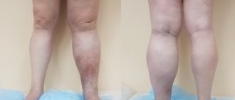
Varicose veins of the lower extremities - what is it? Why does it appear suddenly and progress so quickly? Today, it is considered a common disease in which there is an abnormal enlargement of veins that are visible through the skin and resemble blue or purple nodes. Varicose veins are a pathology that can occur in any part of the body, but is more common on the legs.
This is how varicose veins appear on the legs
Below are the most common signs of the disease. However, each person may only notice some of them.
Innovative treatment of varicose veins at various stages in the center of Moscow
During the consultation, an individual treatment regimen is selected for each patient suffering from varicose veins of the lower extremities, taking into account the stage of the disease. In the early stages, medical conservative treatment with the use of medications, electromyostimulators, compression stockings and sclerotherapy is quite sufficient. Modern treatment of varicose veins in the early stages of pathology can be implemented by gluing veins. For this purpose, one of the best medical techniques used today is sclerotherapy, the essence of which is the removal of dilated veins by introducing into them special drugs, the so-called sclerosants. In advanced stages of varicose veins that are not amenable to conservative treatment, non-surgical treatment is performed ( Venefit™ RFO procedure
or EVLO
).
Treatment of symptoms of varicose veins with thermal methods in our center
These innovative techniques are currently the leading, most advanced and are used in the best phlebological centers around the world, including Moscow and the Moscow region. Good phlebologists often supplement the procedure of radiofrequency (RFO) or laser obliteration (EVLO) of the saphenous veins with miniphlebectomy and scleroobliteration using the Foam-Form method.
Causes of varicose veins and risk factors?
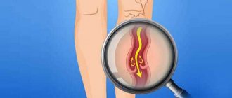
Blood vessels in the body provide a channel for blood flow. In terms of the blood vessels of the lower extremities, the arteries carry high-pressure, oxygenated blood from the heart to the lower extremities. Veins therefore have thinner and less elastic walls than arteries. If pressure rises in the veins for any reason over a long period of time, this can become the main cause of varicose veins of the lower extremities. The disease is also facilitated by malfunction or damage to the valves that help push blood upward.
The functioning of venous valves normally and with varicose veins
About 30% of adults worldwide suffer from the disease. The risk varies greatly by age and gender. The following are risk factors:
- Obesity. Obesity is a major risk factor for the development of varicose veins of the lower extremities. Excessive weight increases pressure in the veins and aggravates their condition.
- Heredity. Heredity plays an important role in determining susceptibility to varicose veins, but specific factors responsible have not been identified.
- Inaction. Prolonged inactivity or, conversely, increased activity increases pressure in the veins.
- Floor. Women are especially susceptible to varicose veins of the lower extremities due to the effect of progesterone on the veins, as well as the effects of pregnancy. Women have a 3 times higher risk of developing the disease.
- Pregnancy. Varicose veins of the lower extremities during pregnancy occur due to hormonal influences. However, enlarged veins often return to normal within one year after giving birth. Pelvic varicose veins may also be observed.
- Age. Varicose veins of the lower extremities usually affect people between 30 and 70 years of age. As we age, the elastic lining of the vein begins to weaken, increasing the chances of enlarged veins.
A risk factor is anything that can increase a person's chances of developing a disease. This could be due to activities such as smoking, diet and many other reasons.
Venous pain - occurrence and treatment
An international group of experts with the participation of the American Venous Forum (AFR), the European Venous Forum (EVF), the International Union of Phlebologists (UIP), and the International Union of Angiologists (IUA) presented a unified terminology characterizing the various manifestations of CVD, recommended for treatment and research work. Thus, it is proposed to distinguish between soreness or pain, a burning sensation, muscle cramps, a feeling of swelling or pulsation, a feeling of heaviness, itching, restless leg syndrome and fatigue [1]. Since all these complaints are not specific, their connection with chronic venous disease is highly likely under the following conditions: direct dependence on the nature of daily activity and the duration of static loads, ambient temperature and hormonal fluctuations in women.
An important sign is the reduction or disappearance of these symptoms after resting or raising the lower extremities above the horizontal level [2]. In addition, a thorough clinical and instrumental examination of the venous system of the lower extremities is necessary.
Specific symptoms of CVD are rarely the subject of epidemiological studies and, as a rule, they are assessed in aggregate. According to various data, the frequency of subjective complaints associated with chronic venous disease occurs in 29-61% of respondents, clearly prevailing in women.
Pain and venous pain: definition and mechanism of development.
As for individual symptoms, in particular venous pain, the range of data is even wider. Thus, Ruckley et al. [3] reported 44% of cases identified by review of medical records. Jawien et al. [4] during a non-specialized examination found venous pain in 75.7% of patients. Langer et al. [5] in a survey of employees at the University of California found pain associated with CVD in 18%. It should be emphasized that pain is often the main reason for patients to see a doctor.
What is venous pain and what is the mechanism of its development? The International Association for the Study of Pain (IASP) defines pain as “an unpleasant sensory and emotional experience associated with actual or potential tissue damage or described in terms of such damage.” In other words, pain is usually more than a sensation associated with existing or possible organic damage, since it is accompanied by an emotional component.
There are acute and chronic pain.
- Acute pain is defined as “pain of short duration of onset with an easily identifiable cause.” Acute pain is a warning to the body about the current danger of organic damage or disease.
- Chronic pain was originally defined as “pain that lasts about 6 months or more.” Now, it is pain that “persistently persists beyond the appropriate length of time during which it should normally end.” Previously, it was believed that pain is not a specific physical sensation, and therefore there are no special receptors that perceive only painful stimulation.
That is, the appearance of a feeling of pain can be caused by irritation of any type of nerve endings, if the force of irritation is strong enough. The existence of specialized bolinocyceptor receptors, characterized by a high perception threshold, has now been proven. They are excited only by stimuli of “damaging” intensity.
All nociceptors are free nerve endings that consist only of the terminal branches of the axial cylinder of the sensory neuron, which is why they got their name, and they are located outside the spinal cord stem, but in the dorsal ganglion. The concentration of nociceptors varies throughout the body. They are mainly distributed over the surface of the skin, in ligaments or joint capsules, and are least often found in deeper tissues and organs. Nociceptors recognize mechanical, thermal and chemical stimuli without adapting to them. That is, different individual pain sensitivity thresholds are associated with the emotional and subjective characteristics of the human psyche.
Nociceptive nerves contain small-diameter primary fibers that have sensory endings in various organs and tissues. There are two main classes of nociceptors, Aδ- and C-fibers, which transmit fast and slow pain sensations, respectively. The class of Aδ-myelinated fibers (covered with a thin myelin coating) conduct signals over a distance of 5 to 30 meters per second, serving to transmit rapid pain. This type of pain is felt within one tenth of a second from the moment the painful stimulus occurs. Activation of Aδ-nociceptors occurs as a result of strong mechanical or thermal effects. Slow pain, transmitted through slower, unmyelinated (“naked”) C-fibers that send signals 0.5 to 2 meters per second, is an aching, throbbing, burning pain—such as venous pain. C-nociceptors perceive mechanical, thermal and chemical stimulation.
There are also so-called “sleeping” nociceptors, which are activated during inflammation. Nociceptors have a certain threshold of sensitivity, that is, a certain minimum level of stimuli is required before they lead to the generation of a signal. In some cases, the excitability of pain fibers becomes excessive, exceeding the actual level of exposure to the painful stimulus, which leads to a condition called hypersensitivity to pain - hyperalgesia. Once the threshold is reached, the signal is transmitted along the nerve axon to the dorsal horn of the spinal cord, where the primary processing of sensory information occurs. Then, nociceptive impulses are transmitted through interneurons to the cells of the anterior and lateral horns, causing reflex motor and autonomic reactions. Another part of the impulses excites neurons, the axons of which form ascending pathways. At the level of the hypothalamus and limbic complex, the formation of emotional and behavioral reactions, autonomic and endocrine shifts that accompany pain occurs. The final analysis of incoming nociceptive information is carried out by the cortex of the parietal, frontal and temporal lobes of the brain.
Along with the nociceptive system, there is its antagonist, the antinociceptive system, which suppresses pain impulses. It includes the structures of the cerebral cortex, the diencephalic level, the periventricular and periaqueductal gray matter (rich in enkephalin and opiate neurons), some nuclei of the reticular formation of the brain stem, in which serotonin and norepinephrine act as neurotransmitters. The participation of serotonin and norepinephrine in the functioning of the antinociceptive system explains the reduction of pain caused by a number of drugs (tricyclic antidepressants, etc.) that suppress reuptake at serotonergic and norepinephrine synapses and thereby enhance the descending inhibitory effect on neurons of the dorsal horn of the brain.
Thus, under normal conditions, there is a harmonious relationship between the intensity of the stimulus and the response to it at all levels of organization of the pain system. An imbalance between the nociceptive and antinociceptive systems forms the sensation of pain either due to the activation of the first, or due to the insufficiency of the second, which gives rise to its pathophysiological changes. In terms of temporal characteristics, this relationship can manifest itself in both acute and chronic pain.
The point, however, is not so much in temporal differences as in qualitatively different neurophysiological, psychological and clinical features of the pain syndrome. Thus, acute pain is always a symptom, but chronic pain can essentially become an independent disease. Chronic pain in its pathophysiological basis may have a pathological process in the somatic sphere, primary or secondary dysfunction of the peripheral system, or the central central nervous system, it can also be caused by psychological factors. According to the IASP, pain is a more general concept that includes both objective (associated with nociception) and subjective experiences that are usually accompanied by nociception, but can also occur without any stimuli.
As noted above, nociception (synonyms: nocireception, physiological pain) is activity in the afferent nerve fibers of the peripheral and central nervous system, excited by a variety of stimuli with “damaging” intensity. Nociception is a neurophysiological concept that refers to the perception, conduction and central processing of signals about harmful processes or influences. That is, this is a physiological mechanism for transmitting pain, and it does not affect the description of its emotional component. It is important that the transmission of pain signals in the nociceptive system itself is not equivalent to perceived pain. In other words, patients with CVD can perceive a nociceptive signal in some cases as pain, and in others as burning, pulsation, a feeling of heaviness and other difficult-to-describe sensations.
Pathogenesis of venous pain.
For a long time there was an opinion that peripheral veins do not have pain receptors. Only relatively recently, using electron microscopy, sensory fibers were discovered in the wall of peripheral veins, the cell body of which is located in the dorsal roots of the spinal cord [6]. Sensory fibers pass along the venous wall and are divided into numerous collaterals. Some of them penetrate the adventitia and end in the venous wall between the intima and media. Others penetrate paravasal tissues and end in demyelinated nerve endings that are in close contact with the vessels of the microvasculature. Subendothelial and paravasal nerve endings are nociceptors that transmit afferent signals generated in the venous wall and paravasal tissues.
That is, the pain syndrome that occurs as a result of pathological processes in the venous wall is associated with irritation of nociceptors. Various agents can act as a trigger. In particular, clinical experiments demonstrate that venous pain can be caused by mechanical stimuli such as venipuncture and catheterization, as well as chemical irritation resulting from the administration of cold (<20°C), hyperosmolar, acidic (pH<4) or alkaline solutions (pH> 11) [7].
In addition, inflammation of the venous wall (phlebitis and thrombophlebitis), as well as thrombus formation, can be an inducer of pain syndrome. In some cases, hypoxia stimulates the synthesis of various proinflammatory mediators, which in turn activate subendothelial and paravasal nociceptors. Such substances are called algogens. Venous pain can be studied in vivo. Thus, in a healthy person, a section of the saphenous vein of the forearm is isolated by applying pneumatic cuffs that compress the adductor and efferent segments. Additionally, local anesthesia is performed to block sensory nerve fibers.
Using this model, it was shown that pain occurs when the diameter of the vein is mechanically increased (using a balloon) by more than 3 times. This phenomenon is also clinically confirmed when angioplasty and stenting of the iliac veins are accompanied by severe pain.
Another interesting fact. Venous pain does not occur if the lumen of the vein is expanded using pharmacological agents, for example, by applying adenosine [8]. In other words, dilatation of healthy veins cannot be the cause of pain. This hypothesis is confirmed by the absence of pain in patients with an arteriovenous fistula for hemodialysis.
Numerous epidemiological studies indicate that there is no correlation between the severity of symptoms and the severity of clinical manifestations of CVD. Although, in the Edinburgh epidemiological study in women, a weak connection was established between varicose veins of the main saphenous veins and a number of symptoms (pain, heaviness and itching), nevertheless, 40% of women had severe varicose veins in the complete absence of physical complaints. In contrast, 45% of patients with typical venous pain had no clinical signs of CVD.
Interestingly, in men there was no connection between pain and the severity of varicose veins [9].
What causes venous pain?
Currently, it is associated with an inflammatory reaction caused by phlebostasis. Inflammation of the venous wall not only causes pain, but also acts as a key mechanism leading to varicose transformation with qualitative and quantitative damage to the cellular and tissue components of the venous wall. The trigger for this mechanism may be regional hypoxia caused by capillary stasis. This confirms a significant decrease in the partial oxygen tension in the vessels of the microvasculature after patients with CVD remained in immobile orthostasis for 30 minutes [10].
In addition, it has been proven that it is hypoxia in the microvasculature that causes activation of endothelial cells, which is manifested by an increase in the concentration of calcium in their cytoplasm, which in turn leads to an increase in the activity of phospholipase A2. Activation of phospholipase A2 is one of the key mechanisms for the synthesis of inflammatory mediators such as bradykinin, prostaglandins E2 and D2, platelet activating factor (PAF) and leukotriene B4. PAF increases local serotonin and histamine activity, causing increased adhesion of neutrophils to the venous endothelium. This process subsequently leads to the infiltration of the venous wall by active neutrophils and their stimulation of the synthesis of leukotriene B4.
Evidence of damage to the venous wall as a result of the inflammatory response, in which neutrophils and endothelial cells are the main participants, has been accumulating over the past 5 years. The presence of neutrophils, monocytes, activated T-lymphocytes, accumulation of macrophages and mast cells, expression of cell adhesion molecules on the surface of leukocytes and endothelial cells (LFA-1, VLA-4, ELAM-1, ICAM-1, VCAM-1), synthesis of cytokines ( IL-1 beta, IL-6, TNF alpha) and prothrombotic factors (von Willebrandt) serve as objective criteria for the inflammatory process in the vein wall.
Many proinflammatory mediators synthesized as a result of hypoxia can act as algogens that activate venous and paravasal nociceptors. In particular, intravenous or paravasal administration of bradykin causes pain in healthy people. A number of studies show that the algogenic effect of bradykinin depends on the synthesis of NO by endothelial and smooth muscle cells of the venous wall, followed by activation of cGMP synthesis [11]. The pain effect of bradykinin is potentiated by prostaglandin E2, which, although itself not an algogen, increases the sensitivity of nociceptors.
In other words, the generation of venous pain occurs as a result of self-accelerating cascade reactions, with the participation of various substances (a kind of “inflammatory mixture”), which not only activate nociceptors, but also disrupt vascular permeability, leading to plasma extravasation with the development of transmural and tissue edema. Over time, this process causes damage to the venous wall with disintegration of its structure and loss of normal elastic properties.
Indirect confirmation of the presented hypothesis is the increase in NO levels and markers of endothelial dysfunction as CVD progresses. Here, it is logical to assume that the severity of inflammation in the vein wall, and, consequently, the level of inflammatory markers will directly correlate with the intensity of the pain syndrome and the degree of varicose transformation.
Meanwhile, this hypothesis was not confirmed in a clinical study, when blood samples were taken from a vein in the dorsum of the foot from 132 patients (C2-C5 according to CEAP) and the level of 12 inflammatory markers was correlated with the intensity of pain assessed on a visual analogue scale [12] . Moreover, it turned out that the pain syndrome does not correlate with the severity of varicose transformation of the saphenous veins.
Thus, there must be some pathophysiological mechanism that induces pain. This can be regional hypoxia, which is provoked by a variety of situations. Indeed, venous pain and other nociceptive disorders usually appear at the end of the working day, after prolonged periods of sitting or standing, as well as during certain phases of the menstrual cycle. Moreover, all these symptoms can occur, not only against the background of visible pathomorphological changes in the venous system in patients with CVD.
In other words, a cascade of chemical reactions that activate venous and paravasal nociceptors is formed long before the pathological remodeling of the venous wall. This position is confirmed by the higher frequency of subjective complaints, including venous pain and heaviness in the legs in patients in the absence of both varicose veins and pathological venovenous reflux. Thus, venous pain and varicose transformation of the venous wall are based on similar biochemical and molecular cellular mechanisms, in particular inflammation. At the same time, the time period required for the development of varicose transformation of peripheral veins, as a rule, is significantly longer in comparison with pain and other nociceptive manifestations in patients with CVD.
How can we explain the significant reduction in pain and other symptoms in the later stages of CVD?
Apparently, this is due to damage to subendothelial and paravasal nociceptors. Damage to sensory nerve endings reflects peripheral neuropathy associated with venous microangiopathy and increased endoneurial pressure. This is confirmed by the fact that in the later stages of CVD the threshold for tactile, vibration and temperature sensitivity increases significantly, which can be explained by the loss of sensory axons. Interestingly, the sensory threshold increases significantly in patients with C5 compared with C2 class CVD [13].
Thus, the decrease in pain in the later stages of the disease can be explained by a decrease in the number of nociceptors.
Pain syndrome is also determined by other components, depending on the individual characteristics of the patient. Such options would include both the reactivity of cellular components (endothelial cells, neutrophils, venous and paravasal nociceptors) and the mechanisms of integration of nociceptive stimuli in the brain. In particular, the amount of different prostaglandins synthesized by endothelial cells is determined by at least 10 individual factors. Similarly, neutrophil activity depends on age and previous sensitivity to various inflammatory signals. In addition, the density of venous and paravenous innervation, as well as the density of ion nociceptive channels that transform a chemical signal into a nerve impulse, has individual characteristics. The resulting chain of these algogenic processes is modulation in the brain, which in turn depends on the effect of endogenous opioids, which vary and depend on genetic factors. An example is the genotype of the enzyme catechol-0-methyl-transferase, which determines the amount of endogenous opioids during a pain impulse and significantly influences pain sensitivity in healthy people [14].
The search for a correlation between the severity of venous pain and other subjective symptoms with the nature of changes in the peripheral venous bed and other factors in patients with CVD is hampered by both the lack of special validated questionnaires and sensitive instrumental methods. In addition, the presence of diverse and poorly controlled endogenous and exogenous factors hinders the study of the exact mechanisms of venous pain and other symptoms of CVD. That is why, the main searches are carried out in experimental studies of the interaction between inflammatory mediators and nociceptors [15,16].
Treatment of venous pain and other symptoms of chronic venous diseases.
Treatment of venous pain and other nociceptive symptoms associated with CVD involves a preliminary objective assessment of not only the state of the venous system of the lower extremities, but also the general clinical status, including psychological, behavioral, and, if necessary, neurophysiological characteristics. In some cases, it is advisable to involve specialized specialists: neurologists and psychoneurologists.
In general, the treatment of symptoms and syndromes that occur with chronic venous disease fits into the general concept of pain syndrome therapy, which includes:
- Elimination of the source of pain and restoration of damaged tissues;
- Impact on peripheral components of pain: elimination of inflammation and swelling, suppression of the synthesis of prostaglandins and other algogens;
- Inhibition of pain impulses along peripheral nerves;
- Impact on processes occurring in the dorsal horns of the spinal cord;
- Impact on the psychological (and at the same time neurochemical) components of pain using pharmacological drugs and psychotherapeutic methods.
The most important component of the pathogenetic treatment of venous pain is compression therapy using bandages or medical knitwear. Its use makes it possible to improve venous blood circulation and microcirculation in the shortest possible time, thereby reducing the degree of excitation of intravenous and paravasal nociceptors.
The prescription of phlebotropic drugs (synonyms: venotonics, phleboprotectors) deserves special discussion. Currently, they are considered as a first-line treatment for the treatment of symptomatic forms of CVD [17,18]. As already discussed above, the most striking nociceptive symptoms appear in the earliest stages of the formation of CVD, when there are no significant disturbances in the venous circulation, and the pathological process affects mainly the microvasculature. Under these conditions, the main task is to suppress the leukocyte-endothelial reaction and its consequences in the form of uncontrolled synthesis of various biologically active substances, many of which act as algogens [19,20].
How do phlebotropic drugs affect venous pain?
RELIEF was one of the first studies where, on a large clinical material (3132 patients, C0S-C4) using a specialized questionnaire (CIVIQ) and a visual analogue scale (VAS), a significant reduction in pain and other symptoms caused by both organic (valvular insufficiency) and ), and functional (phlebopathy) damage to the venous system of the lower extremities [21].
Of interest are the results of a recent double-blind, placebo-controlled study of 592 patients (C3-C4A) with superficial (34%), deep or perforating vein reflux (66%) who had venous pain initially assessed as greater than 4 cm YOUR. Patients in the main group received 2 tablets of Detralex per day, while in the control group - 2 tablets of placebo. Control endpoints were a decrease in pain by at least 3 cm on the VAS scale and an increase in quality of life by at least 20 points on the CIVIQ scale. These results were achieved in 24.6% of patients receiving Detralex and 14.8% of the placebo group (RR=1.67) [22].
What is the optimal duration of pharmacotherapy for pain and other symptoms caused by CVD? To some extent, the answer to this question is provided by a multicenter study by Guillot et al., who recorded a significant decrease in CVD symptoms assessed by a doctor using VAS (values from 0 to 5) every 2 months. All patients took 2 Detralex tablets daily for 12 months. According to the results of the study, a maximum decrease (by approximately 50%) in the severity of CVD symptoms was noted in the first two months. Subsequently, this trend, although less pronounced, persisted, which was noted at each clinical examination [23].
In a high-profile meta-analysis of 5 randomized trials examining the treatment of patients with C6 (open trophic ulcers), Coleridge-Smith et al. [24] showed that the inclusion of Detralex not only promotes faster closure of venous trophic ulcers with an area of up to 10 cm2, but also significantly reduces the severity of pain.
In the context of the prevention of pain in patients with CVD, two more works deserve attention, devoted to the analysis of the features of the perioperative period in patients with C2 who received Detralex. Thus, Veverkova et al. [25] demonstrated a significant reduction in the amount of analgesics required for postoperative pain relief after safenectomy in patients receiving Detralex. Similar data were obtained during the Russian multicenter study DEFANCE [26,27].
Concluding this review, it should be noted that many controversial issues remain regarding both the pathogenesis of chronic venous diseases and methods of their treatment. Only a number of key provisions are beyond doubt:
- Treatment of CVD must begin as early as possible, even when there are no signs of organic damage to the venous bed, but symptoms characteristic of this disease are present.
- It is advisable to use a set of measures, including both conservative methods (normalization of lifestyle, compression, pharmacotherapy, etc.) and surgical interventions, the volume of which should be exactly commensurate with the nature and extent of damage to the venous bed.
- Like any chronic disease, venous insufficiency, regardless of its genesis, tends to recur. That is why all patients with CVD, regardless of whether they have been operated on or not, require lifelong monitoring with, if necessary, therapeutic and preventive measures.
Literature:
- Eklof Bo, Perrin M., Konstantinos T. Delis et al. Update terminology of chronic venous disorders: The VEIN-TERM transatlantic interdisciplinary consensus document. J Vasc Surg 2009; 49:498-501.
- Labropoulos N., Stansby G. Venous and Lymphatic Diseases. Taylor & Francis.2006; 559.
- Ruckley CV, Fowkes FGR, Bradbury AW Venous Disease. Epidemiology, Management and Delivery of Care. Springer.1999; 278.
- Jawien A., Grzela T., Ochwat A. Prevalence of chronic venous insufficiency in men and women in Poland: multicentre cross-sectional study in 40095 patients. Phlebology. 2003; 81: 110-122.
- Langer RD, Ho E, Deneberg JO, et al. Relationship between symptoms and venous disease. Arch Int Med 2005;165:1420-1424.
- Arndt JO, Klement W. Pain evoked by polymodal stimulation of hand veins in humans. J Physiol 1991;440:467.
- Klement W., Arndt JO Pain but no sensation temperature are evoked by termal stimulation of cutaneous veins in man. Neurosci Lett.1991;123:119-122.
- Klement W., Arndt JO Adenosine does not evoke pain from venous and paravascular nociceptors in the human. Cardivasc Res 1992;26:186-189.
- Bradbury A., Evans C., Allan P., et al. What are the symptoms of varicose veins? Edinburgh vein study cross sectional population survey. BMJ.1999;318:353-356.
- Badier-Commandier C., Jacob MP, Michel JB Le remodelage variqueux. Medecine Therapeutique.2006;6:718-723.
- Danziger N. Pathophysiology of pain in venous disease. Phlebolymphology.2008; 15: 107-114
- Howlader MH, Smith PD Symptoms of chronic venous disease and association with systemic inflammatory markers. J Vasc Surg 2003;38:950-954.
- Padberg FT Jr., Maniker AH, Carmel G., et al. Sensory Impairment: a feature of chronic venous insufficiency. J Vasc Surg 1999;30:836-842.
- Zubieta JK, Heitzeg MM, Smith YR, et al. COMT vall58met genotype affects mu-opioid neurotransmitter responses to a pain stressor. Science.2003;299:1240-1243.
- Ramelet AA, Boisseau MR, Allegra C.et all. Veno-active drugs in the management of chronic venous disease. An international consensus statement: Current medical position, prospective views and final resolution. Clinical Hemorheology and Microcirculation. 2005; 33: 309-319
- Bergan J., Scmid-Schonbein G., Coleridge-Smith Ph., et al. Chronic Venous Disease, N Engl J Med. 2006; 355:488-498
- Schmid-Schonbein GW, Granger DN Molecular Basis for Microcirculatory Disorders. Springer. 2003; 640
- Bergan J., Shortell C. Venous Ulcers. Elsevier. 2007; 341
- Nicolaides AN, Allegra C., Bergan J. et all. Management of Chronic Venous Disorders of the Lower Limbs Guidelines According to Scientific Evidence. International Angiology. 2008; 27: 1-59
- Ramelet A.A., Perrin M., Kern P., Bounameaux H. Phlebology, 5th edition, Elsevier Masson. 2008: 565
- Jantet G. RELIEF Study Group. Chronic Venous Insufficiency: Worldwide Results of the RELIEF Study. Angiology. 2002; 53: 245-256
- Pitsch F. Assessment of treatment efficacy on venous symptoms: the example of Detralex. Phlebolymphology.2008; 15: 137-142.
- Guillot B., Guilhou JJ, de Champvallins M., et al. A long term treatment with a venotropic drug: results on efficacy and safety on Daflon 500 mg in chronic venous insufficiency. Int Angiol. 1989; 8: s67-s71.
- Colerige-Smith P., Lok C., Ramelet A.A. Venous leg ulcer: meta-analysis of adjunctive therapy with micronized purified flavonoid fraction. Eur J Vasc Endovasc Surg. 2005; 30: 198-208
- Veverkova L., Jedika V., Wechsler J., et al. Analysis of the various procedures used in great saphenous vein surgery in the Czech Republic and benefit of Daflon 500 mg on postoperative symptoms. Phlebolymphology.2006;13: 195-201
- Savelyev V.S., Pokrovsky A.V., Kirienko A.I., et al. Surgical intervention for varicose veins “under the cover” of micronized diosmin. Angiology and vascular surgery.-2007; 13:47-55
- Saveljev V., Pokrovsky A., Kirienko A., et all. Striping of the great saphenous vein under micronized purified flavonoid fraction (MPFF) protection (results of the Russian multicenter controlled trial DEFANCE). Phlebolymphology. 2008; 15:43-51
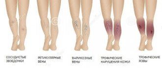
Varicose veins of the lower extremities - stages
- First. Spider veins appear;
- Second. Nodules are visible;
- Third. Swelling of the legs is added;
- Fourth. Skin color becomes darker, almost purple;
- Fifth and sixth. Ulcers form, which may not heal as a result.
This is what varicose veins look like in stage II of the disease
Varicose veins of the lower extremities - diagnosis
In addition to reviewing your medical history as well as a physical examination, testing for vein disease may include the following:
- Duplex scanning is a type of procedure that evaluates the blood flow and structure of the veins in the legs.
- Triplex ultrasound is a procedure similar to duplex ultrasound, which uses color to highlight the direction of blood flow.
- Magnetic resonance venography (MRV) is a diagnostic procedure that uses magnetic resonance technology and intravenous contrast material to visualize veins.
Diagnosis of varicose veins of the lower extremities is carried out with the patient standing
Diagnostics is necessary so that the doctor can prescribe a treatment method that is most suitable for the patient.
Venous trophic ulcer
A venous ulcer is classified as a chronic wound that occurs with a long history of varicose veins and has no tendency to heal. This occurs due to severe structural tissue damage due to impaired venous outflow.
Venous ulcers are caused by long-term chronic venous insufficiency.
V stage of varicose veins: trophic ulcer
Blood pools and stagnates in your legs because defective valves in the veins prevent adequate blood return to the heart.
Symptoms of leg ulcers:
- Painful open wounds on the legs or ankles.
- Itching sensations in the lower extremities.
- Discharge of yellow or green fluid from the ulcer.
- Swelling of the legs.
- Change in skin color around the ulcer.
- Pain in the area of the ulcer.
- Feeling of heaviness in the lower extremities.
Venous trophic ulcers account for up to 80% of all ulcers of the lower extremities. In the vast majority of cases, patients can be helped using modern technologies for treating varicose veins. Yes, most of them are treated. It is enough to contact a good phlebological center in Moscow and a competent specialist.
Many of the listed symptoms and complications of varicose veins are hereditary, so if you have family members with chronic vein disease and you experience any of these symptoms, you are definitely at risk. Factors such as obesity, diet, smoking and alcohol consumption can also have a detrimental effect on the health of your feet.
Varicose veins of the lower extremities - complications
If varicose veins on the legs are not treated, serious problems may arise in the future, both with the veins and with the skin of the legs.
- Trophic ulcers.
- Phlebitis is inflammation of the veins.
- Deep vein thrombosis (DVT), where blood clots form in the deep veins of the legs. This complication can be fatal.
Because of this, the disease can cause significant discomfort and even disability depending on any underlying tasks and severity. Some of these complications can be very life-threatening.
Why you shouldn’t delay treatment when symptoms of varicose veins appear
It should be noted that the degree of overstretching of the veins in each individual case is different, and therefore the consequences of varicose veins are not always possible to predict. Among the most common complications are thrombophlebitis, trophic ulcers, as well as bleeding and thromboembolism with irreparable consequences.
As for surgical intervention, it is usually resorted to in advanced cases. Among all the currently existing surgical techniques, modern miniphlebectomy and innovative endovasal methods of vein obliteration stand out. Much attention should be paid to the prevention of varicose veins. For this purpose, it is better to seek advice from a good phlebologist in Moscow. Do not self-medicate under any circumstances. The consequences can be very sad.
Varicose veins of the lower extremities - drug treatment
It is not always necessary, but if you are concerned about pain, ulcers or just discomfort, then, as a rule, you need to go to the hospital. But do not forget about complications, some of which lead to death, therefore, in order not to worsen the general condition, do not ignore treatment and seek medical help if you have varicose veins of the lower extremities. The doctor will explain conservative treatment and its essence.
One of the most popular venotonic drugs
Varicose veins of the lower extremities, treatment. Operation?
Don't panic if you are diagnosed with varicose veins of the lower extremities. Sclerotherapy treatment may help. This procedure is based on the use of a special solution that is injected into varicose veins. Varicose veins of the lower extremities disappear quite quickly. Sclerotherapy treatment is suitable for treating small veins such as spider veins. Are you afraid of a knife? Varicose veins of the lower extremities can be removed (treatment without surgery). You just need to find a decent doctor.
Varicose veins. Laser treatment in Moscow
Life does not end if you have varicose veins of the lower extremities. Laser treatment will help. The doctor inserts a tiny fiber into the varicose veins through a catheter. The fiber sends out energy that destroys the affected part.
This is a tiny laser fiber that completely eliminates varicose veins
Do varicose veins of the lower extremities remain? Laser treatment, which may seem expensive, is very effective, so the answer is no. Not every patient worries if he has varicose veins of the lower extremities. However, laser treatment leaves only positive reviews. With it, varicose veins of the lower extremities will be defeated! Laser treatment (you will find out the cost at the clinic from the administrator) is short-lived and inexpensive.
Varicose veins. Radio frequency treatment
What to do if you have severe varicose veins of the lower extremities? RFA treatment in Moscow is the safest method, which involves heating the venous wall using radiofrequency energy. Varicose veins of the lower extremities disappear permanently. RFA treatment is very convenient.
How quickly do varicose veins of the lower extremities disappear? RFO treatment in Moscow will convince you of its rapid effect. You will remove varicose veins of the lower extremities. RFO treatment is performed at the highest level. Look at the photos and results - make sure the procedure is effective.
Treatment
Traditionally, all methods of eliminating protruding veins - treating varicose veins - are divided into conservative and surgical (7, English).
Non-invasive methods for treating varicose veins:
- taking medications (phleboprotectors, phlebotonics, disaggregants, anticoagulants);
- physiotherapy;
- compression therapy (wearing specialized compression hosiery);
- exercise therapy;
- Normalization of body weight, correction of hormonal levels, elimination of other provoking factors (physical inactivity, constipation, etc.).
Therapeutic measures are not able to relieve the patient of varicose veins, although they can alleviate his condition, reduce the severity of symptoms and reduce the risk of complications. They are also unable to prevent further progression of the disease.
Classic surgical methods are crossectomy, combined phlebectomy, miniphlebectomy. Flaws:
- traumatic;
- use of general or spinal anesthesia;
- hospitalization;
- discomfort in the early postoperative period;
- long-term rehabilitation.
Currently, radical treatment no longer necessarily means the presence of postoperative scars and long recovery. You can safely get rid of varicose veins using cryosclerotherapy, laser obliteration and RFO.
How to treat varicose veins of the lower extremities with ointments
Today, modern drugs can surprise you with their effectiveness and quick results, however, you should not do anything without the advice of a doctor.
- Anti-varicose leg cream – relaxes and relieves pain. Strengthens the walls of blood vessels.
- Heparin thins the blood and helps reduce the formation of blood clots.
- Lyoton 1000 - relieves swelling, strengthens the walls of blood vessels, but does not completely remove varicose veins of the lower extremities. Treatment (the cost of Lyoton is more than that of other creams) in this way is still incomplete.
Of course, the easiest way is to go to the pharmacy and start treatment yourself, however, you don’t need to flatter yourself with hopes that the ointment or gel will provide you with reliable treatment and will not cause complications. Consult a doctor and do not deceive yourself.
Why are women prone to varicose veins?
Varicose veins are diagnosed in women in approximately half of clinical cases. Reasons (5, English):
- Gender differences in endocrine status. The estrogen-gestagen hormonal background in women of reproductive age is the main predisposing factor to varicose veins.
- Features of the structure of subcutaneous fat tissue.
- The addition of other risk factors: pregnancy, oral contraception, and often hormone replacement therapy.
Female sex hormones affect the condition and tone of the vascular walls, promoting their expansion. Particular importance is attached to progesterone, the hormone of the second phase of the menstrual-ovarian cycle: its relaxing effect on the smooth muscle muscles of not only the walls of internal organs, but also blood vessels has been proven. Promotes venostasis. This is why many women more often complain of heaviness in their legs at the end of the ovarian-menstrual cycle, when the dominant sex hormone is progesterone.
Estrogen also affects the condition of the vein walls, although to a lesser extent; its varicose effect can be both direct and indirect. Estrogens can increase the likelihood of immune complexes being deposited in the walls of the veins, which leads to their thickening and decreased elasticity. They also provoke vasodilation.
The indirect varicose effect of estrogens includes an increased tendency to intravascular thrombus formation.
Combined changes in the hemostatic system include:
- increased fibrinogen levels in the blood;
- increased platelet aggregation even in the absence of violations of the integrity of the vascular endothelium;
- change in the ratio between various coagulation factors.
In healthy women not taking any medications, this tendency to form blood clots is minimal and has no particular clinical significance. The hemostasis system is able to compensate for it. But when other factors affecting hemostasis are added, the natural balance between thrombus formation and thrombolysis is disrupted, and blood viscosity increases. And in women this usually happens faster than in men.
Destabilizing prothrombogenic factors include:
- taking estrogen-based drugs for therapeutic purposes;
- use of COCs (combined oral contraceptives);
- liver diseases;
- various intoxications, often occurring with blood thickening;
- pregnancy.
Another factor that increases the likelihood of developing varicose veins in women is the structural features of subcutaneous fat. It is usually looser than in men, with a small number of connective tissue septa, and is prone to the accumulation of extracellular fluid with the formation of edema.
In most women, the external “framework” around the saphenous veins is not able to restrain their deformation with the appearance of characteristic nodes and curvatures. If there is a concomitant difficulty in lymphatic drainage from the lower extremities, the risk of developing varicose veins increases even more.
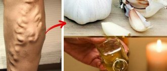
Varicose veins of the lower extremities - treatment at home
How unpleasant varicose veins of the lower extremities are! Our grandmothers still use folk remedies to this day, and knowing the effect of medicinal herbs, you can relieve symptoms and strengthen the walls of blood vessels. But make no mistake - serious varicose veins of the lower extremities, treatment, price, results - everything is interconnected.
- Horse chestnut. Dried flowers (50 g) are poured with vodka (500 ml). Let it brew for 20-21 days and take it orally, before meals, 25-30 drops 2 times a day.
- Garlic ointment. Make a paste of garlic and butter. Rub in before going to bed.
- Apple vinegar. 2 tsp dilute in 200 ml of water. Take before meals. This product has good reviews.
It is important to know that varicose veins of the lower extremities (prices, photos, results are available online) cannot be completely removed with herbs.
Causes of pain syndrome
Long-term research and medical practice indicate a variety of factors leading to painful sensations in the foot. It can be:
- injuries;
- muscle failure;
- autoimmune pathologies;
- uncomfortable shoes;
- lack of calcium;
- hygroma;
- incorrect anatomy.
It should not be ignored if there is pain in the top of the foot or in other parts, swelling, or fever. This may be a sign of serious malfunctions in the body that can lead to disability.
As a rule, discomfort covers the entire foot, which complicates diagnosis and requires high medical skill. Complaints should be addressed to:
- orthopedist;
- traumatologist;
- rheumatologist.
Of course, narrow specialists are available only in specialized clinics. In the clinic, a surgeon is in charge of diseases of bones, cartilage and joint tissue.
How to cure varicose veins permanently?
Unfortunately, if you neglect the disease, both you and your doctor will have to work hard to forget about varicose veins in your legs. At the first stage, the disease responds well to any treatment method, but later stages can upset you. You shouldn’t think about how to treat varicose veins of the lower extremities at home. Contact our center and you will be prescribed the right treatment, thanks to which you can get the desired result.
All you need to do is take better care of your health and adhere to basic preventative measures.
Compression jersey medi
Thanks to the breathable and elastic material, compression jersey provides high wearing comfort. Modern medical compression hosiery is visually indistinguishable from model hosiery, but provides high medical effectiveness when used.
Here you can find more information about medi compression stockings.
The human body
How do veins work?
Vienna
Product Tips
Ideal compression product
Compression hosiery
Frequently asked questions from our patients on the Internet
How to deal with varicose veins on the legs? Daria from Saratov is interested in:
Dear Daria! It is necessary to combat varicose veins in the legs with the help of good specialists. No matter how promising and colorful the sites with guides and advice on self-medication are, remember, they are written by people far from medicine.
How to check your legs for varicose veins? Svetlana from Kazan asks:
Dear Svetlana! As a rule, it is possible to qualitatively check your legs for the presence of varicose veins only in a clinic or medical center specializing in the treatment of veins.
How to diagnose varicose veins? Interested in Lyubov from Stavropol:
Dear Love! You can diagnose your legs for varicose veins at a consultation with any of our phlebologists with an ultrasound duplex scan of the venous system. How easy is it to do this with us? Make an appointment for a consultation at any time convenient for you by phone.
Who most often suffer from leg vein pain?
According to the latest data from the World Health Organization, 60% of working-age residents of developed countries suffer from varicose veins. Moreover, venous disease is one and a half times more common among the fair sex. This is due to the genetic predisposition of women to varicose veins, as well as to the factor of their reproductive function. During pregnancy, women experience some restructuring of the balance of the hormonal system, which causes an increase in body weight, as well as the total volume of blood circulating in the body of the expectant mother. All this, with each subsequent month of pregnancy, negatively affects the vascular wall, as well as the pelvic vessels.
Therefore, if you notice the symptoms of heaviness in the legs described above, it is recommended to immediately make an appointment with a phlebologist and come for an examination and diagnosis.