Cardiac rupture is a violation of the integrity of the walls of the heart. Most often it occurs as a serious complication of myocardial infarction (death of a section of the heart muscle due to the closure of a supply vessel), accompanied by high mortality. Heart rupture is recorded in 2-8% of patients with a heart attack. In most patients, there is a rupture of the wall of the left ventricle, less often of the right ventricle, and even less often of the interventricular septum (the partition between the left and right ventricles of the heart) and the papillary muscles (the internal muscles of the heart that ensure the movement of the valves).
He was treated for the flu, but suffered a heart attack. How to suspect heart problems? More details
Definition:
A cardiac aneurysm is a pathological condition that is accompanied by protrusion of the wall of the heart muscle. It is one of the complications after a myocardial infarction and can develop within several months after its onset. An aneurysm not only negatively affects the functioning of the heart and its contractile functions, but also poses a serious danger to life due to the possibility of sudden rupture.
Is it possible to save a person with a broken heart?
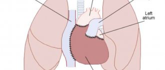
Heart rupture most often ends in death - acute heart failure occurs and the person does not have time to save. However, in some cases, with a slow rupture of the heart, it is possible to perform an urgent operation to strengthen the site of damage using a special “patch” made of synthetic materials. If necessary, additional coronary bypass surgery is also performed, aimed at restoring blood flow and, thus, accelerating scar formation at the site of ischemia and rupture.
Large ruptures are extremely difficult to operate, since the tissues are poorly connected to each other, regeneration slows down, and a significant area of the heart muscle may need to be removed. In these cases, a heart transplant can save the patient, but serious difficulties with its implementation are due to limited time and the lack of a suitable donor.
Those who are at risk must necessarily observe disease prevention - do not overexert themselves, lead a healthy lifestyle, be as nervous as possible and constantly check their heart.
Cause
Usually the cause of the formation of a cardiac aneurysm is a heart attack, most often of the left ventricle of the heart. When they talk about the development of myocardial infarction, the damaged parts of the heart are weakened, and under the influence of blood pressure they can bulge. Blood trapped in them forms a blood clot. Aneurysms occur in patients with atherosclerotic cardiosclerosis and cardiosclerosis of other origins. Much less common are aneurysms that arise due to other reasons: infections, injuries, and congenital defects.
What other reasons can lead to heart rupture?
The causes of cardiac rupture may be the following:
- Heart injury. An accident can provoke a heart rupture if the driver hits his chest on the steering wheel, or a strong blow from an opponent during a single combat
- Anomalies of cardiac development. If there is a congenital thin section in the heart, it can rupture under minor loads
- Tumor lesion of the heart
- Endocarditis - inflammation of the inner lining of the heart - endocardium
- A dissecting aortic aneurysm is a rupture of the largest artery, the aorta, which leads to blood flowing between the layers of the aortic walls and dissecting them further.
- Infiltrative heart diseases. These include sarcoidosis, hemochromatosis, amyloidosis - diseases in which substances accumulate in the heart that should not normally be present.
Early heart attack. Why disaster can threaten young people Read more
Classification
Depending on the time period after myocardial infarction the aneurysm formed, the following forms are distinguished:
- Acute and subacute - develop in the early stages after a heart attack, often complicated by incomplete rupture.
- Chronic - formed after acute due to the replacement of a dead section of the heart muscle with connective tissue. They can be muscular, musculofibrous or fibrous.
Screening and diagnosis of arrhythmia
To diagnose arrhythmias, the doctor usually asks the patient if they have any heart disease and/or thyroid problems. In addition, specific types of medical testing are always performed to detect arrhythmia. This may be a short or long-term passive recording of an electrocardiogram (a day or more), or an attempt to provoke an arrhythmia while simultaneously continuously recording the heart rhythm.
Passive monitoring methods include:
- Electrocardiography (ECG). During an ECG, electrodes attached to specific locations on the arms, legs, and chest record the electrical activity of the heart. An electrocardiogram examines the intervals and duration of each phase of heart contraction.
- Daily ECG monitoring using the Holter method. A portable ECG recorder is installed for a day or more to record the electrical activity of the heart as a person performs his usual daily activities, as well as during sleep.
- Echocardiography. Allows you to use an ultrasound sensor to obtain an image of the chambers of the heart, clarify their sizes, the movement of the walls and valves and other information.
Arrhythmia can be induced using the following tests:
- Tests with physical activity . Some arrhythmias are triggered or worsened by exercise. During the “stress” test, a treadmill or stationary exercise bike is used. During the test, a continuous ECG recording is made. To perform a “stress” test, medications can be used that stimulate the heart in a similar way to physical exercise. Typically, this method is used when it is impossible to perform physical exercise, as well as to establish a diagnosis of coronary heart disease.
- Tilt table test . When a person has unexplained loss of consciousness, slant tests may be helpful. In this case, rhythm and blood pressure are monitored in a horizontal position for 20-30 minutes. Then a special table is moved to a vertical position and rhythm and blood pressure are also monitored for 10 minutes. In this way, the condition of the heart and the specialized nervous system that controls the functioning of the heart when changing body position and moving from a horizontal to a vertical position is assessed.
- Electrophysiological study and mapping . This study is carried out using the thinnest catheters - electrodes, which are carried into the heart cavity. When electrodes are installed in the area of certain areas of the conduction system of the heart, they can be used to study the propagation of an electrical impulse throughout the heart, induce arrhythmia, while studying its localization and mechanism, and also test the therapeutic effect of various medications. This is the most informative and accurate method for diagnosing most arrhythmias. In addition, when performing EPI, it is possible not only to identify, but also to eliminate the arrhythmogenic focus using a special thermal effect called radiofrequency. In this way, most supraventricular and some types of ventricular tachycardias are eliminated. Currently, this method is also used for atrial fibrillation.
Symptoms
Symptoms of acute cardiac aneurysm are weakness, shortness of breath, fever, pulmonary edema, sweating, heart rhythm disturbances, and swelling of the veins in the neck. During the transition to the subacute form, symptoms of coronary insufficiency quickly develop.
With chronic cardiac aneurysm, signs of cardiovascular failure are observed: edema, angina attacks, rhythm disturbances. Complications of the disease can be thromboembolic syndrome with damage to peripheral vessels and arteries of the brain. If you experience such signs, immediately contact the cardiology center.
When an acute aneurysm ruptures, death occurs almost instantly. In this case, the patient experiences severe pallor, hoarse, difficult breathing, cold sticky sweat, congestion of the neck vessels, and cold extremities.
Providing emergency care to a patient
Treatment of cardiac rupture requires emergency surgical care and intensive care in the prehospital and postoperative period.
The following surgical techniques are currently available:
- Suturing a rupture in an open heart;
Installation of an occluder using a catheter;- Prosthetics of valves and vessels (aorta) with donor, animal or artificial materials;
- Heart transplant;
- Pericardial puncture to eliminate cardiac tamponade;
- Coronary artery bypass grafting after the main operation.
Drug treatment is carried out in the intensive care unit in order to maintain vital functions during the recovery period. If a patient is brought to cardiac surgery in a timely manner and in stable condition, his chances of survival increase rapidly.
Patients are often prescribed transfusions of red blood cells, plasma, albumin, and saline solutions to correct the volume of circulating blood and eliminate hypoxia. Blood pressure is monitored, since both hyper- and hypotension can cause rupture of the aorta of the heart to recur and cause damage to vital organs, nullifying the efforts of surgeons.
Treatment
The resulting rupture leads to death. Therefore, the main treatment is to prevent aortic rupture.
If there is an aneurysm, how to behave:
- Avoid intense physical activity.
- Do not allow blood pressure to rise above 140/80.
- Avoid stress.
- Avoid injury.
- Be under the supervision of a doctor and follow all his recommendations.
- If possible, surgery to remove the aneurysm should be performed.
Surgical treatment of aneurysm
There are open operations in which the abdominal cavity is opened, the dilated part of the aorta is removed and a synthetic prosthesis is placed in its place. This operation is technically very complex, traumatic and lengthy. But the result is worth it.
A modern method of treating aneurysms is the installation of a special intravascular endoprosthesis. Through an incision in the thigh and through the femoral artery under X-ray control, the endoprosthesis is brought to the site of the aneurysm. Under the influence of air pumped into it through a catheter, it straightens and closes the aortic defect.
Here is such a complex stent for the spread of an aneurysm to the iliac arteries
Drug treatment
Treatment of patients with dissecting aneurysm and threat of rupture is carried out in the intensive care unit.
In case of dissection, narcotic analgesics (morphine, fentanyl) are prescribed for pain relief. The pain must be relieved. Drugs that reduce cardiac output (beta blockers) and drugs to lower and control blood pressure at a given level (Ebrantil, Enap) are also prescribed. An increase in blood pressure above 180/100 is unacceptable in this situation.
Aneurysm rupture is an extremely serious complication of vascular diseases with a high mortality rate. It is important to diagnose the pathology in time and contact experienced doctors for treatment and surgery.
Signs of a dissecting aneurysm
Main symptoms:
- The pain is intense, localized behind the sternum, in the epigastrium, and can be between the shoulder blades and along the spine. When the dissection passes to the brachiocephalic vessels or to the vessels of the kidneys, it can spread to the head and neck, lower back and legs.
- The nature of the pain is one of the main distinguishing features. The pain is acute, sudden, sharp, and has a migrating nature.
- Vomiting, loss of consciousness, and neurological disorders are possible.
- Hemodynamic disturbances - there may be high or low blood pressure, rapid pulse.
- Signs of heart failure, shortness of breath, swelling.
- Symptoms are caused by the spread of dissection into branches and concomitant acute organ ischemia.
Schematic representation of the delamination
Symptoms may be similar to myocardial infarction, pleurisy, and mediastinal diseases.
Possible complications
The main complications are as follows:
- If a person has a rupture of the heart muscle, then without proper medical care he dies. To prevent this from happening, he needs urgent surgery. If the gap is small, then a person can live only two months without surgery.
- The forecasts, unfortunately, are disappointing, because almost half of the patients may die, even if the operation was performed on time and with proper quality. The thing is that the sutures placed during suturing often come apart.
If you start treatment in a timely manner, it is possible to eliminate complications.
Types of abdominal aortic rupture
Rupture of an abdominal aortic aneurysm can occur in several scenarios:
- Into the abdominal cavity. It manifests itself as a sharp drop in pressure as a result of hemorrhagic and painful shock. Death occurs in a matter of minutes.
- Break through the peritoneum. Characterized by sharp back pain in the lumbar region. The pain is very intense and cannot be relieved with narcotic analgesics. The hematoma can compress the iliac arteries, leading to limb ischemia.
- It can break into the abdominal organs (stomach, small intestine).
- Characterized by hemorrhagic shock, vomiting blood and bloody stools.
Who is at risk of myocardial infarction
Risk factors are:
- hypertonic disease;
- previous heart attack;
- smoking;
- sedentary lifestyle;
- hereditary predisposition;
- increased levels of “bad” cholesterol in the blood;
- obesity;
- diabetes;
- elderly age;
- postmenopausal state (in women);
- regular stress;
- excessive stress (physical and emotional);
- bleeding disorders.
Diagnosis of cardiac aneurysm
In 50% of patients, pathological precordial pulsation is detected.
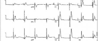
ECG signs are nonspecific - a “frozen” picture of acute transmural myocardial infarction is revealed, there may be rhythm disturbances (ventricular extrasystole) and conduction (left bundle branch block).
ECHO-CG allows you to visualize the aneurysm cavity, determine its size and location, and identify the presence of a parietal thrombus.
The viability of the myocardium in the area of chronic cardiac aneurysm is determined by stress echocardiography and PET .
Using chest x-ray, it is possible to detect cardiomegaly and congestive processes in the blood circulation.
1 ECG - a method for diagnosing cardiac aneurysm
2 ECHO-CG - a method for diagnosing cardiac aneurysm
3 Chest X-ray
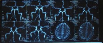
ventriculography, MRI and MSCT are also used to determine the size of the heart aneurysm and detect thrombosis of its cavity.
For the purpose of differential diagnosis of the disease from coelomic pericardial cyst, mitral heart disease, mediastinal tumors, probing of the heart cavities and coronary angiography may be prescribed .
