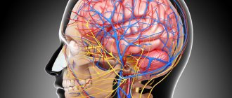
The structure of the heart contains tendon threads called chords. Normally, they are attached to the valve flaps through which blood flows, and prevent them from sagging under the influence of gravity. However, sometimes an additional chord may be detected during diagnosis. This is a structural feature of the heart, which does not always indicate an anomaly. But in some cases it can be dangerous to the health and even life of the patient.
What is a chord
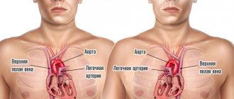
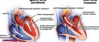
The human heart consists of 4 chambers through which blood regularly passes - 2 ventricles and 2 atria. Blood flow moves through valves, which open in chords. When tensioned, the valve opens the flaps, and when loosened, it closes. Thanks to this, the blood flows only in a given direction and does not move backward.
The valves tension special longitudinal strands - chords. But along with the main ones, additional chords are often identified. They are also attached to certain parts of the heart, but do not take any part in pumping blood. Therefore they are also called false chords.
University
My son is 36 years old. Recently, an ultrasound revealed that he has multiple accessory chords of the left ventricle. Is it good or bad? The clinic didn’t say anything definite... The Rybakov family, Vitebsk region.
I work in the regional center as a cardiologist. I would like to read in the Medical Bulletin about abnormal chords of the heart: how to identify and then manage such patients? The article will be useful for doctors from the outback... Svetlana N., Vitebsk region.
Evgenia Trisvetova, Doctor of Medicine, answers questions from readers and the MV correspondent Sciences, Professor of the 2nd Department of Internal Diseases of BSMU.
— Evgenia Leonidovna, how do experts explain the appearance of additional chords in the heart?
— I would like to note right away that intravital study of the internal structure of the heart became possible thanks to the invention of imaging devices and the development of ultrasound. In 1981, almost 100 years after the anomalous chordae were mentioned, the first intravital description of anomalous cardiac chordae using two-dimensional echocardiography was published.
Abnormal (false) chords are those that are attached not to the valve leaflets, but to the walls of the ventricles. According to one hypothesis, it is a derivative of the inner muscular layer of the primitive heart, arising in the embryonic period when the papillary muscles are unlaced. According to another assumption, abnormal chordae are muscle trabeculae that are drawn into the cavity of the ventricle during its dilatation or the formation of an aneurysm.
The name “chord” reflects the position of the abnormal cord as a geometric body crossing the chamber of the heart. True chords are fibrous cords consisting of collagen fibers. They receive blood supply and innervation from the heads of the papillary muscles, which are fed and innervated through muscle trabeculae located at the base of the same papillary muscles.
Abnormal chordae arise from inherited defects in collagen metabolism; influence of factors that disrupt embryogenesis with genetic predisposition and cause familial non-hereditary forms.
An abnormally located chord (APX), called a moderator band, was first described during a post-mortem examination in 1893. Subsequently, additional cords in the cavity of the left and right ventricles were designated differently: false tendon, false ligaments, additional chords, muscle cords. Modern sources use the term “abnormally located chord” and false tendon.
- How common is this anomaly?
— During ultrasound examination of the heart in children, the frequency of abnormal chordae ranges from 0.2–21.7%. Andrey Korzhenkov, a researcher at the Novosibirsk Research Institute of Therapy, and his colleagues studied the prevalence of abnormal cardiac chords in adults. In 1991, 859 men aged 25–64 years old underwent ultrasound; signs of abnormal cardiac chords were found in 17.1%. According to the results of our echocardiographic study (2002–2004, 2,401 people), in men 18–25 years old, healthy or hospitalized for diseases of the internal organs, abnormal cardiac chords occurred in 8.3% of cases.
The results of the post-mortem study are interesting. Tatyana Domnitskaya, professor of the Department of Functional Diagnostics at the Faculty of Advanced Training of Medical Workers at the Peoples' Friendship University of Russia, examined the hearts of people who died from various diseases. These were 41 men and 46 women, 57–87 years old. Macroscopic examination revealed abnormal cardiac chords in 14% of cases. Other authors pointed to the occurrence of such chords in 17–39% of cases in women and much more often (61%) in men.
In our work (E.L. Trisvetova, O.A. Yudina, 2006 - Author's note), abnormal chords of the heart were identified in 12.1% of cases (578 deaths 15–91 years old (average age 63.1±1 .08), including 295 (51%) men and 283 (49%) women). In the group of deaths with minor cardiac anomalies (n=98), these chords were diagnosed in 71.4% and 2.08±0.59 cases per 100 examined. The ratio of men to women with abnormal cardiac chords was 1:0.64.
— Which parts of the heart “prefer” abnormal chords?
— A. Korzhenkov and co-authors proposed to determine the relationship of the chord to a specific topographical variant using the division of the walls of the left ventricle into 10 segments. According to the classification of J. Botti, transverse, basal and longitudinal chords are found in 3 sections of the left ventricle: apical, midventricular and basal. The most common ARHs of the left ventricle are in 95% of cases, while in the right they are recognized in only 5% of cases; very rarely abnormal chords of the right atrium are detected. Transverse ones occur in 68%, diagonal - in 30%, longitudinal - in 2% of cases.
ARCHs can be single (81.6%) or multiple. Using ultrasound in young men, we detected ARCH in 8.3% of cases. Moreover, in 99.2% of cases, ARCHs were found in the left ventricle. A post-mortem study of the prevalence of ARH showed that in 17–39% of cases they occur in women and much more often (61%) in men. Age does not affect the incidence of ARH.
— How can a specialist detect abnormal chords?
— The most informative method for intravital detection of ARCH is the echocardiographic method. Transthoracic and transesophageal cardiac imaging approaches are used. The sensitivity of transthoracic echocardiography in diagnosing ARCH is 72–87%. The quality of the equipment and the qualifications of the specialist performing the research are important. In practice, both over- and underdiagnosis of ARH are observed. To visualize ARCH, two-dimensional echocardiography is performed from 3 orthogonal projections (sagittal, horizontal, frontal), using the long axis of the left ventricle, the longitudinal axis of chambers 2, 4, 5 and the short axis projection. Parasternal, apical, and subxiphoid approaches are used, allowing one to see all the structures of the heart in real time.
In addition to longitudinal and cross sections, to search for ARH it is necessary to use non-standard approaches and projections. The ARCH criterion is the identification of an echo-dense linear additional formation in the ventricular cavity in 2 mutually perpendicular planes in sectoral scanning mode with confirmation of the results on an M-echocardiogram in the systole and diastole phases. It is necessary to examine the papillary muscles and areas of attachment of the chord to the free walls of the ventricles. The attachment points of the chord are detected in the sectoral scanning mode and the results are confirmed on the M-echocardiogram. An addition to the standard EchoCG protocol has been developed - it is necessary to indicate the main characteristics of the ARCH (localization, topographic variant, echo density in the areas of attachment of the cord, presence/absence of traction of the papillary muscles in systole, thickness and length, changes in the geometry of the left ventricle, the rate of shortening of myocardial fibers in the areas of attachment of the chord ) and other features.
A detailed assessment of the morphometric, topographic and functional signs of ARH is carried out for the differential diagnosis of echocardiographic or clinical manifestations of the abnormality.
The abnormal chordae are located between the following intracardiac structures: the posteromedial papillary muscle and the wall of the left ventricle or the interventricular septum; anterolateral papillary muscle and interventricular septum; papillary muscles; walls of the left ventricle and interventricular septum; walls of the left ventricle. Sometimes abnormal chordae are attached at 3 or more points, forming a membranous structure.
— How should a doctor treat an abnormal finding? Observe the patient, treat?..
— The clinical significance of abnormal chordae has not yet been determined. It is known that people with hereditary and multifactorial connective tissue disorders are characterized by single and multiple abnormal chordae, which are diagnosed along with other minor cardiac anomalies. In this case, clinical manifestations may be caused by multisystem disorders of the structure of connective tissue, including musculoskeletal, skin, ocular, cardiovascular and other visceral signs of dysmorphogenesis, and autonomic disorders.
The actual presence of abnormal cardiac chords may not affect the patient’s subjective sensations and may not bother them. However, if other causes are excluded, they can cause the manifestation of pathological signs. The transverse position of the abnormal chord in the middle third of the left ventricle causes a systolic murmur, which is heard during auscultation of the heart. A long abnormal chord reaching the left ventricular outflow tract is associated with local hypertrophy of the opposite wall of the left ventricle.
Cardiac arrhythmias (ventricular extrasystole, supraventricular/ventricular paroxysmal tachycardia) can occur due to the presence of cells of the conduction system in the abnormal chord or changes in the myocardium at the sites of attachment of the abnormal cord and the appearance of ectopic foci of excitation in the myocardium. The ECG of abnormal left ventricular chordae describes abnormal ventricular repolarization (early repolarization syndrome, nonischemic ST segment depression, flattened or weakly negative T wave), WPW syndrome, and shortened PQ interval.
Short abnormal chords in a transverse position in the cavity of the left ventricle can be injured by the blood flow or torn/ruptured during ventricular diastole. In these places, blood clots are identified, representing a likely source of embolism in the vessels of the systemic circulation. Cases of infective endocarditis that developed as a result of infection of the area of the anomalous chord tear have been described. Changes in the geometry of the left ventricle due to a transverse abnormal chord in the cavity are rare and are compensatory in nature, preventing the expansion of the heart chamber.
Typically, abnormal chordae do not affect the prognosis of life and do not require special treatment. In the case of an echocardiographic “finding,” the patient must be examined to determine whether there are hereditary or multifactorial connective tissue disorders or congenital heart defects.
— Can people with abnormal chordae play sports and have children? Do I need to see a cardiologist?
— If diseases are excluded and there are no clinical symptoms, the abnormal chord is treated as an insignificant feature of the development of the heart. A person leads a normal lifestyle and does not limit himself in physical activity; women plan pregnancy and childbirth. It’s another matter if the doctor diagnosed symptoms of a hereditary or multifactorial connective tissue disorder. Then you need to get specific recommendations from your doctor and follow his advice. If you experience discomfort, shortness of breath, pain in the heart, as well as rhythm disturbances, you need to contact a specialist for additional examination and medical care. Tatyana Skibitskaya Medical Bulletin , February 13, 2015
Why do false chords of the heart appear?
False chords are quite easy to detect already in a child’s heart. Moreover, pediatricians do this while listening to the chest (by the presence of heart murmurs). Typically, additional chordae form during fetal development. The main reasons for their appearance are:
- Heredity - for example, parents or grandparents also had this diagnosis.
- The mother’s unhealthy lifestyle, especially during pregnancy (smoking, drinking alcohol, uncontrolled use of medications, toxic substances), and the presence of chronic or infectious diseases.
- Genetic factor - mutation of genes or chromosomes.
- Unbalanced diet - excess animal fat.
- Heavy workload during pregnancy.
- Unfavorable environmental conditions.
- Constant, severe stress.
- Deterioration of immunity for various reasons.
Patent foramen ovale
This is the opening located between the two upper chambers of the heart (the left and right atria). It functions as a “door” during the development of the child in the womb. Through it, blood is discharged from the fetus from right to left. This is how blood flow exchanges between the small and large circles of blood circulation. An open foramen ovale in a child before birth plays a big role, “helping” the undeveloped lungs cope with blood flow. It is this that ensures the passage of blood to the left atrium and aorta.
When a child is born and his lungs, expanding, begin to work at full strength, the oval window “automatically” closes. A small depression remains in its place. However, there are cases when the oval window does not close after the baby is born. It is important to keep in mind that this anomaly does not belong to heart defects, and it can be detected by echocardiography. Usually, the bubble contrast technique is used for this, and if the baby’s oval window is not closed, air bubbles will move from right to left during examination.
Accessory chord: symptoms
A person can comfortably live their entire life with additional chordae, unaware of their existence. Most often they do not cause any sensation. But sometimes patients may notice the following symptoms:
- fast fatiguability;
- periodic manifestation of fatigue;
- pain in the heart area;
- headache;
- rapid heartbeat (the heart “jumps out”), especially during excitement or conflict.
The described symptoms do not necessarily indicate the presence of a chord, since they may be associated with other causes. Therefore, to accurately determine the cause, it is necessary to undergo professional diagnostics.
Diagnostics
First of all, you need to contact a therapist or children's doctor (pediatrician), who will listen to your heart rhythm using a stethoscope. It is very easy to hear a heart murmur, so the therapist can easily make a preliminary diagnosis after the first examination.
Next, the patient may be referred for additional diagnostics:
- ECG at rest and after exercise;
- blood pressure measurement;
- Holter monitoring.
Thanks to the examinations carried out, it is possible to accurately determine the presence of a chord, the degree of its danger to health, as well as other diseases (if any).
Treatment of the disease
If the false chord is not life-threatening and does not affect health, preventive monitoring by a cardiologist is indicated. Moreover, treatment as such is usually not prescribed - the patient regularly visits a specialist every 3-6 months.
If the situation is dangerous and the chord threatens the functioning of the heart (can lead to cardiac arrest), surgical intervention is indicated to remove it. Such a decision is made if the functioning of the heart is severely impaired, causing regular disruptions in the distribution of blood flow.
It is also allowed to use folk remedies (in consultation with the doctor):
- Take a handful of raisins every day in the morning (on an empty stomach);
- take valerian decoctions;
- drink tea based on peppermint or calendula flowers.
Combating complications
Despite the fact that trabeculae in the cavity of the left ventricle do not pose a threat to the child’s health, with the wrong lifestyle they can go from a benign form (painless) to a malignant one (requiring regular use of medications). Therefore, when a disease is detected, doctors immediately determine prohibitions for the patient. These include:
- Extreme sports, sports career, active gymnastics, excessively active dancing, scuba diving.
- Unauthorized taking of medications. Each drug (even those not related to heart disease), its dosage, must be approved by a specialist. Many pharmacological drugs affect intravascular pressure and increase heart rate, which is unacceptable for this type of pathology.
- Intense physical and emotional stress.
In addition, the doctor issues a certificate prohibiting the child from participating in school physical education competitions and exempting him from certain types of exercises. He develops such a complex of activities so that the child’s additional trabecula of the left ventricle is trained and does not bother him in the future. This shows:
- Easy short run.
- Walking on a tightrope.
- Drill exercises.
- Jumping.
- Gymnastic procedures on a skipping rope, wall bars, bench, with a ball.
In addition to all this, a child with an additional chord will benefit from attending massage courses, eating right, going to slow dances, resting more often and not getting into stressful situations.
The best way to give birth to a healthy baby is to avoid factors that can negatively affect the development of the fetus (for the entire period of pregnancy). If the expectant mother has this disease, it is recommended to take it much more seriously. This way you can reduce the risk of an additional chord. If a child has already been born with such a pathology, you need to carefully monitor the baby’s well-being and listen to every medical recommendation so that the child grows and develops on par with his peers.