Heart defects are represented by a large group of abnormalities. Many are not noticeable for a long period. There are no symptoms. The patient lives fully and has no idea that there is a problem.
Then three scenarios are possible: a sharp actualization of the pathology and the formation of a clinical picture, sudden death, and further asymptomatic progression.
One of the variants of the anomaly is represented by a displacement of the muscular organ relative to the normal anatomical position.
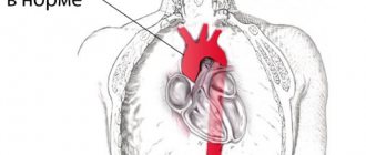
Dextrocardia is a mirror arrangement of the heart. It is not yet formed in the second week of intrauterine development, as a result of ectopia (displacement) of the cardiac tube to the right.
The danger is not the deviation itself, but the accompanying complications. According to statistics, persons with dextrocardia suffer from other defects in 30% of cases.
They are detected almost immediately after birth, since it is impossible not to see the displacement of the organ.
In the absence of a complicated course, treatment of the pathology is not required. Regular outpatient monitoring and standard screening measures are indicated as part of preventive examinations.
Development mechanism
The question of whether dextrocardia is considered a disease has not yet been resolved. More and more doctors are inclined to believe that an uncomplicated process is not considered pathological. How is an anatomical disorder formed?
Conventionally, there are two types of this condition. True, when the anomaly is the result of the influence of perinatal factors, and false, it can be acquired and is always pathological.
In the first case, the main role is given to the genetic factor and random errors. The heart is formed in the second week of gestation and represents the rudiments of a full-fledged organ.
Under the influence of one factor or another, the tube shifts to the right and begins to develop from the opposite side.
The formation of other organs occurs in accordance with the new “configuration”.
There are three options:
- Isolated dextrocardia is an altered location of the heart and normal location of other internal organs.
- Partially combined - reverse arrangement of the heart and lungs (the left lung has three lobes, and the right lung has two).
- Complete - a mirror arrangement of the heart and the same transposition of the internal organs.
If a genetic defect is to blame, other defects also develop. Including those not related to cardiac structures.
A false form is the result of gross violations. Their influence occurs already in the second or third trimester.
There are known cases of organ displacement in older age. The classic variant is a lung tumor, usually malignant.
As it grows, it provokes a shift of the muscular heart to the other side, pushes it aside, and causes tissue compression.
In such a situation, doctors talk about dextroposition or false vice. We are talking about a disease, not a developmental feature.
Large neoplasias can cause significant displacement. Therefore, during auscultation (listening to tones), the heart will be detected on the other side.
The same effect is produced by a previous inflammatory disease (rarely), lung pathologies, or accumulation of fluid in the chest.
How does the anomaly form?
Read more: Treatment of heart defects in children
It is believed that the fetal heart tube is formed already in the early stages of pregnancy (in the first 10 weeks). Its curvature to the right side leads to displacement, formation of the heart and great vessels on the right.
The fetus may simultaneously have other abnormalities of organs and systems associated with genetic mutations. The most studied mutations are the ZIC3Shh, Pitxz, HAND, and ACVR2 genes. Hereditary transmission of the anomaly is assumed.
The exact mechanism of dextrocardia has not been proven. In most cases, the heart functions normally. The child grows and develops without noticing the defect. Pediatricians observe such children at risk for the possibility of developing cardiac pathology.
Possible complications and dangers
Cases of unfavorable current dextrocardia should be taken into account. Possible consequences:
Death
It is unlikely if there are no gross abnormalities in the functioning of the cardiovascular or respiratory systems. The main causes of death are acute organ dysfunction and asphyxia (suffocation).
No doctor can give a prognosis. This can happen at any time.
Children with a complicated course of dextrocardia live from a year to 12-18 years, rarely longer. Even competent treatment does not guarantee an increase in the average.
Frequent pneumonia
Almost all children with this anomaly have a history of Kartagener syndrome. This is a violation of the functional activity of a special ciliated (ciliated) epithelium located in the respiratory tract.
When foreign particles or agents penetrate, tissues become irritated, trying to push out foreign objects. A cough begins.
In children with dektrocardia and Kartagener's syndrome, the reflex is greatly weakened. Therefore, bacteria with dust and particles of saliva settle in the lungs, causing constant inflammatory processes that can become fatal.
Reproductive dysfunction
It is the result of circulatory disorders in the pelvic organs. This is a consequence of insufficient contractility of the myocardium.
The filling of the cavernous bodies with blood is weak, therefore a high-quality erection does not occur. In addition, the testicles also suffer from ischemia. Spermatogenesis becomes impossible, hence infertility.
Correction of heart function leads to an improvement in reproductive function, but complete recovery in severe patients cannot be achieved.
Intestinal disorders
Also associated with insufficient trophism (blood supply). This time the digestive tract suffers.
Subjectively, the process is manifested by abdominal pain of unknown origin and localization, flatulence, constipation, and diarrhea. The periods alternate.
Septic shock
Develops as a result of generalized inflammation. The result of a long course of an infectious process, most often the same pneumonia.
Immunity weakens, resistance to aggressive environmental factors decreases.
Mental and mental development disorders
Children with dextrocardia mature mentally later than their peers. No gross defects are observed; full compensation is possible with age. But this is not an axiom.
Retarded physical development
Since the blood circulation of all tissues is impaired, hypoxia (oxygen starvation) is observed. Bones grow more slowly, and there is a delayed increase in muscle mass. Puberty does not begin upon reaching 10-11 years of age, sexual development is poor.
In the remaining 70% of cases (possibly less, the data varies, unified statistics are not presented), the right-sided location of the heart is not accompanied by pronounced deviations in the functional activity of other organs and systems. Everything remains normal.
Combination with other features
Rarely does an isolated “simple” anomaly occur. Dystopia (disorder of arrangement) also affects other organs. The most common combinations are found in childhood in the form of:
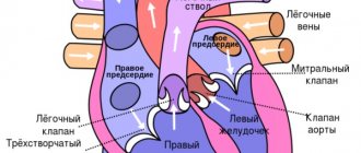
- tetralogy of Fallot - dextroposition of the aorta, pulmonary stenosis or complete occlusion, ventricular septal defect, significant hypertrophy of the right ventricle;
- reverse position of the main arteries;
- ventricular and atrial septal defect;
- pulmonary stenosis;
- valvular endocardial defects;
- double ventricular outlet;
- two- or three-chamber heart.
During transposition, the aorta emerges from the right ventricle, and the pulmonary artery emerges from the left.
A great danger to the life of a newborn is represented by “blue” defects, which appear in the first hours after birth. The vital activity of internal organs depends on the speed of surgical intervention to eliminate the defect.
With “white” defects, oxygen deficiency manifests itself in preschool age. Therefore, there is time for examination and preparation for surgery. The combination with pathology of the digestive and respiratory organs is expressed in:
- heterotaxic syndrome - there is no spleen or there are several underdeveloped spleens, practically non-functional;
- primary ciliary dyskinesia - the pathology consists of underdevelopment of the ciliated epithelium of internal organs, occurs in ¼ of patients with dextrocardia, is accompanied by multiple bronchiectasis, impaired anatomical structure of the bronchi, trachea, larynx, infertility in men due to low sperm motility;
- genetic mutations on the thirteenth pair of chromosomes (trisomy) - manifested by multiple malformations (Patau syndrome), defects of the nervous system (brain), eyes (microophthalmia, congenital cataracts), extra fingers, cleft palate and lip, changes in the urinary and genital organs, causes intrauterine fetal death, born children rarely live more than five years.
During diagnosis, a combination of dextrocardia with other features of the development of internal organs may be detected.
20% of people with dextrocardia are diagnosed with Kartagener Syndrome, a hereditary disease. It is characterized by the following clinical manifestations:
- immobility of the cilia of the respiratory tract, which help move mucus, which leads to the development of chronic bronchitis;
- chronic form of otitis;
- rhinitis;
- sinusitis;
- male infertility.
The next anomaly is dextroversion. This is the location of the apex of the heart behind the sternum on the right side, the left ventricle is in front of the right. The left parts of the heart structure are on the left side, and the right parts are on the right. This is clearly visible with ECG (electrocardiography).
This anomaly may be caused by diseases of the pleura, diaphragm or kidneys. If the patient has undergone surgery or his muscles have been damaged or deformed, this can also provoke dextroposition.
Among the rare abnormal conditions of the heart, transposition of large vessels is noted, when they are connected in a completely opposite direction. This is due to the fact that the location of the heart chambers has changed.
For unknown reasons, an anomaly such as congenital transposition of the great arteries appears. It is caused by the unconventional location of the lower half of the heart (on the opposite side).
Reasons for the mirror arrangement of the heart
The exact components of the development of dectrocardia are not known. It is assumed that a genetic factor is of primary importance.
Heredity as an unfavorable factor requires constant screening during pregnancy. If there is at least one person in the family with this pathology (relatively speaking), the risk is 10-15%.
Regular monitoring during gestation will help detect an anomaly. After birth, the baby is carefully looked after. In complicated cases, symptoms appear almost immediately or on the second day.
In neonatology, there are cases of death of small patients a few hours after birth.
Another significant factor is the negative impact on the mother’s body during pregnancy. What points require special control:
Smoking
The consumption of tobacco products should be eliminated at least 2-3 months before conception. Moreover, one should not show weaknesses at the time of gestation.
In the first trimester, the tissues and cells of the embryo are most sensitive to malnutrition. Tars, nicotine, and cadmium compounds cause intoxication, poisoning, and a decline in the trophism of organ primordia.
Dextralgia is a possible option, one of many. Whether you are lucky or not depends on the ability of the mother’s body to cleanse the blood of harmful substances.
Alcohol consumption
It works in the same way. Ethanol provokes a drop in blood flow speed. Reduced oxygen supply to the embryo. Avoid alcohol completely. There is nothing useful in such drinks, it is a myth and an advertising ploy.
Ecology
We are talking about impurities in the air and the quality of water in the place where you live. An excess of calcium, magnesium, and iodine salts in liquids and foods plays a huge role.
Climatic conditions are less important. Statistics show that the incidence of dextrocardia is approximately the same in hot and cold regions. There is no geographical or racial or national conditionality.
Radiation background
The higher the level of ionizing radiation, the greater the risks. The problem lies in the formation of free radicals, oxidation of cell walls and the formation of spontaneous mutations.
Children in regions with high levels of radiation are more often born with heart and vascular defects and developmental disabilities. Dextrocardia is also possible.
Herpetic infection
The strain doesn't really matter. 1 - simple, 2 - genital. They provoke pathological processes relatively rarely.
Epstein-Barr virus or type 5 is much more dangerous, like Varicella-Zoster (causes chickenpox). Viral agents have high mutagenic activity. Therefore, treatment of all such conditions begins long before pregnancy. Still at the gestation planning stage.
Attention:
It would not be superfluous to take a karyotype test and consult a geneticist to rule out incompatibility between partners and future parents.
Damage to pyogenic flora
Mainly Staphylococcus aureus. It is resistant (resistant) to antibiotics and drugs in general. Its waste products have mutagenic potential and are constantly produced.
A complete cure does not occur, but there is a good chance of transferring the agent to a latent, depressed state.
Supervision is carried out before pregnancy, several months. During gestation, taking medications is contraindicated, except in extreme cases.
Poor nutrition
An abundance of fried and smoked food in the diet is unacceptable. Dishes prepared using this method contain carcinogenic substances. These are mainly products of heat treatment of vegetable fats.
Even cooking with your own hands will not save you. Not to mention the dubious quality of purchased products.
Salt is consumed in amounts up to 7 g, more - provokes excess fluid in the body, increased blood volume, disruption of the heart and kidneys. Consequently, ischemia of the embryo, and in the future of the fetus.
The diet should be balanced. Protein, a minimum of fat and quickly digestible carbohydrates. It is better to discuss the menu features with a specialized specialist.
Uncontrolled use of medications
Antibiotics, hormonal agents (synthetic progesterone substitutes, the same Duphaston, which doctors in Russia and the countries of the former Soviet Union prescribe without regard to the indications and condition of the pregnant woman).
Antiviral and fungicides are no less dangerous. Especially antihelminthic medications. Psychotropics (neuroleptics, tranquilizers, antidepressants).
If there are grounds for admission, a consultation with a treating specialist is indicated. In some cases, a revision of therapy is required to ensure a safe pregnancy.
It could also be a coincidence. However, nothing just happens. We are talking about a complex combination of factors that cannot be identified.
Dextroposition of the heart is not considered a true form of the anomaly. It is extremely rarely congenital. Development factors include tumors, lung development disorders, fluid accumulation in the chest.
Are there preventive measures?
As already noted, in order to feel good with this disease and eliminate the likelihood of complications developing, it is necessary to adhere to the preventive methods prescribed by a specialist. The main prevention of complications of such a disease, of course, can be considered supportive therapy, which may consist of taking medications and undergoing useful procedures.
There is no special prevention for dextrocardia. To prevent any defects in the child, the mother must be prepared before pregnancy begins. Therefore, obstetricians-gynecologists advise a woman to plan a pregnancy, get examined and treat chronic diseases. It can be difficult to leave work in hazardous working conditions, but this significantly reduces the risk of pathology in the fetus.
Care should be taken when referring future parents who have relatives with developmental anomalies to genetic counseling.
Cardiac dextroposition is not a disease, but an anatomical feature. If it is detected, you must follow the doctor's recommendations. In adulthood, the patient must be warned about the developmental defect if an examination or operation is prescribed. Life activities may be limited only by concomitant anomalies or diseases.
Symptoms
Outside of the complicated course, there are no manifestations at all. If third-party defects are connected, signs of heart failure appear. They develop incrementally, not all at once.
Sample list:
- Paleness of the skin. The dermal layers become bluish, and the vessels become visible through them. The child looks like a wax figure.
- Cyanosis of the nasolabial triangle. Blue discoloration of the area around the mouth and lips.
- Moodiness. The child becomes restless and refuses to eat. Frequent regurgitation is possible. The opposite is likely. Lethargy, long periods of sleep, short periods of wakefulness.
The child is not able to talk about his own condition. Therefore, the main role is given to objective diagnostic measures.
Adult patients and older children complain of deterioration in health:
- Chest pain. Burning, pressing. Medium intensity. They manifest themselves sporadically, inconsistently.
- Dyspnea. Against the background of minimal physical activity. And then completely at rest.
- Reduced tolerance to mechanical loads. The so-called tolerance.
- Ascites, accumulation of fluid in the abdominal cavity.
- Pain in the right hypochondrium. The liver suffers.
- Yellowing of the skin. As a result of the release of bilirubin into the blood. This is a worrying sign.
As it progresses, cerebral symptoms may occur. Headache of moderate intensity. Vertigo, the patient cannot orient himself in space. This is a temporary phenomenon. It indicates cerebellar ischemia. Subsequently, regular fainting.
Attention:
When the brain is involved in a pathological process, the prognosis worsens sharply. The further course and outcome depend on the therapy performed.
Diagnostics
Detection of the disorder is indicated immediately after birth. Auscultation reveals an anomaly, the heart is on the right. In the absence of complications, 1 and 2 tones are clear.
To confirm or refute the pathology, an objective instrumental study is performed.
- If there is an opportunity to complain about the condition, the symptoms are assessed. Outside - they look at objective signs, such as cyanosis, pallor of the skin.
- Measuring blood pressure and heart rate. Deviations are interpreted in favor of the presence of pathology.
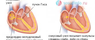
- Electrocardiography. ECG with dextrocardia is normal. In some cases, there are signs of arrhythmia such as ventricular fibrillation or intensification of the sinus node.
- Echocardiography (ECHOCG). Shows organic defects on the tissue side. Visualization allows you to identify all violations.
- MRI in controversial cases. Chest tomography is usually prescribed for adult patients. To detect dextroposition of an organ, tumors of the anatomical region.
In the future, regular examinations are indicated. Once every 3-6 months. In the absence of pronounced violations, the frequency of preventive examinations is reduced. A variant of the physiological norm is stated.
The speed and adequacy of the child’s physical and mental development are also taken into account. Respiratory activity and the functioning of the digestive tract may be disrupted later.
The maximum likelihood of severe complications occurs before 2 years of life.
Diagnosis of a pathological disease
Most cases of dextrocardia are diagnosed using an electrocardiogram (ECG) and chest x-ray.
An ECG with inverted or reversed electrical waves usually indicates dextrocardia.
Once a doctor suspects dextrocardia, they may use computed tomography (CT) or magnetic resonance imaging (MRI) to confirm the condition.
Cartagena syndrome
Cartagena syndrome is when dextrocardia situs inversion is accompanied by primary ciliary dyskinesia, an inherited condition in which the cilia that help move mucus become immobile.
Approximately 20% of patients with dextrocardia also suffer from Cartagenere syndrome.
Dextroversion
Dextroversion occurs when the heart is incorrectly positioned further to the right and rotates to the right. The right ventricle usually follows the left, while the left remains left. This complication is diagnosed using an electrocardiograph (ECG).
Dextroposition
Dextroposition occurs when the heart becomes dislocated or moved to the right. Unlike dextrocardia, other organs are not mirrored.
This disease is usually associated with acquired diseases of the lungs, diaphragm, or pleura (the membranes surrounding the lungs).
Surgery and muscle damage or strain can also contribute to the development of dextroposition.
Doctors often use an ECG test to distinguish dextroposition from dextrocardia [KG3].
Large Vessel Rearrangement (TGA)
TGA occurs when the main vessels of the heart flow backward due to reversal of the heart chambers. This is a fairly rare disease.
Congenital corrected transposition of the great arteries (CCTGA).
This condition occurs when the lower half of the heart unfolds, especially the lower ventricles and connected valves.
The condition is even rarer than TGA, and the cause is still unknown. About 0.5 to 1 percent of all children born with heart disease have CCTGA, according to the Adult Congenital Heart Association.
Tricuspid atresia
This is a congenital defect in which the tricuspid valve does not develop. The tricuspid valve prevents blood from the right ventricle from flowing back into the right atrium.
Single-, double- and bicuspid ventricles or bicuspid ventricles
These cases occur when the aorta, which normally transports oxygenated blood from the left ventricle to the rest of the body, and the pulmonary artery, which carries deoxygenated blood from the right ventricle to the lungs, join in the same (left or right) ventricle.
pulmonary stenosis or atresia
It occurs when the heart's pulmonary (pulmonary) valve, which allows blood to leave the heart, becomes narrowed (stenosis) or fails to develop (atresia).
Heterotaxis
Heterotactic syndrome occurs when mirrored internal organs do not develop or function properly. Depending on the organs involved and the severity of the disease, heterotaxy can be life-threatening.
Endocardial cushion defect
This is a congenital heart defect in which the tissues separating the chambers of the heart do not develop, interfering with blood flow. This condition is also known as atrioventricular canal defect.
When the first signs of abnormal heart development appear, the doctor must prescribe a series of diagnostic procedures. The most common methods for diagnosing this disease include:
- general examination of the patient, auscultation and percussion. These methods make it possible to listen to the beats of the upper part of the heart on the right side. When palpating internal organs, it is necessary to remember that organs can also be located symmetrically;
- chest x-ray, which can be used to observe the abnormal position of the heart;
- ECG of the heart. An ECG with dextrocardia gives a picture as if the diagnostician made a mistake and installed the electrodes incorrectly. That is, the first lead will give a mirror image of the first, and the second and third will seem to be swapped. That is, the second lead will correspond to the third and vice versa;
- Ultrasound, CT, MRI;
- if necessary, the doctor may also prescribe an angiocardiogram or cardiac catheterization.
Treatment
It is not required as such. With dextroposition, surgical treatment of the underlying disease is indicated: total excision of the tumor, drainage of the chest, removal of fluid, restoration of the anatomical integrity of the lungs.
Actually, the congenital mirror arrangement of the muscular organ without symptoms does not imply correction.
Heart failure is treated with drugs based on glycosides, antiarrhythmics. A highly qualified specialist is required. Because many medications are contraindicated for young children.
It is mandatory to correct breathing disorders, liver function and digestive tract problems in general. Cardioprotectors are used to restore metabolic processes in the myocardium, hepatoprotectors.
In the vast majority of cases, no special treatment is needed. Regular examinations for dynamics are indicated.