Diagnostics
Postthrombotic disease of the lower extremities is detected based on an external examination by a doctor, using instrumental examination methods and anamnesis data.
In the latter case, the patient is interviewed and the history of the previous illness is studied - if the patient was treated for thrombosis, the likelihood of PTFS is very high. Using duplex scanning, the condition of the venous wall, the speed of blood flow, the evacuation of blood and its outflow from the extremities are revealed. Also, ultrasound, passing through hard and soft tissues, provides information about the presence or absence of blood clots.
As an addition to the diagnosis of PTFS, the patient may be prescribed an x-ray using a contrast agent. Once the disease is confirmed, appropriate treatment is prescribed.
Etiology and pathogenesis
Postthrombotic disease develops after thrombosis, since the veins can no longer fully recover and irreversible consequences occur, provoking the development of pathology. As a result, the vessel is deformed, the venous valves are damaged - their function is reduced or completely lost.
The main reasons for the development of PTPS cannot be described point by point, since the formation of post-thrombophlebitis syndrome leads to one persistent disorder - thrombosis of a venous vessel. This disease leads to blockage of the vein and disruption of blood flow. As the treatment continues, after a few days the blood clot begins to gradually dissolve, and the damaged vessel fills with blood again.
But at this stage there is one peculiarity - after restoration, the vein is no longer able to fully perform its functions - it is deformed, its walls are not so smooth, and the valve apparatus functions poorly. All this leads to stagnation and the development of insufficient pressure in the venous system of the extremities. Blood is not discharged through perforating veins from deep vessels to superficial ones - therefore postthrombophlebitis syndrome affects all vessels of the lower limb.
Over time, the subcutaneous and internal veins expand, a compression drop in pressure, a slowdown in blood flow and the appearance of new clots. As a result, the disease becomes chronic and constant signs and symptoms appear that bother the patient.
How blood clots block veins and damage valves
Blood clots form in a vein just as a dam forms in a river bed. Just like water floods fields during a flood. Swelling of the leg also occurs due to thrombosis, and just like fields, it can only be drained through streams outside the main riverbed. Over time, changes occur and these small venous channels enlarge and protrude through the skin, increasing blood flow. This explains why a person who has had deep vein thrombosis has dilated saphenous veins. In some cases, these veins are mistaken for varicose veins, and if the valves in these veins work normally and they are removed, the condition of the leg may worsen. That is why any person needs an ultrasound duplex scan before undergoing surgery for varicose veins after deep vein thrombosis. Since the small outflow channels are enlarged, leg swelling decreases. At the same time, several mechanisms occur in the thrombosed vein and the blood clots are broken down through a process called thrombolysis.
Thrombosis and thrombolysis
This process can be divided into several stages:
- To prevent blood loss, a blood clot forms on the damaged vascular wall, partially or completely blocking the lumen of the vessel (the size of the clot depends on the nature of the damage);
- partially or completely blocked blood flow reduces the load on the vessel and makes it possible to fully restore damaged tissues;
- after healing of the damaged area, the thrombolysis mechanism is launched, which is necessary to restore vascular patency;
- under the influence of blood-thinning enzymes, the blood clot dissolves and full blood flow is restored.
These mechanisms are typical for a healthy person when he or she receives an injury, but sometimes this is also possible during the initial stages of thrombosis. Natural thrombolysis during thrombosis can occur by changing the diet (including blood thinning products in the menu), but only if the formation has a loose blood structure.
But in most patients, the formations that narrow the vascular lumen consist not only of platelets; atherosclerotic deposits are additionally attached to them and fibrinous threads are deposited on them. The body can no longer destroy such a dense structure on its own, and medications are used for this.
Indications and contraindications for the treatment method
- Acute arterial embolism - blockage of an artery by a blood clot brought from other places. The most common procedure is open embolectomy with a Fogarty probe through an approach below or above the blockage.
- Arterial thrombosis against the background of atherosclerotic plaques (atherothrombosis). It is dangerous to use a Fogarty probe in this case, as the artery wall may collapse. Rotarex allows you to remove thrombotic masses from an artery affected by atherosclerosis without damaging the vascular wall. In addition, soft atherosclerotic plaques are cut off. After removing the thrombus, angioplasty and stenting of the affected arteries are necessary.
- Removal of blood clots from vascular prostheses. Can be performed from open approaches using various modifications of Fogarty catheters. Most often, blood clots are removed from large vascular prostheses (aorto-femoral) as part of repeated operations.
- Removal of blood clots from previously installed stents. Stent thrombosis often requires repeated interventions. It is not possible to use a Fogarty balloon to remove blood clots from stented areas of arteries, as it will rupture against the stent or damage the arterial wall. For such thromboses, it is much more effective and safer to use Rotarex technology.
Surgical recanalization
The following types of surgical recanalization are performed:
- Removal of a blood clot is carried out minimally invasively, using endovascular therapy methods. An incision is made under local anesthesia, a catheter is inserted into the damaged vessel and, under the supervision of the operating physician, it is brought to the location of the thrombus. Next, the clot is captured and removed from the vein.
- Bypass surgery is used when the clot cannot be removed. A bypass path of blood flow is formed. The material used is your own vessels - veins taken for plastic surgery, or synthetic analogues.
- Ligation involves applying a ligature above and below the site of the clot, blood flow is redistributed among small arteries and veins.
- Stenting is the insertion of a balloon that dilates the vessel. Blood circulation improves and platelet aggregation on the affected wall decreases, but such an operation is advisable only if thrombus formation is gradual.
Surgery is not always performed to restore blood flow. Recanalization of the umbilical vein is performed to provide access to the liver and gallbladder in case of their pathology. Infusion solutions are administered through a catheter; in case of purulent damage to organs, antibacterial agents are delivered to the site.
Recanalization of a thrombus is often a lengthy process that requires the attention of not only doctors, but also patients. To achieve a better result, as well as to prevent further thrombus formation, the patient must reconsider their lifestyle, follow a diet and promptly consult a doctor at the first symptoms of the disease. Advances in modern medicine can significantly increase the likelihood of a full recovery.
Advantages of treatment at the ISC
Our clinic uses both traditional technologies and minimally invasive modern methods to remove blood clots.
- Fogarty catheter thrombectomy is a traditional method of removing blood clots from an artery using a balloon probe. The principle is to pass the probe in a folded state through the thrombus, followed by inflating the balloon and pulling it out along with the thrombotic masses.
- Rotarex is a technology for removing chronic blood clots from the arteries. The principle is based on the splitting of blood clots by a special probe head rotating at high speeds and its simultaneous suction by a special spiral mechanism (presented in the film). The technology opens up unlimited possibilities for treating severe vascular pathology without incisions.
- Aspirex is a similar technology for removing fresh blood clots from arteries and veins. Suitable for the thinnest vessels, does not damage venous valves. A special spiral suction atraumatically removes all blood clots from the vessel if the thrombosis period does not exceed 2 weeks.
Vitamins and nutrition to strengthen the walls of blood vessels
Have you been struggling with HYPERTENSION for many years without success?
Head of the Institute: “You will be amazed at how easy it is to cure hypertension by taking it every day...
In addition, it is useful to eat porridge from various cereals - oatmeal, buckwheat, corn and rice. It would be better to replace pasta with porridge.
How can we strengthen the walls of blood vessels? For this, it is also useful to eat legumes such as peas, beans, lentils, and soybeans. Soy is an indispensable product for keeping the walls healthy, since it contains all the minerals and compounds required by the body that help remove cholesterol from the body.
There are also vitamins to strengthen the walls of blood vessels. Vitamin P is very important, which is extremely effectively absorbed by the body in combination with vitamin C. Vitamin Z reduces fragility and restores elasticity to the walls. It is for this reason that your daily diet must include foods that are rich in this vitamin compound.
Especially invaluable plant products for strengthening the walls of blood vessels are onions, garlic and eggplants. They rid blood vessels of excess fatty deposits and free the walls from fragility. The active substances contained in cucumbers are also effective.
As for fruits, grapefruit is the leader among citrus fruits; among berries it is worth noting red and black currants, as well as chokeberry. If it is impossible to eat fresh fruits and vegetables, it is better to include green tea, chokeberry and rose hip decoctions in your diet.
In order to strengthen it, it is recommended to harden it with a contrast dousing. The temperature difference and water pressure on the walls of the vessel trains the cardiovascular system well and develops a normal response to climatic, seasonal and weather changes. In addition, water procedures have a positive effect on the nervous system. There are also drugs that strengthen the walls of blood vessels.
See the continuation of this article here: strengthening blood vessels part 2
Recanalization of fallopian tubes and umbilical vein
Recanalization of the fallopian tubes is the restoration of their patency by removing connective tissue adhesions in their lumen.
This is a mechanical obstacle that often causes female infertility.
The process is asymptomatic and develops after previous infectious processes, abortions, or prolonged use of spirals.
This manipulation can be performed on an outpatient basis. A catheter is inserted and, under camera control, adhesions are dissected.
Through the restored umbilical vein it is possible to administer infusion solutions for a long time during operations on the liver and biliary tract.
For purulent diseases and abscesses, antibiotic solutions are administered.
The technique is contraindicated in case of local inflammatory processes, kidney tumors, compressing the portal vein.
Determination of vein recanalization and options for solving the problem
When they first contact a specialist and are offered to carry out recanalization, patients are overtaken by fear due to a lack of understanding of what will be done to them. You need to know that recanalization of veins after thrombosis is the restoration of vascular patency, which can be achieved in various ways. There are 3 main methods of restoring venous patency:
- Independent, or natural.
- Conservative or medicinal.
- Operative or surgical.
Under normal conditions, the formation of a blood clot is a protective reaction of the body, which is aimed at preventing the development of bleeding.
Under certain conditions: blood thickening, increased activity of the coagulation system, hereditary predisposition, impaired venous outflow, this process becomes pathological.
The resulting thrombus can partially or completely block the lumen of the vessel; when it breaks off, an embolism develops, which can cause death. If the blood clot is overgrown with connective tissue, it will not resolve on its own.
With a balanced functioning of the blood coagulation system, immediately after the formation of a blood clot, the fibrinolytic system is activated. Its activities are aimed at disorganizing and resolving the blood clot using special substances. This type of thrombus destruction is possible in the initial stages, while it has a loose structure.
Medical recanalization is carried out in the absence of a threat of blood clot rupture or partial blockage of the vessel, which cannot lead to loss of function of a vital organ. There are several groups of drugs used separately or in combinations
The list of groups, main representatives and the effect they have are presented in the table.
| Group of drugs | Name | Action provided |
| Anticoagulants of direct and indirect action | Unfractionated heparin, low molecular weight heparin, warfarin | They have little effect on the resorption of an existing blood clot, but prevent its growth and increase in diameter and length |
| Antiplatelet agents | Aspirin, Curantil | Inhibits the process of platelet aggregation, that is, gluing them together |
| Fibrinolytic agents | Streptokinase, Alteplase | Quickly dissolve a blood clot |
| Angioprotectors | Detralex, Venarus | They do not take part in the resorption of a blood clot, but inhibit thrombus formation |
In situations where there is no serious danger to life, direct-acting anticoagulants - heparins - are more often used.
The duration of therapy reaches a year or more. Additionally, antispasmodics, antioxidants, and drugs that improve blood rheology are prescribed.
What is this – surgical recanalization of veins? Surgical methods are used in cases where it is necessary to quickly eliminate the problem, with complete occlusion of the vessel, with a high risk of blood clot rupture and the development of embolism. Several methods are used:
- thrombectomy – removal of a blood clot from the lumen of a vessel. In 1946, the first successful operation of this type was performed, and today they are performed in all major surgical centers in the world;
- bypassing - creating bypass paths;
- stenting – installation of a special stent (a device that widens the lumen of a vessel), which restores normal blood flow.
The choice of surgical treatment method is carried out by a vascular surgeon based on the results of additional research methods. The location of the thrombus, its prevalence, and the condition of other vessels are taken into account.
What can happen if postthrombotic syndrome occurs?
Changes in the skin occur, the addition of induration, eczema, hyperpigmentation and, as a finale, trophic ulcers are sometimes very large, and the leg may look like an inverted bottle of champagne. What can help? The best thing is to prevent possible deep vein thrombosis. But it’s like the proverb: “It’s better to be rich and healthy than...”. If you have postthrombotic syndrome, only three treatment methods can help: compression
(compression stockings, tights, bandages, in combination with specially designed physical therapy),
drug therapy
(phlebotonics and disaggregants) and valve surgery (various options
for restoring the valve apparatus
with preventing the discharge of blood through incompetent perforating veins). No other methods have yet been invented. In order to help patients with post-thrombotic syndrome, knowledge, patience and modern treatment methods are needed - this is, first of all, a method of restoring the valve apparatus through a low-traumatic operation without the use of incisions. We are ready to help you.
Vein recanalization. What is this Classification?
Recanalization is the process of restoring the patency of a vessel whose lumen is closed by a thrombus. Vein recanalization occurs in one of three ways:
- natural;
- medicinal;
- surgical
Natural recanalization is a physiological process. It occurs under the influence of aseptic fibrinolysis. The clot resolves on its own in almost half of the cases. In addition to the destruction of the blood clot, its revascularization is possible: it grows with microvessels and collagen structures. The patency of the vessel is restored and the destruction of the clot is accelerated. Independent removal of blockage is possible at the stage of a “loose” blood clot. When it is overgrown with connective tissue, fibrinolysis is difficult.
However, sometimes the body cannot cope with the dissolution of a blood clot: blood thickening, increased clotting activity, genetic predisposition, and impaired venous circulation complicate this process. The resulting clot blocks the lumen of the vessel and can cause embolism, a life-threatening condition. In these cases, medical recanalization is indicated.
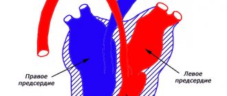
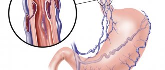
Deep vein thrombosis of the lower extremities
The deep venous system of the lower extremities plays a major role in the outflow of venous blood from the lower extremities, normally providing the outflow of 80-90% of the blood.
When deep vein thrombosis occurs in the lower extremities, the outflow of most of the blood from the lower extremities is hampered. Deep vein thrombosis is a condition that occurs as a result of the formation of a blood clot in the lumen of the veins of the lower extremities.
This condition is life-threatening due to the possibility of a blood clot breaking off and migrating through the bloodstream to the pulmonary artery, which in this case leads to pulmonary embolism.
Deep vein thrombosis is one of the main causes of pulmonary embolism. In turn, approximately a third of all cases of sudden death are caused by pulmonary embolism.
Causes of deep vein thrombosis
Various reasons lead to the occurrence of deep vein thrombosis, including limb injuries, recent surgical interventions, cancer, chemotherapy treatment, prolonged immobilization and physical inactivity, hematological diseases, varicothrombophlebitis of the superficial veins, taking hormonal contraceptives, pregnancy and the postpartum period, obesity, old age and etc.
Symptoms of thrombosis
The clinical picture is directly proportional to the extent of the thrombotic process. In the initial stage of thrombosis, pain in the lower limb, swelling, and fever occur.
In cases of total thrombosis of the deep system with a transition to the iliac veins, blue phlegmasia may develop, which is characterized by a violation of the general condition, high temperature, a change in the color of the skin to a purple hue, an increase in the circumference of the limb several times and severe pain.
There are several stages of the thrombotic process: the stage of thrombus formation, the stage of organization and the stage of recanalization. Thrombosis is most dangerous in the initial stage, when thrombotic masses are not yet fixed to the vein wall. During this period, the likelihood of thromboembolism is highest.
Subsequently, the blood clot is organized and fixed to the wall of the vein. In certain cases, a floating thrombus occurs - a freely “dangling”, not fixed top of the thrombus, posing a threat of detachment.
After time, recanalization of the thrombus occurs - resorption of the thrombus with restoration of the lumen of the vein to one degree or another. But, unfortunately, the valves that regulate unidirectional blood flow permanently die, which leads to the development of postthrombophlebitis syndrome.
Treatment methods for deep vein thrombosis
Treatment methods depend on the stage of the disease, the state of the thrombotic masses and the extent of the thrombotic process. Treatment is predominantly conservative; in some cases, surgical treatment is indicated.
The earlier treatment is started, the more favorable the prognosis - the risk of thromboembolism is greatly reduced, further spread of the blood clot stops, recanalization (restoration) of the vessel lumen occurs to a greater extent, and therefore the manifestations of postthrombophlebitic syndrome are minimized for the rest of life.
https://www.youtube.com/watch?v=9R8tIqtxOPM
If there are complaints, clinically and with the help of instrumental research methods, it is possible to make a correct diagnosis and prescribe a course of necessary treatment, and in some cases, save the patient’s life.
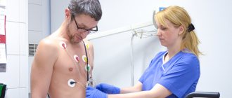
At ACMD-Medox you will be consulted by a vascular surgeon; if necessary, you will undergo ultrasound diagnostics of blood vessels (duplex scanning of blood vessels) and other instrumental studies.
Remember! Early contact with a vascular surgeon contributes to more effective treatment and a better long-term prognosis.
The valve apparatus is not restored
The prevalence of acute venous thrombosis is very high and amounts to about 2%. Treatment of any deep vein thrombosis of the lower extremities ends in 80% of cases with post-thrombotic changes in the vein, loss of the valve apparatus and the so-called post-thrombotic syndrome. The patient is concerned about various sets of complaints. And above all, swelling, pain, trophic disorders - skin induration, trophic ulcers that do not heal for years. The main cause of these disorders is the loss of valve apparatus in the veins, which prevents blood from flowing back through the veins. It would seem that the lumen of the vein is restored after the treatment, the process of recanalization occurs, but the valve apparatus is not restored. Dynamic hypertension develops, causing all of the above complaints. For many years, surgeons have been trying to find a way to restore normal venous blood flow after acute venous thrombosis and vein recanalization, offering various surgical interventions and conservative treatment methods. But until now, none of these methods have been able to significantly improve the quality of life of this category of patients and achieve long-term remission. Now, finally, we have seen a method that allows us to get the result. This is a method of restoring the valve apparatus through a low-traumatic operation without the use of incisions. In order to understand what happens after deep vein thrombosis, it is necessary to understand how the outflow of blood occurs through the venous system of the limb.
Methods for diagnosing vascular damage
They resort to the use of laboratory and instrumental diagnostic methods. For laboratory analysis, venous blood is used, a coagulogram, or hemostasiogram, is examined. It reflects the activity of the coagulation and anticoagulation systems. The level of fibrinogen, thrombin, prothrombin, prothrombin index and activated partial thromboplastin time (aPTT) are determined. Each of these indicators is important and diagnostically valuable.
In routine practice, non-invasive, that is, not requiring damage to integrity, instrumental diagnostic methods are used. This is vascular ultrasound with Doppler sonography, vascular angiography with contrast agent, and, if necessary, MRI with contrast.
How the treatment method works
Removal of blood clots using a Fogarty probe
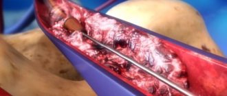
An open thrombectomy using a Fogarty probe is performed through the incision. The incision is usually made below the site of thrombotic blockage of the artery, since the removal of blood clots is easier through the blood flow. If the patient has thrombosis of the aorta or iliac arteries in the abdomen, then the incision is made in the groin area; if thrombosis is in the popliteal artery, then the access is made on the lower leg or foot. After isolating and assessing the condition of the artery, a 3-5 mm incision is made on it, into which the Fogarty balloon probe is inserted in a compressed state. For better elasticity, a metal conductor is installed in the probe, which is removed after passing the probe above the blood clot. A syringe is attached to the probe, through which the balloon is inflated. After this, the surgeon removes the inflated balloon, which pushes the thrombotic masses towards the cut artery. Pressure of the blood above the clot helps the process of removing the clot. After removing the clot, the procedure is repeated to ensure complete removal. Vascular clamps are placed on the artery and the hole in it is sutured with a vascular suture. After this, the wound is sutured layer by layer.
This simple operation helps well with acute blockage of intact arteries by thromboembolism (a blood clot brought from the heart or from larger arteries), however, if thrombosis of arteries affected by atherosclerosis occurs, then thrombectomy with a Fogarty probe becomes impossible or even dangerous, since the probe easily breaks on sharp edges atherosclerotic plaques or can get caught on the plaque and tear it. Therefore, for such cases, we use other methods of removing blood clots.
Removing blood clots with a Rotarex Straube probe
The Rotarex Straube probe allows you to clear the artery of blood clots without resorting to a surgical incision, through a puncture of the artery. At the same time, this probe does not damage the walls of the artery, carefully removing thrombotic masses and freeing the lumen of the vessel. The principle of operation is to crush the clot with a rotating probe head and suck its particles into the lumen of the probe and remove them from the lumen of the vessel into a special bag. The use of Rotarex Straube is possible even in cases of chronic thrombosis due to atherosclerotic and diabetic arterial lesions. The Rotarex Straube probe clears blood clots from the vessel, but does not remove atherosclerotic plaques, therefore, to restore the lumen of the vessels, angioplasty and stenting of narrowed arteries of the leg are performed if necessary. The Rotarex Straube endovascular thrombectomy procedure is performed without incisions, only through a puncture of the artery wall, so the main method of pain relief is local anesthesia. Probe thrombectomy is indispensable when removing blood clots from intestinal arteries with mesenteric thrombosis. Considering the severe general condition of patients with this pathology and the difficulties with access to the mesenteric arteries, thrombectomy with a probe from the initial parts of the superior mesenteric artery is the preferred method of restoring blood circulation in the intestine.
Drug recanalization
If there is no life-threatening condition (risk of blood clot rupture or blockage of vital vessels that has already occurred), then conservative treatment is first applied. Depending on the location of the thrombus formation, a medication is selected.
It can be:
Fibrinolytics. Drugs of this group (Alteplase, Streptokinase) are administered intravenously and promote rapid thrombolysis, but their use causes many adverse reactions. The need for rapid thrombolysis appears when vital arteries are blocked (heart attack or PE - pulmonary embolism).
- Angioprotectors. They hardly participate in the lysis process, but prevent further thrombus formation. These include drugs such as Detralex and Aescusan.
- Antiplatelet agents. Agents that reduce the ability of platelets to aggregate (stick together). The most famous drug is Aspirin, which is used in cardiology to prevent thrombotic complications. This group also includes Curantil, Thrombo-Ass, Tirofiban, etc. (the list of drugs is very long).
- Anticoagulants. Drugs that promote active blood thinning. The most famous representatives of this group are Warfarin and Heparin.
But if there is no life-threatening condition, the recanalization process occurs slowly and takes about six months (sometimes maybe more). Additionally, patients are prescribed vasodilators and medications that improve blood flow.
In most cases, if the disease was detected in a timely manner, the walls of the artery are completely cleared and restore their tone, and on the veins, a slightly expanded and slightly deformed area may indicate the presence of the disease after successful treatment.
Prognosis and complications
The prognosis for postthrombophlebitic vein damage is relatively favorable in cases where the patient adheres to the doctor’s basic recommendations - does not violate the treatment program and follows the basic rules for preventing relapses of the disease. With this approach, it is possible to achieve a maintaining optimal state for a long time.
If the rules of the health program are violated, the patient experiences complications in the form of poor circulation in the extremities, which can lead to gangrene, requiring amputation. The second serious complication is infarction of the brain or internal organs when a blood clot enters the general bloodstream.
Treatment and symptoms of thrombophlebitis of the deep veins of the lower extremities
The formation of blood clots in the lumen of the veins is quite common and is called acute deep vein thrombosis of the lower extremities. With this disease, 27–35% of patients experience thromboembolism of the arteries in the lungs.
Thrombophlebitis of the veins of the lower extremities occurs relatively rarely in healthy people, and the number of diseases increases every year
Therefore, the development and application of conservative treatment of the disease is an important task in vascular surgery.
Thrombophlebitis of deep veins
Vein thrombi occur for various reasons and develop with a normal epithelial layer on the vessel wall. Their formation begins in the veins of the leg - on their valves, where blood clotting factors accumulate due to the vortex of blood flow through the valve leaflets and in the area of the vein division.
Platelets provoke early thrombus formation by settling on the valves of the veins and at points where the integrity of the epithelial layer is compromised.
They attach to the endothelium, or the exposed collagen layer on the vein walls. The subsequent event is platelet aggregation, release of thromboplastin from the tissues and the appearance of a red thrombus.
The latter is retractable, capable of aseptic lysis and consists of fibrin, erythrocytes and platelets.
Subsequently, the behavior of the thrombus is influenced by the processes of fibrinolysis and coagulation.
The action of fibrinolysin leads to lysis within three to four days, most of the thrombus is destroyed, fragmented, dislodged and can move into the pulmonary arteries.
Subsequently, the formation may resolve without affecting the venous wall or be replaced by connective tissue if the thrombus is of significant size and the area of adhesion to the wall is quite extended.
With extensive varicose veins, the thrombus settles in the overlying veins or spreads to the perforating veins and deep veins of the affected surface of the lower limb. Deep venous thrombosis involves the spread of the thrombus to the femoral and popliteal veins, and the discharge of blood from them can stop thrombosis along the ascending line.
Clinic of the disease
Symptoms of thrombophlebitis of the deep veins of the lower extremities depend on the location of the thrombus and the degree of its spread and changes in venous patency (obstruction or stenosis of the lumen), the occurrence of collaterals. Clinical manifestations are varied - from an asymptomatic course of the disease to severe pain and extensive swelling, sometimes it ends in gangrene of the extremities.
Asymptomatic progression is typical for the case when venous outflow occurs without obstacles, while the situation is difficult to recognize and the indicators are characteristic of only one limb. Sometimes the first noticeable sign is only thromboembolism of the arteries in the lungs. Manifestations of the disease occur quickly - within two to three hours to two days from the appearance of a blood clot:
- the ankle, foot, and distal part of the lower leg swell;
- pain is felt when light pressure is applied to the lower leg muscles;
- pain appears in the calf when the foot is dorsiflexed and calms down at rest;
- at the site of the lesion, the lower leg becomes hot due to inflammation and increased blood flow;
- dilated superficial veins are observed;
- There is a difference in circumferential size between the affected and normal limbs.
What problems does scarring of veins cause?
First of all, this leads to leg pain and swelling. If the valves in the deep veins are damaged, blood flows in the opposite direction - to the foot. This causes an increase in pressure in the vessels at the ankle level, which leads to swelling. Many people with incompetent deep vein valves after venous thrombosis (the valves don't work) have pain when standing up. And this pain intensifies during the day and goes away if the person lies down again. Some people experience a feeling of blood overflowing in the lower leg due to incompetence of the deep veins.