The color of the hematoma can be different - from bright red to purple, most often it is heterogeneous - its edges are darker, bluish in color, and the inside of the hematoma is red. Below in the article you will find the causes of the disease; the doctors who treat him; necessary medical procedures for treatment; as well as general information about the disease, its localization, features of diagnosis of diseases and their treatment. However, we advise you to consult a doctor, because self-medication in 90% of cases is fraught with the disease progressing to the chronic stage with extremely unpleasant complications
Make an appointment and consultation
General information
For some reason, almost all people are sure that arterial blood is the type that flows in arterial vessels. In fact, this opinion is wrong. Arterial blood is enriched with oxygen, which is why it is also called oxygenated. It moves from the left ventricle to the aorta, then goes through the arteries of the systemic circulation. After the cells are saturated with oxygen, the blood turns into venous and enters the veins of the BC. In a small circle, arterial blood moves through the veins.
You may be interested in: Aromatase inhibitors: purpose and list of drugs
Different types of arteries are located in different places: some are deep in the body, while others allow you to feel the pulsation.
Venous blood moves through the veins into the CD and through the arteries into the MC. There is no oxygen in it. This liquid contains a large amount of carbon dioxide, decay products.
Function
Blood has specific and general functions. The latter include:
- nutrient transfer;
- transport of hormones;
- thermoregulation.
Venous blood contains a lot of carbon dioxide and little oxygen. This difference is due to the fact that oxygen enters only the arterial blood, while carbon dioxide passes through all vessels and is contained in all types of blood, but in different quantities.
Hematoma. general information
Hematoma is a condition characterized by the accumulation of liquid or coagulated blood inside the body, which occurs as a result of rupture of blood vessels and is localized in soft tissues. Hematomas can be small in size, or they can compress soft tissues and nearby organs. Hematomas form under the skin, mucous membranes, deep within the muscles, in the walls of internal organs, and in the brain.
When treating Hematoma, doctors at the BIOSS clinic use both time-tested and the latest developments and proprietary techniques.
Our clinic employs the best doctors in Moscow who have extensive experience in treating Hematoma
There are several classifications of hematomas:
Color
Venous and arterial blood have different colors. In the arteries it is very bright, scarlet, light. The blood in the veins is dark, cherry-colored, almost black. This is related to the amount of hemoglobin.
When oxygen enters the blood, it enters into an unstable combination with the iron contained in red blood cells. Once oxidized, iron colors the blood bright red. Venous blood contains many free iron ions, which is why it becomes dark in color.
WHEN YOU SHOULD SOUND THE ALARM, THE FIRST SYMPTOMS OF HEMATOMA
A hematoma gives its symptoms and signs almost immediately after injury.
- Firstly, the skin at the site of the hematoma is sharply painful.
- After a short period of time, the site of injury begins to swell, the tumor can spread significantly and interfere with movement (for example, with a hematoma on the ankle, the swelling may be such that it is impossible to move independently or step on the affected leg).
- After swelling, the site of hemorrhage quickly turns red. Patients feel internal tension in the area of the hematoma; it is hard to the touch.
The color of the hematoma can be different - from bright red to purple, most often it is heterogeneous - its edges are darker, bluish in color, and the inside of the hematoma is red.
Blood movement
When wondering what the difference is between arterial blood and venous blood, few people know that these two types also differ in their movement through the vessels. In arteries, blood moves away from the heart, and through veins, on the contrary, towards the heart. In this part of the circulatory system, blood circulation is slow as the heart pushes fluid away from itself. Valves located in the vessels also affect the reduction in movement speed. This type of blood movement occurs in the systemic circulation. In the pulmonary circle, arterial blood moves through the veins. Venous - through the arteries.
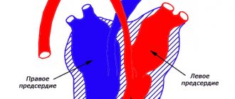
In textbooks, on schematic representations of blood circulation, arterial blood is always colored red, and venous blood is always colored blue. Moreover, if you look at the diagrams, the number of arterial vessels corresponds to the number of venous ones. This image is approximate, but it fully reflects the essence of the vascular system.
The difference between arterial blood and venous blood also lies in the speed of movement. The arterial is ejected from the left ventricle into the aorta, which branches into smaller vessels. Then the blood enters the capillaries, nourishing all organs and systems at the cellular level with useful substances. Venous blood collects from capillaries into larger vessels, moving from the periphery to the heart. When a fluid moves, different pressures are observed in different areas. Arterial blood pressure is higher than that of venous blood. It is ejected from the heart under a pressure of 120 mm. rt. Art. In the capillaries the pressure drops to 10 millimeters. It also moves slowly through the veins, since it has to overcome the force of gravity and cope with the system of vascular valves.
Due to the difference in pressure, blood for testing is taken from capillaries or a vein. Blood is not taken from the arteries, since even minor damage to the vessel can provoke extensive bleeding.
TYPES OF HEMATOMA AND THEIR TREATMENT
There are several approaches to the classification of hematomas.
Hematomas are distinguished depending on:
- the nature of bleeding - they can be arterial, venous and mixed;
- localizations – subcutaneous, intramuscular, intracranial, etc.;
- clinical signs - encysted, pulsating, simple.
In addition, situational hematomas are distinguished in the treatment, which require a special approach, for example, hematomas during childbirth, hematomas during pregnancy, etc.
An arterial hematoma is a hematoma that contains arterial blood in the cavity. As a rule, such hematomas are bright red, they are often diffuse - with a wide distribution on the surface. Venous hematoma occurs when there is compression and disruption of the integrity of the vein. These hematomas are bluish-violet in color, they are inactive and hard to the touch. The most common hematomas are mixed, when both arterial and venous blood enters the cavity.
A subcutaneous hematoma forms under the layer of skin and looks more like a bruise. They can form both due to injuries and due to various diseases - tuberculosis, syphilis, scarlet fever, lupus erythematosus. Often such hematomas form in people suffering from hemophilia. At the slightest damage to the vessel, spots appear on the skin. Subcutaneous hematomas can be of three degrees.
With a mild hematoma, its symptoms appear prolonged - approximately a day after the injury, while it absolutely does not interfere with the functioning of the organ on which it appeared. Painful sensations are weak and sometimes do not occur at all. If the hematoma is not complicated by anything, then it goes away on its own without any treatment. A moderate hematoma forms within three to four hours. In this case, the hematoma can partially disrupt the functioning of the organ on which it originated. A slight swelling and swelling of the soft tissues forms around such a hematoma. Apply ice and a pressure bandage to the site of the hematoma and contact a medical facility. A severe hematoma can occur with serious injury. In this case, the presence of a hematoma disrupts the functioning of organs. Hemorrhage forms quickly - literally within an hour you can notice a blue spot at the site of damage. Most often this is a subcutaneous hematoma, which is visible to the naked eye. Over time, the hematoma intensifies and can become intramuscular. In this case, the patient will feel numbness and soreness in the muscles. Such a hematoma requires a mandatory examination by a doctor and further treatment. If a hematoma is not treated, it can cause serious harm to the human body.
Intramuscular hematoma is characterized by the accumulation of blood in the muscles. In this case, the patient feels significant pain in the area of damage. Muscle function is impaired. In order to cure such a hematoma, it is necessary to consult a doctor. You may need to surgically open the hematoma and drain the cavity.
of intracranial hematomas - epidural, intracerebral, subdural, intraventricular.
Epidural hematomas are a collection of blood between the meninges and the skull bone. Most often, such hematomas occur near the temple; their development is associated with a traumatic moment (a blow to a stone, a blow to the head with a blunt object). With an epidural hematoma, the artery most often suffers, so such a hematoma is also arterial. At the site of the rupture, up to one hundred and fifty milliliters of blood quickly accumulates. The appearance of such a hematoma leads to compression of the brain. In this case, the patient loses consciousness for a short time, and then regains consciousness, but feels a headache, weakness, and vomiting. After several hours of improvement, a sharp deterioration occurs. Depending on the size of the hematoma, a state of coma can quickly occur. Heart contractions slow down, blood pressure drops, and the eyes stop responding to stimuli (except for the pupils). When such a hematoma is diagnosed, emergency surgery is prescribed to eliminate it.
Subdural hematomas are hemorrhages between the arachnoid and dura mater of the brain.
Venous blood collects in such hematomas, so they are also venous. Quite often, such hematomas are bilateral - the first occurs at the site of the impact, and the second - at the counter-impact. These hematomas have a larger area than epidural ones and can sometimes contain up to three hundred milliliters of blood. In the presence of such a hematoma, crisis phenomena in the patient may increase over the course of two days - hemiparesis, breathing problems, epilepsy, and bradycardia occur. The surgical treatment of such a hematoma consists of excision of the hematoma itself, restoration of the integrity of the bone and revision of the brain. In some cases, drainage is applied.
Intracerebral hematoma is very difficult to diagnose. Hemorrhage may occur slowly, increasing in volume day by day. Symptoms of intracerebral hematoma gradually appear. In some cases, immediate bruising may appear some time after the injury. The symptoms of such a hemorrhage depend on where it occurred - there may be hearing, speech, vision impairment, loss of consciousness, memory impairment, loss of sensitivity, or vice versa, hypersensitivity. Most often, such a hematoma is treated with special drugs that help it resolve. They are used if the volume of spilled blood is less than thirty milliliters. Otherwise, surgery may be necessary.
Intraventricular hematoma occurs when hemorrhage occurs in the ventricles of the brain. Treatment of such a hematoma depends on its size. If it is impossible to treat such a hematoma conservatively, then they resort to surgical intervention.
Bleeding
When providing first aid, it is important to know which blood is arterial and which is venous. These species are easily identified by their flow patterns and color.
With arterial bleeding, a fountain of bright scarlet blood is observed. The liquid flows out pulsatingly and quickly. This type of bleeding is difficult to stop, which is the danger of such injuries.
When providing first aid, it is necessary to raise the limb and compress the damaged vessel by applying a hemostatic tourniquet or pressing it with finger pressure. In case of arterial bleeding, the patient must be taken to the hospital as quickly as possible.
Arterial bleeding may be internal. In such cases, a large amount of blood enters the abdominal cavity or various organs. With this type of pathology, a person suddenly becomes ill, the skin turns pale. After some time, dizziness and loss of consciousness begin. This is due to lack of oxygen. Only doctors can provide assistance with this type of pathology.
With venous bleeding, dark cherry-colored blood flows out of the wound. It flows slowly, without pulsation. You can stop this bleeding yourself by applying a pressure bandage.