Phlebectasia is a disease of the veins associated with their expansion due to an inflammatory process. The risk of complications depends on the location of the pathology and the neglect of the process. Typically, the pathology does not lead to the development of severe consequences and is a cosmetic defect (in the case of damage to the jugular vein (JV), however, in severe cases, circulatory disorders may occur. In the early stages, the disease responds well to treatment, so it is important to consult a doctor in a timely manner and carry out a diagnosis.
What it is
The disorder is an expansion of the vein. In this case, the condition can be either congenital or acquired. If the first is diagnosed after birth, then the second can be caused by pathologies of the vein valves, reflux of blood from deep veins to the superficial, as well as disruption of the blood circulation process caused by tumors or scars.
The disease is accompanied by disruption of the valves and dilation of the veins. Literally from Latin the name is translated as “extension”.
Causes
Having analyzed what phlebectasia is, you can draw your own conclusions about what leads to the development of pathology. Doctors call the main reasons for the development of the disease:
- disruption of normal valve operation;
- problems with blood flow;
- presence of tumor or scars.
Phlebectasia of the esophagus occurs as a result of problematic blood flow in the hepatic veins. The pathology is provoked by cirrhosis of the liver or blood clots in the organs.
Why is this happening
Phlebectasia disrupts the functioning of valves and blood vessels. The regulation of venous blood flow is suspended. Clots appear. With a large number of such formations, dysfunction of the entire venous network develops.
If the jugular vein is dilated even slightly, it is manifested by the following symptoms:
- swelling of the cervical vessels, their enlargement;
- the appearance of a blue sac on the upper section of the vein;
- swelling of the neck;
- a feeling of tightness that occurs when turning the head;
- breathing problems;
- pain when touching the neck;
- loss of voice.
Signs of pathology depend on the stage:
- Swelling of blood vessels in the neck. The patient does not feel any discomfort. A sign of pathology is identified during a visual examination.
- Drawing pain. The patient's intravenous pressure increases if he makes rapid and sudden head movements.
- Acute pain of high intensity. The man's voice is hoarse. Breathing is difficult.
When the left or right internal jugular vein expands, disturbances in the activity of the circulatory system occur.
Phlebectasia can occur at any age. Probable reasons:
- Injuries to the ribs on the left or right, neck, spinal column, which leads to stagnation of unpurified blood.
- History of concussion.
- Osteochondrosis in a patient.
- Pathologies of the cardiovascular system. Phlebectasia affects people with heart failure, ischemia, and hypertension.
- Endocrine pathologies.
- Prolonged work at the computer.
- Benign and malignant tumors.
The disease takes time to develop. Even if a person has predisposing factors, this does not mean that he is already sick. It is necessary to monitor your health more carefully.
Predisposing factors include:
- insufficient development of connective tissue cells;
- hormonal changes in the body;
- back injuries, including fractures;
- intervertebral hernia;
- staying in an uncomfortable position for a long time;
- wrong diet.
Hormonal causes of pathology are more common in women. During puberty and pregnancy, there is a risk that the veins will swell.
Other factors include depression and stress. The jugular veins have nerve endings. If all is well, these endings form venous vessels of high elasticity. But when a person is stressed, intravenous pressure increases, which impairs the elasticity of the veins.
Other unfavorable factors include:
- alcohol abuse;
- smoking;
- eating foods with toxins;
- increased stress on the body - at the physical and mental levels.
Classification
Classified according to the location of the disease:
- phlebectasia of the jugular vein;
- esophagus;
- lower extremities.
By origin they are divided into:
- congenital phlebectasia;
- acquired.
Doctors distinguish stages of development based on the clinical picture.
- The first degree is detected during endoscopy by chance - the expansion does not exceed 3 millimeters, the lumens are not darkened, ectasia is not present or is small.
- The second degree is characterized by venous adjustment - strong expansion, for example, if the problem concerns the esophagus, then their volume occupies about a third of the organ cavity.
- The third stage is determined by enlargement and swelling of the veins, the presence of nodule formations, occupies more than half the volume of the esophagus, and there is a deterioration in the condition of the mucosa.
- The fourth stage threatens with thinning of the walls of the mucous membrane, and in other organs there are cluster-shaped formations and bleeding.
The prognosis is favorable in the first 3 stages. The neglected fourth can be fatal, especially phlebectasia of the IJV.
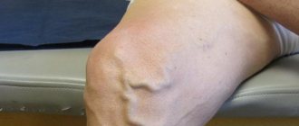
Phlebectasia of the jugular veins
With phlebectasis of the jugular vein, one of the branches is affected: internal, external or anterior. As a rule, the pathology is not dangerous, but is a cosmetic defect. However, with significant damage to the internal jugular veins (IVVs), which drain blood from the skull, complications may develop.
Causes of phlebectasis of the jugular veins:
- injury to the neck, skull, spine with the development of inflammation;
- violation of sterility when installing a catheter or performing injections;
- inflammation in tissues located near the jugular vein;
- penetration of drugs (for example, calcium chloride) into the tissues surrounding the vessels;
- venous congestion in injuries of the back and chest;
- being forced to be in an uncomfortable position;
- cardiovascular pathology associated with decreased elasticity of the vascular wall;
- the presence of neoplasms compressing the vessel.
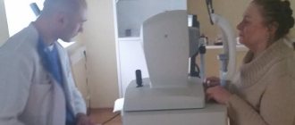
If symptoms of the disease appear, you should seek medical help. The doctor will refer the patient for additional examination to confirm the diagnosis.
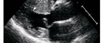
Diagnosis of phlebectasis of the jugular veins:
- Ultrasound;
- MRI and CT;
- phlebography;
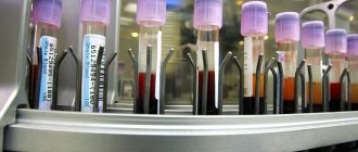
- lab tests.
Based on the results of additional diagnostics, treatment is prescribed. Non-advanced stages of the disease are amenable to conservative therapy. Anti-inflammatory drugs in the form of ointments (Ibuprofen, Indomethacin), antihistamines (Suprastin), and broad-spectrum antibiotics in case of bacterial infection are used.
In most cases, surgery is not required, but the patient must visit a doctor periodically to monitor the condition. In case of severe pathology and the risk of complications, one type of operation is performed:
- circumferential resection of extension;
- longitudinal resection;
- strengthening the vascular wall with a polymer mesh;
- resection with vein grafting.
Possible complications
The disease can either manifest itself or occur in a closed, latent form. Tumor formations appear, which increase in size over time and become tense. The skin underneath becomes thinner, and ulcerative processes begin.
This leads to varicose veins, and when squeezing or lifting the limbs upward, the unpleasant symptoms decrease. Phleboliths - round formations - are present in the later stages. The disease progresses slowly, pain, motor dysfunction and trophic problems are possible.
With intestinal pathology, pain occurs in the stomach and liver. As the disease progresses, the symptoms intensify, and the walls of the esophagus and stomach become thinner. This is fraught with disruption of the gastrointestinal tract.
Manifestations
Pathology of the esophagus occurs at stages 1-2 asymptomatically. The patient is not bothered by pain. Sometimes there is a burning sensation in the chest and shoulder blades, and belching with a sour taste. As the disease progresses, pain symptoms are observed in the chest, esophagus, stomach, and liver.
At stage 4, vomiting of blood occurs, and purple blood clots are present in the stool. Swallowing will be impaired. Constant loss of blood leads to anemia, a person feels weak, cannot work or move normally, loses weight, feels short of breath and has a strong heartbeat.
Phlebectasia in a child is usually diagnosed as congenital. Enlarged and tortuous venous ducts are noticeable and can be localized anywhere.
It appears either from birth or in the first months of life. The color of the skin changes, the integument becomes cyanotic. Next, tumor formations appear.
Phlebectasia of the esophagus
Venous stagnation is the main cause of the development of phlebectasis of the esophagus. Among the etiological factors, there are 2 main groups: hepatic and extrahepatic. Phlebectasia is provoked by cirrhosis, hepatitis, fatty degeneration of liver cells due to metabolic disorders, complications of infectious diseases (tuberculosis, syphilis), parasitic lesions, and hereditary pathologies. In addition, bulging of the esophageal veins occurs against the background of disruption of the cardiovascular system, increased blood clotting, compression of blood vessels, inflammation of the lymph nodes near the liver, and connective tissue pathology.
The peculiarity of phlebectasis of the esophagus is the difficulty of making a timely diagnosis. In the early stages, the disease has an asymptomatic course. As it progresses, the elasticity of the venous walls is impaired, they become brittle, resulting in the formation of ruptures, which leads to bleeding.
Phlebectasia of the esophagus goes through several stages:
- first stage: symptoms are not expressed, however, during instrumental examination it is possible to detect an expansion of the venous lumen by several millimeters, foci of ectasia are either absent or appear sporadically;
- the second stage is characterized by the appearance of the first symptoms of phlebectasia; instrumental diagnostics reveal tortuosity of the veins, the walls are enlarged in the lower sections, the mucous membranes are not changed, the vascular lumen is expanded to 1 cm;
- third stage: the veins are thickened by more than 1 cm, fill the cavity of the esophagus by two-thirds, the nodes are detected visually, reflux develops;
- the fourth stage is the most severe, bleeding develops, the mucous membranes are damaged, the nodes are located in clusters, there is no lumen of the esophagus.
The first and second degrees are the initial stages of the development of pathology, which are detected only during an instrumental examination, the third and fourth stages are characterized by distinct signs, requiring immediate initiation of treatment.
Symptoms of esophageal phlebectasis:
- discomfort and chest pain;
- heartburn, belching;
- difficulty swallowing dry food;
- shortness of breath not associated with physical activity.
As the disease progresses, without medical assistance, a dangerous complication may develop - bleeding. Its presence is indicated by the red tint of the vomit (in some cases they take on the color of coffee grounds), feces may become liquid and black, shortness of breath intensifies, the patient feels fatigue, fatigue, heart function is disrupted, and a decrease in the concentration of iron in the blood is detected.
The presence of such symptoms allows one to suspect a pathology of the esophagus, but additional diagnostic tests are necessary to confirm the disease.
Diagnosis of phlebectasis of the esophagus:
- laboratory tests can detect signs of bleeding: a decrease in hemoglobin, the number of red blood cells, occult blood when performing a coprogram;
- with the help of instrumental methods it is possible to identify the cause of the development of phlebectasia: ultrasound of the abdominal organs, esophagoscopy, MRI and CT, radiography of the esophagus with contrast;
After confirmation of the diagnosis, all treatment measures are aimed at preventing bleeding or stopping it. In addition, it is important to eliminate the cause of phlebectasia. The main directions of treatment: surgical and medicinal. In addition, the doctor recommends changing your diet.
If the disease is diagnosed at stages 1-3, conservative therapy is carried out; at stage 4 of the pathology, surgery is indicated.
Drug therapy:
- beta blockers to lower blood pressure;
- vitamin therapy;
- colloidal solutions;
- hemostatic agents;
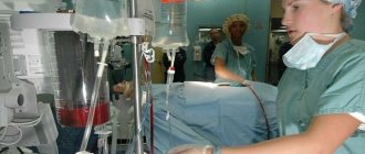
The operation is performed in severe cases when complications have developed and drug treatment is ineffective. One of the surgical techniques is selected:
- electrocoagulation – removal of damaged areas of a vessel using current;
- installation of a bandage - special rubber discs over the damaged vein;
- endoscopic doping - ligation of blood vessels using special attachments or nylon threads;
- sclerotherapy - injection of a special solution into the vessel that glues damaged elements;
- bypass surgery - installation of a stent connecting blood vessels;
- balloon tamponade - compression of blood vessels using a probe.
Diagnostics
If symptoms of pathology are found, contact a physician. Based on anamnesis collection, palpation, and examination, he will make a primary diagnosis and determine the localization of varicose veins.
Laboratory tests include general and biochemical blood tests. It is carried out as prescribed by the therapist.
But it is not always possible to establish an accurate clinical picture and select the correct course of treatment based on laboratory tests and anamnesis.
They also resort to instrumental diagnostic methods:
- endoscopy;
- x-ray;
- scanning of the vascular network;
- esophagoscopy.
Naturally, the principle of collecting tests, prescribing, and complex of examinations change depending on the localization of tumor processes. For consultation, if problems are found in the gastrointestinal tract, contact a gastroenterologist, lower extremities - a phlebologist. In any case, pathology requires a comprehensive examination, which is carried out by several doctors.
First aid for attacks is to stop bleeding. The doctor selects the optimal treatment plan to not only stop it, but also to prevent the next one, which is very likely to happen.
Then drugs that relieve inflammatory processes are included. The purpose depends on the area in which the dilated veins are localized.
Choose:
- antacids, which reduce acidity in the stomach;
- diuretics;
- vasoconstrictors;
- hemostatic drugs.
Beta blockers and vitamin kits are used as therapy. To restore blood flow and blood components, solutions are taken. In some cases, especially if the disease progresses and is at stages 3-4, a blood or plasma transfusion will be required.
In case of severe damage to the venous network, surgical intervention is resorted to. It is carried out using various methods, in particular, electrocoagulation, bandaging, and ligation. In case of cirrhosis, bypass surgery is chosen.
After surgical intervention on the organs of the gastrointestinal tract, the patient is prescribed a lifelong diet that prevents an increase in the acidity of gastric juice.
Features of the disease
Depending on the location, dilatation of the veins of the lower extremities, rectum (hemorrhoids), esophagus and jugular veins are distinguished. Vascular damage occurs under the influence of various factors, including: inflammatory changes, dysfunction of valves, changes in blood flow.
When exposed to one or more pathogenetic factors, an inflammatory reaction is triggered in the vascular wall, phlebitis, periphlebitis, and thrombophlebitis develop. In the presence of microorganisms in the lesion, an infectious process occurs; in their absence, an aseptic process occurs (this form is more often provoked by the influence of drugs).
The expansion of the jugular vein does not depend on the gender and age of the patient; in addition, it can be congenital. Men over 50 years of age are more susceptible to damage to the vessels of the esophagus.
Who's at risk
Adult men (over 50 years old) are at risk. According to statistics, women suffer from phlebectasia 3-4 times less often than men.
Phlebectasia at stage 1 is not dangerous, but at stage 3-4 it leads to consequences. The symptoms are not pronounced and can be confused with other diseases. If there is any suspicion, contact a therapist.
Preventive measures include limiting physical activity, choosing the right diet and taking vasoconstrictor medications.
The material was prepared specifically for the website venaprof.ru, edited by pharmacist M.N. Aleksandrova.