When we talk about ultrasound of the vessels of the lower extremities (ultrasound Dopplerography), we mean Dopplerography, Doppler scanning, Doppler scanning. Most people do not understand and do not see the difference in duplex scanning of veins and arteries. In this article I want to briefly provide information for those interested in this research. All vessels in the human body are divided into veins and arteries. Arteries are vessels that carry blood from the heart to the organs. Veins are vessels that carry blood from organs to the heart. Briefly about the structure. Arteries consist of three layers: internal, intermediate and external. These layers help maintain elasticity under high pressure. Veins also consist of several layers: internal (soft (arteries do not have it)) and due to which they are elastic, and external (elements of muscle tissue).
Based on different anatomical structures, different diseases arise. Using ultrasound, the following main diseases can be identified: -Atherosclerosis; - Varicose veins; -Deep vein thrombosis; - Thrombophlebitis.
When performing an ultrasound scan of the veins of the lower extremities, the doctor sees the following veins: iliac, subcutaneous, popliteal, femoral, tibial, plantar, etc.
When performing an ultrasound scan of the lower arteries: all branches of the large arteries, femoral, iliac, tibial and foot.
During the examination, the doctor evaluates the shape of the vessels and their location, the diameter of the lumen, the presence of blood clots and their density, size, structure and flotation (free movement through the blood flow). The speed of blood flow, the uniformity or disturbance of the main blood flow are also measured. The presence or absence of reflux (reverse current movement), vascular valves - their condition, the immediate condition of the vascular walls. The presence or absence of plaques in atherosclerosis. And the presence of connections between veins and arteries.
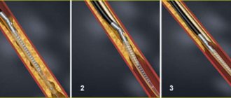
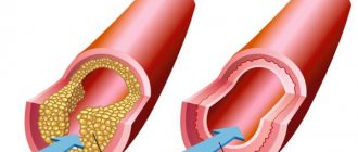
The main disease of the arteries of the lower extremities is atherosclerosis. Atherosclerosis is most often a chronic disease associated with the deposition of cholesterol and lipoproteins in the lumen of blood vessels.
Ultrasound of leg vessels: the essence of the method
The idea of using ultrasound to study blood vessels was introduced after the substantiation of the Doppler effect, named after the Austrian physicist. It means a change in the frequency of waves perceived by the observer. Ultrasound of the vessels of the lower extremities is an informative, accessible and safe method. Therefore, it is used to identify numerous problems.
Types of research
Ultrasound is a non-invasive diagnostic method that can visualize small details and see structural changes. It has virtually no contraindications and is painless. To determine vascular diseases, three types of studies are carried out: two-dimensional Doppler sonography, duplex angioscanning, triplex scanning.
Two-dimensional Doppler ultrasound is an outdated technique that allows you to assess the condition of superficial and deep veins. It is used to identify primary signs of vascular diseases. The study is prescribed for people who experience fatigue when walking and increased sensitivity to cold. A diagnosis is necessary if swelling occurs in the legs and varicose veins develop.
Two-dimensional Doppler sonography is used to determine the patency of veins and assess the condition of the valves. During the study, the doctor does not receive direct information. He can judge the presence of diseases approximately, based on the nature and speed of blood flow, blood pressure indicators, frequency of the pulse sinusoid, the presence of tortuosity and kinks. The advantage of standard Doppler ultrasound is that the studies can be performed at the patient's bedside. The equipment is compact in size and easy to transport.
With the advent of informative and more accurate imaging methods, two-dimensional Doppler sonography has faded into the background. In modern medical centers, duplex angioscanning is performed. This is an effective technique, thanks to which phlebologists obtain reliable results. Using this method, doctors carefully examine the patency of the superficial and deep veins, assess the speed of blood flow and identify pathological areas.
Duplex angioscanning is a study that combines the advantages of classical ultrasound and energy mapping. When using color Doppler, doctors distinguish blood clots that are not visible under normal conditions. The research takes a long time. The specialist studies the structure of blood vessels in detail and draws conclusions. A color image appears on the monitor, allowing you to determine the speed and direction of blood flow, the condition of the venous valves, and the elasticity of the vessel walls.
During the study, functional tests are performed. This is an additional measure due to which abnormalities and vascular changes are detected. Duplex angioscanning is indicated for varicose veins, impaired blood outflow, and decreased lumen of blood vessels. This technique does not pose a danger to humans. During the examination, the specialist makes an accurate diagnosis and prescribes treatment.
Triplex scanning is an innovative method used to determine the condition of the vessels of the lower extremities. It combines the advantages of the previous two methods. Vascular examination using advanced equipment has significant advantages:
- high information content - the phlebologist identifies all kinds of defects: pathological tortuosity of blood vessels, blood clots, narrowing of the walls, aneurysms. The doctor determines the speed of blood flow and evaluates the structure of blood vessels
- efficiency - scanning devices operate in three modes. Therefore, experts determine the important anatomical features of the vascular system of the lower extremities: degree of tortuosity, patency, structure of the walls
- effectiveness - using triplex scanning, vascular pathologies are diagnosed in the early stages and an adequate treatment method is determined. It is often performed before surgery to help create a surgical plan. This way you can avoid complications and guarantee the best outcome.
After diagnosis
Having completed the study, the doctor gives the patient the opportunity to wipe off the gel, while he himself fills out the study protocol. Ultrasound of arteries is a highly informative method. In the absence of significant pathology, or during a follow-up examination, such a diagnosis is final and the patient is released. If an ultrasound scan reveals surgical pathology of the arteries of the lower extremities, the patient is referred for additional research methods (MSCT of the arteries with contrast or MRI in angio mode). In case of critical or acute ischemia after ultrasound, the patient can be sent immediately to hospitalization and additional examinations are carried out in the hospital.
The ultrasound angioscanning protocol must be preserved, since the doctor may need to observe changes in the ultrasound picture over time.
Preparing for an ultrasound
Ultrasound examination does not require special preparation. Before the procedure, you do not need to limit yourself in food. Canceling medications that a person is taking also does not make sense. Medicines do not have a negative effect on ultrasound results.
Step by step procedure
The study of the vessels of the lower extremities takes place in several stages:
- the patient is freed from clothing below the waist (pants, shorts, tights, skirt)
- The doctor applies a small amount of gel to the legs, which increases the throughput of ultrasound.
- the person lies down on the couch with his stomach down and then turns over. The specialist examines the surface of the thighs and lower legs. At the final stage, the doctor examines the vessels in a vertical position. In this way, the blood flow of the vascular system is assessed
An ultrasound examination lasts more than half an hour. The procedure is absolutely painless. It does not cause any discomfort. During the diagnosis, the phlebologist examines the condition of the deep veins, arteries, venous valves, and junctions of large vessels. During the examination, information is displayed on the monitor. The specialist selects the frequency of ultrasonic waves, depending on the depth of the vessels. The doctor examines the clinical picture and makes the necessary conclusions.
Ultrasound of the veins of the lower extremities
Ultrasound detection of deep vein thrombosis
Types and purposes of ultrasound of the veins of the lower extremities
In recent decades, ultrasound diagnostics has become a simple, inexpensive, and most importantly, accessible research method.
The quality and capabilities of ultrasonic devices are constantly being improved, their classification is constantly expanding. The great advantage of the technique is its non-invasiveness. Vein ultrasound is no exception. Modern ultrasound scanners allow a thorough diagnosis of the venous system.
The main goals of ultrasound of the veins of the lower extremities are to assess the condition of the vessels (their diameter, course, relationship with other structures, wall condition), examine blood flow parameters, and identify blood clots. The article will talk about the types and purposes of ultrasound examination of blood vessels, as well as how ultrasound of the veins of the lower extremities is performed.
Due to the constant improvement of devices, many terms have appeared to denote various components of ultrasound examinations and their combinations.
Let's try to figure out what ultrasound, DS, ultrasound, ultrasound of veins are.
- Doppler ultrasound (Doppler ultrasound) is a method based on the Doppler effect. This effect consists of changing the frequency of an ultrasonic wave when reflected from a moving object. In the lumen of the vessel, such objects are formed elements of blood, for example, red blood cells. Computer processing of reflected signals allows one to obtain a curve of the speed characteristics of blood flow. Currently, ultrasound scanning in its pure form is not used, but is included in the structure of other studies.
- DS (duplex scanning (USD)) - allows you to visually evaluate the vessel - its wall, course, lumen, as well as conduct an ultrasound scan of any part of the vessel - i.e. in addition to examining the vessel, show the characteristics of blood flow at any point. Modern devices also use coloring of blood flows in blue and red colors, based on the Doppler effect (the so-called color Doppler or color Doppler mapping (CDC)). Combining duplex scanning with color flow is also called triplex scanning.
- USAS (ultrasound angioscanning) - a synonym for DS, is the same.
- Ultrasound of veins (ultrasound examination of veins) - this term refers to the entire combination of the above methods. Modern devices provide all the components of the study - ultrasound, DS, and color Doppler mapping (CDC).
Currently, when a doctor prescribes an ultrasound scan of the veins of the lower extremities, an ultrasound scan of the veins of the lower extremities, he means that the patient will undergo an ultrasound scan of the veins according to all possible parameters.
Indications for the study
Many factors negatively affect the functioning of the vessels of the lower extremities. These include heredity, a sedentary lifestyle, prolonged sitting and heavy physical activity. An important indicator that causes varicose veins is excess weight. Therefore, if you are overweight, the risk of developing diseases of the circulatory system increases.
Ultrasound of the vessels of the lower extremities is performed in the presence of the following problems:
- fatigue in the legs even after a short walk
- a feeling of heaviness that does not go away for a long time
- tingling in the lower extremities
- signs of varicose veins: formed nodules, mesh
- swelling of the legs (especially in the afternoon)
- numbness of fingers
- high blood sugar
- night pain in the lower extremities
Pregnancy is a special condition in which a woman rapidly gains weight. This negatively affects her health. Swelling of the legs, fatigue, networks of dilated vessels are the main symptoms indicating the presence of varicose veins. During pregnancy, the pressure of body weight on the legs constantly increases. Therefore, a woman should wear compression tights. These are products widely used in medicine. They prevent deep vein thrombosis, reduce swelling and provide a compressive effect.
Pregnant women should undergo ultrasound examination if vascular disease is present or suspected. The procedure does not have a negative effect on the fetus. After the diagnosis, the doctor identifies inflamed areas and assesses the condition of the blood vessels. If thrombophlebitis is diagnosed, medications that thin the blood are prescribed.
Ultrasound of the vessels of the lower extremities has no strict contraindications. Even children are allowed to do it. Timely examination helps to identify impaired blood supply, atherosclerosis, varicose veins and other diseases.
Who is indicated for ultrasound diagnostics of veins?
Despite the fact that ultrasound is an absolutely safe diagnostic technique, it requires certain indications.
Ultrasound is prescribed if the patient has certain complaints:
- unpleasant (pain, burning) sensations along the vessels
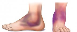
- Changes in skin color (redness, pallor) along the vessels. If the skin on the legs changes color (if the skin becomes bluish, purple or burgundy, this indicates the presence of diseases of the veins of the lower extremities)
- spasmodic contractions in the calf muscles
- pain and swelling of the lower extremities
- the appearance of spider veins on the legs
- Tingling and numbness in the legs
- Trophic ulcers
In addition, ultrasound is also indicated for the purpose of prevention. A possible risk group includes people suffering from obesity, diabetes mellitus and those who are engaged in hard work and whose professional activities involve standing for long periods of time (salespeople, security guards, etc.).
Ultrasound diagnostics are periodically performed for patients suffering from diabetes mellitus, chronic venous insufficiency and thrombophlebitis.
Ultrasound equipment
To conduct research, devices are used that are designed for visual diagnostics of the human body. Ultrasound rooms are equipped with multifunctional equipment designed to meet stringent requirements. Modern models of devices have ultra-precise focusing and compact dimensions. They provide high quality images.
To improve performance, the devices are equipped with various modes. For example, two-dimensional visualization, as well as color and power Doppler. Many models are equipped with glare-free monitors that rotate freely around an axis and tilt in the desired direction. It is possible to connect several sensors simultaneously. The received information is recorded on the hard drive and stored in a database.
When developing ultrasound machines, new technologies are being introduced: grain suppression, image detailing, automatic image optimization, wireless information transfer. The scanners are easy to operate. If necessary, a specialist can adjust the settings. The image is instantly transferred to the monitor. The doctor examines the details and assesses the situation.
There are two types of equipment on sale: stationary ultrasound and portable ultrasound. Stationary devices are installed in rooms designed for ultrasound examinations of leg vessels. They are impressive in size and provide reliable diagnostics. Portable devices are portable and small in size. Such devices are designed to conduct research in any place: ward, ambulance, emergency department. They are indispensable in cases where it is necessary to urgently determine a person’s condition and take the right actions.
How to prepare for ultrasound examination of leg vessels?
The procedure usually does not require special preparation and does not require adherence to strict diets, however, specialists at the Istok Health Clinic claim that in order to obtain the most accurate results, certain restrictions must be observed
- Postpone active physical activity one day before ultrasound examination.
- Avoid drinking any alcoholic beverages 24 hours before the test;
- On the day of ultrasound examination, do not drink coffee, cocoa, strong tea or energy drinks.
- Do not smoke tobacco products at least 3 hours before your appointment.
- Before going to the clinic, do not rub or warm your feet under any circumstances.
Decoding and results
Ultrasound is an affordable imaging method that does not use harmful radiation. During the diagnosis, the doctor determines the thickness of the vascular membranes, pulsation index, and vascular resistance. The minimum and maximum speed of blood movement is also determined.
After conducting the study, the specialist receives accurate data that indicates the condition of the vascular system. It defines a number of important parameters and reflects them in the protocol. Based on such information, the doctor gives an opinion, according to which the vascular surgeon develops treatment tactics.
Using ultrasound examination of the vessels of the lower extremities, various diseases are detected:
- stenosis is a term that refers to the narrowing of blood vessels in the circulatory system. The development of such a pathology leads to undesirable consequences: muscle atrophy, ulcerative formations, pain. Stenosis appears in people who lead a sedentary lifestyle and are overweight. Diabetes mellitus also leads to changes in the structure of blood vessels
- Varicose veins are a serious disease accompanied by impaired blood flow. The trigger for the development of pathology is considered to be disruption of the venous valves with the subsequent occurrence of blood reflux. The main symptom of the disease is dilated saphenous veins. First, a person is bothered by a feeling of heaviness in the legs and a burning sensation, and then swelling appears. Pregnant women and people who spend a lot of time in one position (office workers, salespeople, drivers, bank tellers) are predisposed to varicose veins.
- Phlebitis is a disease in which the vein wall becomes inflamed. Its main symptoms include soreness, a slight increase in temperature, and redness of the skin. Phlebitis can be chronic or acute. The appearance of the disease is provoked by varicose veins, infection, and mechanical damage to the vessel. When diagnosing phlebitis, the doctor prescribes a set of therapeutic agents
- thrombosis - the formation of blood clots inside blood vessels. This is a pathological process because the free flow of liquid connective tissue through the system is limited. The key signs of the disease are severe swelling, pain and induration. Thrombosis may be asymptomatic. In such cases, the disease is detected by ultrasound. This problem cannot be ignored, because it leads to serious consequences.
Ultrasound of the vessels of the lower extremities is a study with which you can detect the disease in time and prescribe effective treatment. To carry it out, equipment with wide functionality is used. This is a guarantee that the results will be reliable.
Thrombophlebitis of superficial veins
This disease often begins with minor pain in the legs.
This pain goes along the course of the saphenous veins and the skin over the thrombosed veins turns red, becomes warmer than normal skin to the touch, with possible inflammation. There is a slight increase in body temperature. After 5-6 days, the temperature returns to normal or remains at a slight increase. Thrombophlebitis of the lower extremities may occur without an increase in body temperature.
A concomitant sign of thrombophlebitis is swelling of the leg where the blood clot has formed. The skin along the veins becomes inflamed in the form of “stripes”. The function of the lower limb suffers, walking is accompanied by pain and lameness.
Subsequently, compacted areas of skin of various sizes begin to appear. Their size depends on the diameter of the thrombosed veins. These hardened areas can be easily felt and are actually blood clots. But not always compacted areas can show the border of the pathological process. In order to identify the severity and extent of the disease, it is necessary to conduct an ultrasound scan of the veins of the lower extremities with consultation of a cardiovascular surgeon or phlebologist.
Dopplerography of the vessels of the lower extremities
Doppler is often used when examining blood vessels. Dopplerography allows you to assess the condition and functioning of blood flow. This research method shows the speed of blood flow.
Dopplerography is mandatory for the following complaints:
- With regular leg cramps;
- With severe swelling;
- With a tendency to hematomas.
- For reliable results, the skin of the legs must be smooth. Therefore, it is necessary to get rid of vegetation on the lower extremities.