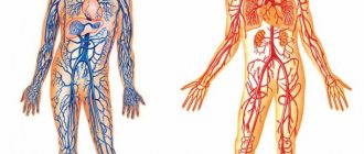
Both arteries and veins are types of blood vessels in the cardiovascular system
An artery carries blood away from the heart, and a vein brings blood back to the heart.
Blood vessels are necessary to transport blood throughout the body. Blood carries oxygen and nutrients to various tissues in the body, allowing them to function.
The heart and blood vessels make up the cardiovascular system. This system contains a complex network of vessels with different structures and functions.
In this article, we discuss the differences between arteries and veins. We also highlight the different types of blood vessels and how they work as part of the cardiovascular system.
Arteries and veins: definitions
Arteries and veins are types of blood vessels that transport blood throughout the body.
Blood vessels form two systems, leading to and from the heart. These two systems form the circulatory system.
The systemic circulation supplies oxygen and other vital substances to organs, tissues and cells.
The systemic arteries transport oxygen-rich blood from the left ventricle to the rest of the body. The low-oxygen blood then collects in the systemic veins and is sent to the right atrium.
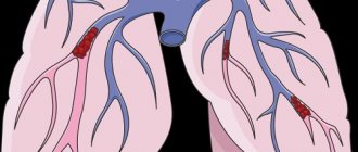
The pulmonary circulation is where fresh oxygen enters the blood.
The pulmonary arteries transport low-oxygen blood from the right ventricle to the lungs. The pulmonary veins then transport oxygen-rich blood back to the heart through the left atrium.
Capillaries are the third type of blood vessel in the body. They supply blood directly to the organs.
What are arteries
These are vessels that transport oxygen from the heart to the internal organs. By contracting the myocardium, blood circulation is ensured at a speed of 20 cm/s. Purified blood, full of oxygen and nutrients, is necessary for metabolism.
Passing through organ tissue saturates it with carbon dioxide, excreted through venous hematopoiesis.
They are divided into three types:
- diameter;
- structural features;
- topographic principle.
The diameters are:
- large;
- small.
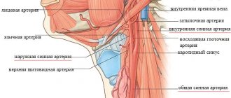
Large in diameter, unlike other components of the vascular system, are: aorta, carotid and subclavian.
The aorta arises from the left ventricle of the heart along the spinal column, dividing into the left and right iliac branches. It begins a large circle of blood circulation, supplying oxygen to the organs and tissues of the body.
General sleep supports the functioning of the brain, providing it with oxygen and microelements necessary for metabolism.
The subclavian vessel supplies blood to the occipital parts of the brain, medulla oblongata, cerebellum and cervical spine. The left arch departs from the aorta, bending around the pleura and, passing through the upper aperture of the chest, exits to the neck and lies in the space of the first rib.
Arterioles are small in diameter. Their task is to regulate blood flow in the SMC.
- Why do the veins in the arms hurt in the elbow bend?
Arteriolar tone determines peripheral resistance, which, along with the stroke volume of the heart, affects blood pressure.
There are three types:
- elastic;
- muscular;
- mixed.
The first type includes mainly the aorta. Its structure is characterized by the predominance of elastic fibers over muscle fibers.
The muscle type contains smooth muscle fibers and is characterized by a weak outer elastic membrane. An example is arterioles.
The muscular-elastic type is distinguished by the presence of muscle and elastic fibers in the structure of the vessel.
Types of arteries
There are three types of arteries:
Elastic arteries
Elastic arteries are large vessels leaving the heart. For example, they include the pulmonary artery and aorta. The aorta is the main artery that carries blood away from the heart.
The heart forcibly pumps out blood to move it throughout the body. Elastic arteries must be flexible to cope with the rush of blood. They expand when the heart pumps out blood.
Elastin is a protein found in many tissues that provides flexibility to organs, including elastic arteries.
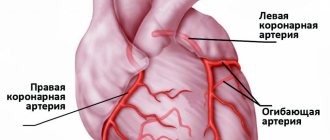
Muscular arteries
Elastic arteries carry blood to muscular arteries such as the femoral or coronary arteries.
Smooth muscle fibers make up the walls of muscle arteries. Muscles allow these arteries to expand and contract. These changes in size control how much blood moves through the arteries.
Arterioles
Arterioles are the smallest type of arteries. They distribute blood from larger arteries through networks of capillaries.
The outer layer of arterioles also contains smooth muscle, which regulates expansion and contraction.
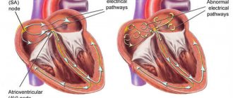
Movement of blood through vessels
The difference between arteries and veins is clearly demonstrated by the mechanism of blood flow. When the heart muscle contracts, blood is forced into the arteries. In the largest of them, the aorta, pressure can reach 150 mm Hg. Art. In the capillaries it decreases significantly to 20. In the vena cava the pressure is minimal and amounts to 3-8 mm Hg. Art.
Types of Veins
Arteries and veins are designed roughly the same, but veins are thinner and have less muscle, which allows them to hold more blood. Veins typically contain about 70% of the body's blood at one time.
Venules are the smallest type of vein. They have very thin walls in order to hold a lot of blood. They pump low-oxygen blood through capillaries from the arteries directly into the veins. The blood then returns to the heart through a series of veins of increasing size and muscle.
There are two main types of veins: pulmonary and systemic.
Systemic veins are further classified into:
- Deep veins : These veins usually have a corresponding artery nearby and are found in muscle tissue. These veins may have a one-way valve to prevent blood from flowing backwards.
- Superficial veins : These veins do not have an artery of the same name nearby and are found close to the surface of the skin. They may also have a one-way valve.
- Connecting veins : These small veins allow blood to flow from the superficial veins to the deep veins.
Different functions - different structure.
The highest blood pressure will be at the blood outlet from the heart (in the left ventricle), slightly lower pressure will be in the arteries, even lower in the capillaries, and the lowest in the veins and at the entrance of the heart (in the right atrium).
The arteries carrying the oxygenated blood pushed out by the heart must resist the high pressure in the circulatory system. That's why they have an elastic membrane. In addition, they must also change their lumen in order to vary the level of blood flow in different organs in response to the actions of the autonomic nervous system - for this they have a well-developed layer of smooth muscle tissue. Therefore, the walls of the arteries are much thicker than the venous ones, they are much more elastic and contain a large number of muscle elements.
The walls of the veins, in turn, are thin and pliable, contain virtually no muscle elements, and ensure the return of blood to the heart. The veins in the lower body have valves that prevent blood from flowing back. Thus, the vascular bed adapts to changing load levels mainly due to changes in the lumen of the arteries.
Anatomy
Veins and arteries consist of three layers:
- Tunica adventitia : The outer layer of a blood vessel, composed of collagen and elastin. This layer allows the vessel to expand or contract, depending on the type of vein or artery. This function is important for controlling blood pressure.
- Tunica media : This is the middle of the blood vessel. Elastin and muscle fibers form the shell of the carriers. The amount of elastin or muscle varies depending on the type of blood vessel. For example, elastic arteries contain few muscle fibers in their membranes.
- Tunica Intima: This name refers to the inner layer of a blood vessel. It mainly contains elastic membranes and tissues and may include valves that help blood move in the right direction.
The cardiovascular system
The cardiovascular system connects the heart and blood vessels together. The system forms a closed circuit of vessels that transport blood throughout the body.
The cardiovascular system is essential for maintaining life. This is the first major network of organs to develop in embryos.
All body tissues need oxygen and nutrients to survive. They also require the removal of waste, which is a byproduct of metabolism.
Blood is needed both to provide oxygen and nutrients and to remove waste from tissues.
The heart pumps blood throughout the body. It must work constantly and with sufficient force so that all tissues of the body receive enough blood to function. Cardiovascular disorders can have serious consequences.
Cardiovascular diseases are a group of disorders that affect the heart and blood vessels, such as coronary heart disease.
These diseases are the leading cause of death worldwide, accounting for approximately 17.9 million deaths in 2021.
Arteries and veins: conclusions
Arteries are a type of blood vessel that transports blood away from the heart. Veins carry blood back to the heart. Along with capillaries, these blood vessels are responsible for moving blood to the tissues around the body.
The heart pumps blood through a complex system of blood vessels. There are several types of arteries and veins with different functions. For example, some contain more muscle to change the amount of blood they carry.
The cardiovascular system is essential to life. Changes in the heart or blood vessels can be serious and sometimes fatal.
Differences between venous and capillary blood analysis
For most tests, which are prescribed by doctors of various specializations to diagnose diseases and monitor the progress of their treatment, the biomaterial is venous blood.
Capillary biomaterial is sometimes used to perform OAC. In any case, donating blood from a vein is preferable, since in this case the research results will be more informative. When performing OAC with different types of biomaterial, the results may differ. Therefore, to obtain reliable information about the patient’s health status over time, it is recommended to submit the same type of biomaterial. Capillary blood analysis
The main advantage of testing blood taken from a finger is the ease of taking biomaterial. This is why this type of test is often prescribed to young children. Research on this biomaterial has a significant number of limitations and disadvantages. Most laboratory tests do not use capillary blood. The research results are not highly accurate. There is a high probability of their distortion due to the formation of microclots, tissue fluid entering the blood, and platelet aggregation. Features of the procedure for taking biomaterial also lead to a decrease in the information content of research results. If questionable results are obtained, the study is usually repeated, and venous blood is used as a biomaterial.
Venous blood analysis
Blood from a vein is used for a variety of studies (for example, hormonal). Drawing blood by an experienced nurse takes a few minutes and does not cause significant discomfort. The results of such studies are characterized by high accuracy and great information content. To obtain the most accurate results and reduce the risk of their distortion by external factors, it is important to strictly follow the recommendations for preparing for the study. Preparation rules depend on the type of analysis. In addition, the attending physician can, if necessary, give the patient individual recommendations.
Preparing to donate blood
Before taking the test, it is important to exclude all factors that could affect the result. Such factors include physical activity (for example, climbing stairs, brisk walking), significant emotional arousal, and stress. Therefore, before donating blood, it is recommended to sit near the office or in the waiting room for 15-20 minutes. It is advisable to avoid heavy physical work and intense training a few days before the test.
If for any reason a patient cannot visit a clinic or private medical center to donate biomaterial, it is necessary to use the services of a laboratory that provides home testing services.
In most cases, blood is donated before starting medication or 2 weeks after finishing it. If it is necessary to evaluate the concentration of the drug in the blood, their use is continued in accordance with the doctor’s recommendations. To avoid misinterpretation of the results, it is important to inform your doctor about taking any medications, as well as dietary supplements.
If the patient has undergone X-rays, physical therapy, or a rectal examination, blood donation is usually delayed for some time. The final decision on the time of testing is made by the doctor for each patient individually.
It must be taken into account that in girls and women of reproductive age, the results of hormonal studies are significantly influenced by physiological factors. Blood must be donated for testing strictly at the phase of the cycle recommended by the doctor.