Glioma is a brain tumor that originates from neuroglial cells. Neoplasms are the most common type of malignant pathogenesis of varying degrees of aggressiveness. Symptoms of glioma in adults and children will depend on where it forms. The main diagnostic methods are CT and MRI; the most accurate result is obtained by cytohistological analysis of the selected material (biopsy). The main treatment method for brain glioma is surgical removal of the pathological tumor. Treatment is always comprehensive; in addition to surgery, chemotherapy and radiotherapy are prescribed. Recovery and life expectancy after tumor removal depend on many factors: the adequacy of treatment, the degree of malignancy of the brain tumor, the stage of the disease, the general condition of the patient and many other factors. Benign gliomas have the best prognosis, while grade 3 and 4 gliomas have the worst prognosis. For example, with glioblastoma, life expectancy is usually no more than two years.
Origin of brain gliomas and characteristics
There is still no consensus on which cells give rise to glial tumors. The classical view is based on the fact that gliomas develop from mature glial cells (oligodendrocytes and astrocytes). Recently, they have become more inclined to believe that they are formed in the so-called “malignancy gaps.” This means that brain tumors of this type are formed from immature, slowly proliferating cells that gradually become malignant (degenerate into cancer). How exactly the neoplasm will develop depends on transformations in the genome, for example, astrocytomas (a type of glioma) are formed due to mutations in the TP53 gene,
The share of gliomas in the general population of brain tumors is about 60%. Most often, neoplasms form in the brain and are primary in nature. Brain stem glioma is diagnosed relatively rarely. The color of neoplasia ranges from dark red to soft pink, the shape in most cases resembles a circle or spindle, but can also have irregular outlines. The size depends on the stage of the disease: from a few millimeters to 10-14 cm.
The most common location of the tumor is the area of the chiasmata or cerebral ventricles. Benign glioma, as a rule, does not spread to the bone structures of the skull and does not grow into the membranes of the brain, however, this cannot be excluded. Malignant glioma grows slowly at first, but as carcinogenesis progresses, its growth accelerates. Infiltrative growth is characteristic, i.e. the ability to grow into neighboring tissues, which during diagnosis does not allow defining clear boundaries of the pathogenic focus.
Note. Brain gliomas are not characterized by metastasis, however, they lead to degeneration of neighboring tissues.
Possible complications
{banner_banstat9}
Consequences are relatively rare. Years pass from the development of microangiopathy to the final outcome. However, you cannot relax.
Possible problems include:
- Stroke. Acute cerebrovascular accident. Provokes the death of nerve tissue. Severe disability with neurological deficit. The death of a person is possible and even probable.
- Encephalopathy. The process is similar, but there is no destruction of cerebral structures yet.
- Vascular dementia. Dementia due to insufficient brain nutrition.
Complications develop spontaneously. Treatment is the only way to prevent them.
Causes and risk factors
Cancer and benign dysplasia can form in the brain at any age, but the likelihood of developing pathology increases as you get older. The overall peak incidence occurs in the seventh decade. The tumor process develops more often in men than in women.
The main causes of brain glioma are the following:
- exposure to ionizing radiation (radiation is the main risk factor);
- genetic prerequisites due to heredity;
- damage by oncogenic viruses;
- head and cervical brain injuries;
- bad habits.
Important. There is no specific prevention against brain cancer. Minimizing risk factors, a healthy lifestyle, quality nutrition, regular exercise and a calm emotional background reduces the likelihood of developing a tumor process.
Demyelination
Demyelinating brain disease according to ICD-10 has codes G35, G36 and G77. The process caused by damage to the nervous tissue negatively affects the functioning of the entire organism as a whole. Certain nerve endings are covered with a myelin sheath, which performs important functions in the body. For example, myelin ensures the rapid transmission of electrical impulses and, accordingly, if this process is disrupted, the entire system suffers. Myelin consists of lipids and protein compounds in a 70/30 ratio.
Demyelinating disease is not only multiple sclerosis, it is also neuromyelitis optica and acute disseminated encephalomyelitis. These diseases have no cure, but their progression can be slowed down. In general, doctors give a favorable prognosis when treating these pathologies. The diagnosis of “multiple sclerosis” is now made more often, but the disease itself is milder than 30-40 years ago.
Make an appointment
Classification
Depending on the type of brain cells that degenerated and gave rise to pathogenesis, the following types of tumors are distinguished:
- astrocytomas are the most common type, their share in the general population is about 50%;
- oligodendrogliomas – the share in the general population of tumors of this type is up to 10%;
- Ependymomas are the rarest forms (occurrence less than 7%).
According to the WHO classification (which is generally accepted), neoplasia is ranked depending on the degree of malignancy.
Benign gliomas
These are tumors of the first degree of malignancy, for example, astrocytomas: giant cell, pilocytic, juvenile subependymal. They are benign because they grow slowly, have no signs of cancer, and are easy to treat. The prognosis for life with benign glioma is favorable. After tumor removal, patients live 10 years or longer.
Low grade glioma
Second degree of malignancy. It is also classified as a benign neoplasm, but with a borderline degree of malignancy. The growth of pathological tissue is slow (low-grade), they are well differentiated, as a rule, there is only one sign of cancer (cell atypia). Tumors of this type can degenerate into cancer and easily progress to the third and fourth degree of malignancy. These include diffuse and fibrillary astrocytomas. Treatment is complex: surgery to remove atypical tissues, additionally radio and chemotherapy.
Malignant gliomas
These include gliomas of grade 3 and 4:
- Third degree of malignancy . There are all signs of malignancy, except for tissue necrosis. Tissues lose clear differentiation, tumor growth accelerates (high-grade), the boundaries are unclear, and growth into nearby tissues is typical. The most striking example is anaplastic astrocytoma, which most often develops in middle-aged and older people. Treatment is hampered by the lack of clear boundaries of the tumor; life expectancy depends on how much atypical tissue is removed, as well as the stage of carcinogenesis at the beginning of treatment.
- The most dangerous is the fourth degree of malignancy (glioblastoma). It develops between the ages of 40 and 70 years. In this case, all the signs of malignancy are present, including necrosis. The tumor grows quickly, penetrates other tissues and has no clear boundaries. This significantly complicates therapy. The prognosis is unfavorable.
Neoplasia is divided into two types, depending on growth characteristics:
- Tumors of nodular growth . As a rule, these are benign neoplasms with clear contours. They are characterized by formation anywhere in the brain and the presence of cysts. Examples: pleomorphic xanthoastrocytoma and piloid astrocytoma.
- Diffuse type formations . There are no obvious boundaries, the size can reach significant sizes, neoplasia grows into adjacent brain tissue, which complicates the removal of dysplasia. These are often malignant tumors, such as glioblastoma or anaplastic astrocytoma, or those that can quickly degenerate.
Symptoms
Clinical manifestations are divided into general and focal. The latter manifest themselves depending on which part of the brain the pathological formation is formed.
General symptoms
General signs of brain cancer (or a benign tumor) arise due to increased pressure inside the skull, compression of neighboring tissues by the tumor body and the negative influence of its metabolites. Common symptoms include:
- dizziness, headache, discomfort in the head;
- dyspeptic manifestations not related to food intake or poisoning;
- loss of appetite, weight loss;
- blurred vision;
- changing the psycho-emotional behavior habitual for a person;
- memory problems, weakening of mental activity;
- signs of epilepsy.
It's important to understand. That general symptoms are not specific. If they bother you regularly, you need to urgently visit a doctor.
The pathological process in the early stages, as a rule, occurs latently or its signs are weak. Often neoplasms are found by chance, when examining a patient for another disease or for preventive purposes.
Focal symptoms
These are specific signs that arise as a result of damage to the cerebral structures of the central nervous system. The clinical picture depends on the location of the pathological focus:
- Glioma of the optic nerve is formed from cells of its trunk. Tissue growth is slow, accompanied by infiltration, but does not grow into the dura mater. The neoplasm is considered benign; it can form along the entire length of the nerve, but is more often located in the orbital part. Symptoms of optic nerve glioma: first, visual acuity decreases (as the nerve and elements of the eye are damaged), then bulging eyes develop.
- Diffuse glioma of the brainstem is highly malignant. Since diffuse dysplasia is formed in the area where the nerves responsible for the functioning of the limbs are located, when they are damaged, the coordination of the arms and legs is disrupted, up to paresis and paralysis. Clinical signs of diffuse pontine glioma are significant even with small sizes of dysplasia. There are many nerve cords passing through the bridge, so the clinic can be more diverse.
- Glioma of the frontal lobe of the brain is manifested by apathy, mental changes, the person becomes nervous, hot-tempered, and sudden mood changes are characteristic. The later stages of the disease are characterized by the onset of paralysis.
- Glioma of the temporal region causes impaired coordination of movements, memory deteriorates, and diction suffers. In addition, temporal lobe glioma negatively affects the sense of smell.
- Glioma of the cerebellum and cerebellar vermis leads to movement abnormalities. The gait is disturbed, it seems that the person is drunk. This is because it becomes difficult for the patient to maintain balance, his arms and legs do not obey, and maintaining the body in space becomes problematic.
- Glioma of the parietal lobe is accompanied by disturbances in fine motor skills, so a person’s handwriting changes, and over time it becomes difficult to write. Tactile sensations become dull.
- Glioma of the quadrigeminal plate or quadrigeminal plate causes visual disturbances, abnormalities in eye movement, and hearing impairment.
- Glioma of the corpus callosum is accompanied by visual hallucinations (the same ones occur in the presence of a tumor in the occipital lobe), violations of logic, as well as many other signs: changes in sensitivity, muscle weakness, and so on.
- Pontine glioma (diffuse or common) leads to disruption of conduction functions, since the nuclei of the cranial nerves are localized in this area. Therefore, the clinical picture will be quite broad. A violation of sensory perception, movements, and so on is recorded.
- Glioma of the cerebral ventricle leads to an increase in intracranial pressure due to disruption of the outflow of cerebrospinal fluid. Hydrocephalus may develop.
Chronic cerebral ischemia
Cerebrovascular diseases are one of the main problems of modern medicine. It is known that in recent years the structure of vascular diseases of the brain has changed due to the increase in ischemic forms. This is due to an increase in the proportion of arterial hypertension and atherosclerosis as the main cause of cerebrovascular pathology. When studying individual forms of cerebral circulatory disorders, chronic ischemia ranks first in prevalence.
Chronic cerebral ischemia (CHI) is a special type of vascular cerebral pathology caused by a slowly progressive diffuse disorder of the blood supply to the brain with gradually increasing various defects in its functioning. The term “chronic cerebral ischemia” is used in accordance with the International Classification of Diseases, 10th revision, instead of the previously used term “dyscirculatory encephalopathy”.
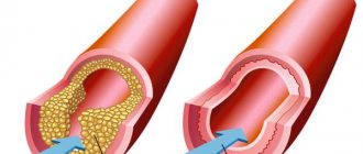
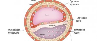
The development of chronic cerebral ischemia is promoted by a number of reasons, which are commonly called risk factors. Risk factors are divided into correctable and non-correctable. Uncorrectable factors include old age, gender, and hereditary predisposition. It is known, for example, that a stroke or encephalopathy in parents increases the likelihood of vascular diseases in children. These factors cannot be influenced, but they help to identify in advance those at increased risk of developing vascular pathology and help prevent the development of the disease. The main correctable factors in the development of chronic ischemia are atherosclerosis and hypertension. Diabetes mellitus, smoking, alcohol, obesity, insufficient physical activity, poor nutrition are the reasons that lead to the progression of atherosclerosis and the deterioration of the patient’s condition. In these cases, the blood coagulation and anticoagulation system is disrupted, and the development of atherosclerotic plaques is accelerated. Due to this, the lumen of the artery decreases or is completely blocked (Fig.). At the same time, the crisis course of hypertension is especially dangerous: it leads to an increase in the load on the blood vessels of the brain. Arteries modified by atherosclerosis are unable to maintain normal cerebral blood flow. The walls of the vessel gradually become thinner, which can ultimately lead to the development of a stroke.
| Drawing. MR angiogram: occlusion of the right middle cerebral artery |
The etiology of CCI is associated with occlusive atherosclerotic stenosis, thrombosis, and embolism. A certain role is played by post-traumatic dissection of the vertebral arteries, extravasal compression due to pathology of the spine or neck muscles, deformation of the arteries with permanent or periodic disturbances in their patency, hemorheological changes in the blood (increased hematocrit, viscosity, fibrinogen, platelet aggregation and adhesion). It must be borne in mind that symptoms similar to those that occur with chronic ischemia can be caused not only by vascular, but also by other factors - chronic infection, neuroses, allergic conditions, malignant tumors and other reasons with which a differential diagnosis should be made . If the described disorders are supposed to have a vascular origin, instrumental and laboratory confirmation of damage to the cardiovascular system is necessary (ECG, Doppler ultrasound of the main arteries of the head, MRA, MRI, CT, biochemical blood tests, etc.).
To make a diagnosis, one must adhere to strict diagnostic criteria: the presence of cause-and-effect relationships (clinical, anamnestic, instrumental) of brain lesions with hemodynamic disturbances with the development of clinical, neuropsychological, psychiatric symptoms; signs of progression of cerebrovascular insufficiency. The possibility of subclinical acute cerebral dyscirculatory disorders, including small-focal, lacunar infarctions, which form symptoms characteristic of encephalopathy, should be taken into account. For the main etiological reasons, atherosclerotic, hypertensive, mixed, and venous encephalopathies are distinguished, although other causes leading to chronic cerebral vascular insufficiency (rheumatism, vasculitis of other etiologies, blood diseases, etc.) are also possible.
The pathomorphological picture of CCI is characterized by areas of ischemically altered neurons or their loss with the development of gliosis. Small cavities (lacunae) and larger lesions develop. With multiple lacunae, the so-called “lacunary state” is formed. These changes are predominantly observed in the area of the basal ganglia and have a typical clinical expression in the form of amyostatic and pseudobulbar syndromes, dementia, described at the beginning of the twentieth century. French neurologist P. Marie. The development of status lacunaris is most characteristic of arterial hypertension. In this case, changes in blood vessels are observed in the form of fibrinoid necrosis of the walls, their plasmatic impregnation, the formation of miliary aneurysms, and stenoses.
The so-called criblures, which are dilated perivascular spaces, are distinguished as changes characteristic of hypertensive encephalopathy. Thus, the chronic nature of the process is pathomorphologically confirmed by multiple zones of brain ischemia, especially its subcortical regions and cortex, accompanied by atrophic changes developing against the background of corresponding changes in the cerebral vessels. Using CT and MRI, in typical cases, multiple microfocal changes are detected, mainly in the subcortical zones, periventricularly, often accompanied by cortical atrophy, dilation of the cerebral ventricles, and the phenomenon of leukoaraiosis (“periventricular glow”), which is a reflection of the demyelination process. However, similar changes can be observed during normal aging and primary degenerative-atrophic processes of the brain.
Clinical manifestations of CCI are not always detected by CT and MRI studies. Therefore, the diagnostic significance of neuroimaging methods cannot be overestimated. Making a correct diagnosis for a patient requires an objective analysis of the clinical picture and instrumental examination data from the doctor.
The pathogenesis of cerebral ischemia is caused by insufficiency of cerebral circulation in its relatively stable form or in the form of repeated short-term episodes of discirculation.
As a result of pathological changes in the vascular wall, developing as a result of arterial hypertension, atherosclerosis, vasculitis, etc., autoregulation of cerebral circulation is disrupted, and there is an increasing dependence on the state of systemic hemodynamics, which also turns out to be unstable due to the same diseases of the cardiovascular system. Added to this are disturbances in the neurogenic regulation of systemic and cerebral hemodynamics. Brain hypoxia itself leads to further damage to the mechanisms of autoregulation of cerebral circulation. The pathogenetic mechanisms of acute and chronic cerebral ischemia have much in common. The main pathogenetic mechanisms of cerebral ischemia constitute the “ischemic cascade” (V.I. Skvortsova, 2000), which includes:
- decreased cerebral blood flow;
- increase in glutamate excitotoxicity;
- calcium accumulation and lactic acidosis;
- activation of intracellular enzymes;
- activation of local and systemic proteolysis;
- emergence and progression of antioxidant stress;
- expression of early response genes with the development of depression of plastic proteins and a decrease in energy processes;
- long-term consequences of ischemia (local inflammatory reaction, microcirculatory disorders, damage to the BBB).
A condition called “oxidative stress” plays a major role in damage to brain neurons. Oxidative stress is an excessive intracellular accumulation of free radicals, activation of lipid peroxidation (LPO) processes and excessive accumulation of lipid peroxidation products, aggravating overexcitation of glutamate receptors and enhancing glutamate excitoxic effects. Glutamate excitotoxicity is understood as hyperstimulation by mediators of excitation of NDMA receptors of N-methyl-D-aspartate, provoking dilatation of calcium channels and, as a consequence, massive influx of calcium into cells, with subsequent activation of proteases and phospholipases. This leads to a gradual decrease in neuronal activity, a change in the neuron-glia ratio, which causes a deterioration in brain metabolism. Understanding the pathogenesis of CCI is necessary for an adequate, optimally selected treatment strategy.
As the severity of the clinical picture increases, pathological changes in the vascular system of the brain intensify. If at the beginning of the process stenotic changes in one or two main vessels are detected, then most or even all of the main arteries of the head turn out to be significantly changed. Moreover, the clinical picture is not identical to damage to the great vessels, due to the presence in patients of compensatory mechanisms of autoregulation of cerebral blood flow. The condition of intracranial vessels plays an important role in the mechanisms of compensation for cerebral circulatory disorders. With well-developed and preserved collateral circulation pathways, satisfactory compensation is possible, even with significant damage to several great vessels. On the contrary, individual structural features of the cerebral vascular system may be the cause of decompensation (clinical or subclinical), aggravating the clinical picture. This may explain the more severe clinical course of cerebral ischemia in middle-aged patients.
Based on the main clinical syndrome, several forms of CCI are distinguished: with diffuse cerebrovascular insufficiency; predominant pathology of the vessels of the carotid or vertebrobasilar systems; vegetative-vascular paroxysms; predominant mental disorders. All forms have similar clinical manifestations. In the initial stages of the disease, all patients complain of headache, non-systemic dizziness, noise in the head, memory impairment, and decreased mental performance. As a rule, these symptoms occur during a period of significant emotional and mental stress, requiring a significant increase in cerebral circulation. If two or more of these symptoms are often repeated or exist for a long time (at least the last 3 months) and there are no signs of an organic nature, instability when walking, or damage to the nervous system, a presumptive diagnosis is made.
The clinical picture of CCI has a progressive development and, according to the severity of symptoms, is divided into three stages: initial manifestations, subcompensation and decompensation.
Stage 1 is dominated by subjective disorders in the form of headaches and a feeling of heaviness in the head, general weakness, increased fatigue, emotional lability, dizziness, decreased memory and attention, and sleep disturbances. These phenomena are accompanied, although mild, but quite persistent objective disorders in the form of anisoreflexia, discoordination phenomena, oculomotor insufficiency, symptoms of oral automatism, memory loss and asthenia. At this stage, as a rule, the formation of distinct neurological syndromes (except for asthenic) has not yet occurred, and with adequate therapy, it is possible to reduce the severity or eliminate both individual symptoms and the disease as a whole.
The complaints of patients with the 2nd stage of CCI more often include memory impairment, loss of ability to work, dizziness, instability when walking, and manifestations of an asthenic symptom complex are less often present. At the same time, focal symptoms become more distinct: revival of reflexes of oral automatism, central insufficiency of the facial and hypoglossal nerves, coordination and oculomotor disorders, pyramidal insufficiency, amyostatic syndrome, increased mnestic-intellectual disorders. At this stage, it is possible to identify certain dominant neurological syndromes - discoordination, pyramidal, amyostatic, dysmnestic, etc., which can help in prescribing symptomatic treatment.
At the 3rd stage of CCI, objective neurological disorders in the form of discoordination, pyramidal, pseudobulbar, amyostatic, and psychoorganic syndromes are more pronounced. Paroxysmal conditions—falls, fainting—are more common. In the stage of decompensation, cerebral circulation disorders are possible in the form of “small strokes” or prolonged reversible ischemic neurological deficit, the duration of focal disorders in which ranges from 24 hours to 2 weeks. At the same time, the clinical picture of diffuse insufficiency of blood supply to the brain corresponds to that of moderate encephalopathy. Another manifestation of decompensation may be a progressive “complete stroke” and residual effects after it. This stage of the process with diffuse damage corresponds to the clinical picture of severe encephalopathy. Focal symptoms are often combined with diffuse manifestations of brain failure.
In chronic cerebral ischemia, there is a clear correlation between the severity of neurological symptoms and the age of the patients. This must be kept in mind when assessing the significance of individual neurological signs that are considered normal for elderly and senile people. This dependence reflects age-related manifestations of dysfunction of the cardiovascular and other visceral systems, affecting the state and functions of the brain. To a lesser extent, this dependence is observed in hypertensive encephalopathy. In this case, the severity of the clinical picture is largely determined by the course of the underlying disease and its duration.
Along with the progression of neurological symptoms, as the pathological process develops in the neurons of the brain, an increase in cognitive disorders occurs. This applies not only to memory and intelligence, which are impaired in the 3rd stage to the level of dementia, but also to such neuropsychological syndromes as praxis and gnosis. Initial, essentially subclinical disorders of these functions are observed already in the 1st stage, then they intensify, change, and become distinct. The 2nd and especially the 3rd stages of the disease are characterized by pronounced impairments of higher brain functions, which sharply reduces the quality of life and social adaptation of patients.
In the picture of CIM, several main clinical syndromes are distinguished: cephalgic, vestibulo-ataxic, pyramidal, amyostatic, pseudobulbar, paroxysmal, vegetative-vascular, psychopathological. A feature of the cephalgic syndrome is its polymorphism, inconstancy, lack of connection in most cases with specific vascular and hemodynamic factors (excluding headaches during hypertensive crises with high blood pressure), and a decrease in the frequency of occurrence as the disease progresses.
The second most common syndrome is vestibulo-ataxic syndrome. The main complaints of patients are: dizziness, instability when walking, coordination disorders. Sometimes, especially in the initial stages, patients, complaining of dizziness, do not notice coordination problems. The results of otoneurological examination are also insufficiently indicative. In later stages of the disease, subjective and objective discoordination disorders are clearly interrelated. Dizziness and unsteadiness when walking may be partly due to age-related changes in the vestibular apparatus, motor system and ischemic neuropathy of the vestibulocochlear nerve. Therefore, to assess the significance of subjective vestibulo-ataxic disorders, their qualitative analysis during a patient interview, neurological and otoneurological examination is important. In most cases, these disorders are caused by chronic circulatory failure in the blood supply of the vertebrobasilar arterial system, so it is necessary to rely not on the subjective sensations of patients, but to look for signs of diffuse damage to the parts of the brain that are supplied with blood from this vascular system. In some cases, in patients with stages 2–3 CCI, ataxic disorders are caused not so much by cerebellar-stem dysfunction, but rather by damage to the frontal-stem pathways. There is a phenomenon of frontal ataxia, or apraxia of walking, reminiscent of hypokinesia in patients with parkinsonism. A CT examination reveals significant hydrocephalus (along with cortical atrophy), i.e., a condition similar to normal pressure hydrocephalus occurs. In general, the syndrome of circulatory failure in the vertebrobasilar system is diagnosed with CCI more often than insufficiency of the carotid system.
A feature of the pyramidal syndrome is its moderate clinical manifestation (anisoreflexia, facial asymmetry, minimally expressed paresis, revitalization of oral automatism reflexes, hand symptoms). A clear asymmetry of reflexes indicates either a previously existing cerebral stroke or another disease occurring under the guise of CCI (for example, large intracranial processes, consequences of traumatic brain injury). Diffuse and fairly symmetrical revival of deep reflexes, as well as pathological pyramidal reflexes, often combined with a significant revival of oral automatism reflexes and the development of pseudobulbar syndrome, especially in old age, indicates multifocal vascular damage to the brain (subject to the exclusion of other possible causes).
In patients with clinical manifestations of circulatory failure in the vertebrobasilar system, paroxysmal conditions are often observed. These conditions may be caused by a combined or isolated effect on the vertebral arteries of vertebrogenic factors (compression, reflex), which is associated with changes in the cervical spine (dorsopathies, osteoarthritis, deformities).
Mental disorders are quite characteristic and varied in form at different stages of CCI. If in the initial stages they are of the nature of asthenic, asthenodepressive and anxiety-depressive disorders, then in the 2nd and especially in the 3rd stage they are joined by pronounced dysmnestic and intellectual disorders, forming the syndrome of vascular dementia, which often comes first in the clinical picture .
Electroencephalographic changes are nonspecific for CCI. They consist of a progressive decrease in the β-rhythm, an increase in the proportion of slow θ- and δ-activity, accentuation of interhemispheric asymmetry, and a decrease in EEG reactivity to external stimulation.
CT characteristics undergo dynamics from normal indicators or minimal atrophic signs in the 1st stage to more pronounced small-focal changes in the brain substance and atrophic (external and internal) manifestations in the 2nd stage to sharply defined cortical atrophy and hydrocephalus with multiple hypodense foci in the hemispheres - in the 3rd stage.
A comparison of clinical and instrumental characteristics in patients with atherosclerotic, hypertensive and mixed forms of CCI does not reveal any clear differences. In severe cases of hypertension, a faster rate of increase in psychoneurological disorders, early manifestation of cerebral disorders, and a greater likelihood of developing lacunar stroke are possible.
Treatment of CCI should be based on certain criteria, including the concepts of pathogenetic and symptomatic therapy. To correctly determine the pathogenetic treatment strategy, one should take into account: the stage of the disease; identified mechanisms of pathogenesis; the presence of concomitant diseases and somatic complications; age and gender of patients; the need to restore quantitative and qualitative indicators of cerebral blood flow, normalize impaired brain functions; the possibility of preventing recurrent cerebral dysgemia.
The most important direction of CCI therapy is the impact on existing risk factors, such as arterial hypertension and atherosclerosis. Treatment of atherosclerosis is carried out according to generally accepted regimens using statins, in combination with correction of the diet and lifestyle of patients. The selection of antihypertensive drugs and the order of their prescription is carried out by a general practitioner, taking into account the individual characteristics of patients. Complex therapy for CCI includes the prescription of antioxidants, antiplatelet agents, drugs that optimize brain metabolism, and vasoactive drugs. Antidepressants are prescribed for severe asthenodepressive manifestations of the disease. Antiasthenic drugs are prescribed in the same way.
An important component of the treatment of CCI is the administration of drugs with antioxidant activity. Currently, the following drugs of this series are used in clinical practice: Actovegin, Mexidol, Mildronate.
Actovegin is a modern antioxidant, which is a deproteinized extract of the blood of young calves. Its main effect is to improve the utilization of oxygen and glucose. Under the influence of the drug, the diffusion of oxygen in neuronal structures significantly improves, which makes it possible to reduce the severity of secondary trophic disorders. There is also a significant improvement in cerebral and peripheral microcirculation against the background of improved aerobic energy exchange of vascular walls and the release of prostacyclin and nitric oxide. The resulting vasodilation and decrease in peripheral resistance are secondary to the activation of oxygen metabolism of the vascular walls (A. I. Fedin, S. A. Rumyantseva, 2002).
In case of CCI, it is advisable to use Actovegin, especially in the absence of effect from other treatment methods (E. G. Dubenko, 2002). The method of application consists of drip administration of 600–800 mg of the drug for 10 days, followed by switching to oral administration.
A constant in the treatment regimen for CCI is the use of drugs that optimize cerebral circulation. The most commonly used drugs are: Cavinton, Halidor, Trental, Instenon.
Halidor (bencyclane) is a drug that has a multidirectional mechanism of action due to phosphodiesterase blockade, antiserotonin action, and calcium antagonism. It inhibits the aggregation and adhesion of platelets, prevents the aggregation and adhesion of erythrocytes, increasing the elasticity and osmotic resistance of the latter. Halidor reduces blood viscosity, normalizes intracellular metabolism of glucose and ATP, affects phosphokinase and lactate dehydrogenase, and enhances tissue oxygenation. It has been proven that the use of this drug for 8 weeks eliminates the clinical manifestations of chronic cerebral vascular insufficiency in 86% of patients. The drug has a positive effect on a person’s emotional environment, reduces forgetfulness and absent-mindedness. Halidor is prescribed in a daily dose of 400 mg for 6–8 weeks.
Instenon is a combined drug with neuroprotective action, including a vasoactive agent from the group of purine derivatives, a substance that affects the state of the ascending reticular formation and cortical-subcortical relationships, and, finally, an activator of tissue respiration processes under hypoxic conditions (S. A. Rumyantseva, 2002; B V. Kovalchuk, 2002).
The three components of instenon (etophylline, etamivan, hexobendine) jointly act on various parts of the pathogenesis of ischemic brain damage.
Etophylline, a vasoactive component of the purine series, activates myocardial metabolism with an increase in stroke volume. The transition from a hypokinetic type of blood circulation to a normokinetic one is accompanied by an increase in cerebral blood flow. An important effect of the component is an increase in renal blood flow and, as a consequence, dehydration and diuretic effects.
Etamivan has a nootropic effect in the form of a direct effect on the processes of memory, attention, mental and physical performance as a result of increased activity of the reticular formation of the brain.
Hexobendine selectively stimulates metabolism based on increased utilization of oxygen and glucose, due to increased anaerobic glycolysis and pentose cycles. At the same time, the physiological mechanisms of autoregulation of cerebral and systemic blood flow are stabilized.
Instenon is used intramuscularly 2.0 ml, course - 5-10 procedures. Then oral administration of instenon-forte continues, 1 tablet 3 times a day for a month (S. V. Kotov, I. G. Rudakova, E. V. Isakova, 2003). A clear regression of neurological symptoms is observed by the 15th–20th day of treatment. A particularly good effect is observed with the combined use of Actovegin (drops) and instenon (intramuscular injections or oral administration). Instenon therapy has a positive effect on cognitive functions, especially on the regulation of mnestic activity and psychomotor functions.
In the complex therapy of CCI, much attention is paid to nootropic drugs that increase the resistance of brain tissue to various adverse metabolic influences (ischemia, hypoxia). The actual “nootropic” drugs include derivatives of piracetam (nootropil, lutetam), encephabol.
Piracetam increases the synthesis of high-energy phosphates (ATP), enhances aerobic metabolism under hypoxic conditions, facilitates impulse conduction, normalizes the ratio of phospholipids of cell membranes and their permeability, increases the density and sensitivity of receptors, improves interaction between the cerebral hemispheres, improves metabolic processes in the central nervous system, facilitates neuronal transfer.
Piracetam improves microcirculation due to its disaggregant properties, facilitates the conduction of nerve impulses, and improves interaction between the hemispheres of the brain. The drug normalizes the ratio of phospholipids of cell membranes and increases their permeability, prevents the adhesion of red blood cells, reduces platelet aggregation, reduces the levels of fibrinogen and factor VIII, and relieves spasm of arterioles. The drug is prescribed in a daily dose of 2.4–4.8 g for 8–12 weeks.
Encephabol is a derivative of pyritinol. The drug increases the density and sensitivity of receptors, normalizes neuroplasticity. It has a neuroprotective effect, stimulates learning processes, improves memory, memorization and concentration. Encephabol stabilizes the cell membranes of neurons by inhibiting lysosomal enzymes and preventing the formation of free radicals, improves the rheological properties of blood, increases the conformational ability of red blood cells, increasing the ATP content in their membrane. For adults, the average daily dose is 600 mg for 6–8 weeks.
Antiplatelet drugs include acetylsalicylic acid and its derivatives (cardiomagnyl, thrombo ACC). Given the presence of contraindications when prescribing acetylsalicylic acid, other drugs with antiplatelet activity (Curantil, Tiklid, Plavix) are often used.
Symptomatic therapy for CCI includes the prescription of drugs that reduce the manifestations of various symptoms of the disease. For all patients with stages 2–3 of the disease, it is advisable to prescribe anti-anxiety or antidepressant drugs. Benzodiazepine drugs are the safest for long-term use.
Grandaxin is an atypical benzodiazepine derivative, a selective anxiolytic. The drug effectively eliminates anxiety, fear, and emotional stress without sedation or muscle relaxation. The drug has a vegetative-corrective effect, which makes it possible to use it in patients with severe vegetative-vascular syndrome.
In neurological practice, a daily dose of 50–100 mg is used, the duration of use is determined individually for each patient.
The prevalence of chronic vascular pathology of the brain, the progression of its course, and the high degree of disability of patients determine the social and medical significance of the problem of CCI therapy. Currently, in clinical practice there is a trend towards increasing the use of non-drug treatment methods. This is due to the absence in patients of the phenomenon of addiction to medicinal substances with a long period of therapeutic aftereffect.
Considering the complexity of the pathogenetic mechanisms of CCI, during therapy it is necessary to achieve normalization of systemic and cerebral circulation, adjust the metabolism in the brain tissue, and the state of hemorheology. Currently, the possibilities for pharmacological correction of the manifestations of CCI are quite extensive; they allow the use of various drugs that affect all parts of the pathogenesis of post-ischemic and post-hypoxic damage to nervous tissue.
Thus, recognizing the causes, identifying risk factors and, therefore, the real possibility of effective targeted treatment and preventing the development of chronic cerebral vascular pathology requires accurate knowledge of the structural, physiological and clinical features of the manifestation of the disease. This becomes possible thanks to a systematic approach to the study of etiology, pathogenesis, clinical picture and modern methods of therapy.
M. V. Putilina , Doctor of Medical Sciences, Professor
RGMU, Moscow
Diagnostics
After an oral conversation and a physical examination (coordination, mental state, work of analyzers and other external signs are studied), the patient is examined using:
- tomography (CT, MRI, PET);
- ultrasound diagnostic methods (echoencephalography);
- EEG (electroencephalogram);
- angiography of cerebral vessels;
- ophthalmological examination;
- scintigraphy and others.
The most reliable method is biopsy (tissue collection is performed when cancer is removed or stereotactic biopsy). Histological analysis of tissue samples gives a complete picture of the type of neoplasm and all its features.
Treatment of brain glioma
Surgical removal of brain glioma is the main method of choice. Treatment is always complex. The results of the operation are supported by the administration of radiotherapy and chemotherapy (before and after the operation). As a rule, surgery to remove gliomas gives a good result only in the case of benign neoplasias that are at the first stage of pathogenesis. At this time, it is possible to completely remove the abnormal tissue.
In other cases this is difficult or impossible to do. Pathological tissues grow into neighboring ones, their boundaries are clearly indistinguishable, so the contour of the tumor cannot be identified, for example, in diffuse types of cancer. Regardless of the method of performing the operation: radical (trepanation or resection of the skull) or minimally invasive (stereotactic techniques and others), their meaning remains the same - it is important to try to remove all the pathological tissue (total removal) or most of it (subtotal).
More often, these tumors are detected at a progressive stage of development, which makes it impossible to perform total removal. In this case, radiotherapy becomes the main method of treatment, for example, this is how diffuse brainstem glioma is treated. Contraindications to surgery may be the presence of other types of cancer, the patient’s poor health or advanced age, the proximity of the cancer to vital structures, the growth of neoplasia in both hemispheres, or a location with difficult access.
Who is at risk
Any disease has its own risk groups. People belonging to such groups should closely monitor their health and immediately consult a doctor at the first suspicious symptoms. With focal pathologies, this group includes patients:
- Hypertension, hypotension.
- Diabetes.
- Atherosclerosis.
- Rheumatism.
- Obesity.
- Sensitive, emotional people living in constant stress.
- Leading a sedentary life.
- Elderly people, regardless of gender (starting from 55-60 years).
They also provoke the development of vascular pathologies:
- Meteor dependence.
- Alcohol abuse.
- Osteochondrosis.
- Addiction.
- Arrhythmia.
- Aneurysms of cerebral vessels.
Recovery period and life prognosis
The recovery period after removal of a brain glioma will depend on many factors. The most important of them are: the quality of the tumor, its stages, the success of surgical treatment and the general condition of the patient. Generally speaking, the prognosis for life with glioma is unfavorable. With subtotal tumor removal, there is a high risk of relapse, so life expectancy after surgery for brain glioma with partial removal is low. Complete removal is possible at the first stage of a benign tumor. About 80% of patients live longer than five years, provided that the tumor of the first degree of malignancy is completely excised. With relapse of grade 3 or 4 glioma, the prognosis does not exceed two years.
Causes of GM damage
Focal lesions can be caused by a variety of reasons.
- How to check the blood vessels of the brain, what methods are used
In case of focal lesions of the brain of a dyscirculatory nature, the cause may be a lack of nutrients caused by circulatory disorders (with ischemic heart disease, stroke and other pathologies). Often the cause of the disease is a neoplasm. It has a negative effect on neighboring areas of the brain, causing various changes in them, including the death of brain cells.