All types of circulatory disorders in the human body also affect the substance of the brain, which ultimately affects its integrity and the ability to function normally.
And “starvation” of cells, which is provoked by a violation or complete cessation of blood supply (in medicine, this process is called ischemia), causes a change in the brain substance of a dystrophic nature. That is, degeneration, and sometimes, although very rarely, even the disappearance of tissues and a significant deterioration in their function.
We will talk more about this pathological condition in the article.
At-risk groups
Typically, focal changes in the white matter of the brain of a dystrophic nature most often occur in old age. Most lesions appear during life and as a result of hypoxia and ischemia. People who lead a sedentary and unhealthy lifestyle are also susceptible to the disease. Genetic predisposition also plays a role. The risk group includes people suffering from high or low blood pressure, diabetes, rheumatism, obesity and atherosclerosis. In addition, emotional individuals prone to stress are at risk of developing pathology.
You may be interested: Inhalations with Miramistin in a nebulizer
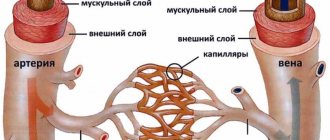
The white matter of the brain coordinates all human activity. It is what connects different parts of the nervous system. White matter is necessary for the two hemispheres to work together.
Classification
Demyelinating disease of the nervous system is classified into different types, which are based on the destruction of the myelin sheath. In this regard, the pathology under consideration is divided accordingly into multiple sclerosis, Marburg disease, Devic disease, progressive multifocal leukoencephalopathy and Guillain-Barré syndrome.
Multiple sclerosis
Multiple sclerosis is characterized as a severe chronic and immunodegenerative disease of the central nervous system, prone to progression. In most cases, the disease occurs at a young age and almost always leads to disability. This demyelinating disease of the central nervous system is assigned code G35 according to ICD-10.
Currently, the causes of the development of multiple sclerosis are not fully understood. Most scientists are inclined to the multifactorial theory of the development of this disease, when genetic predisposition and external factors are combined. The latter include:
- infectious diseases;
- state of chronic intoxication;
- taking certain medications;
- change of place of residence with a sharp change in climate;
- lack of fiber and high-calorie diet.
The progression of multiple sclerosis is characterized by the development of the following symptoms:
- sensory disturbances (goosebumps, numbness, tingling, burning, itching);
- visual impairment (impaired color rendering, decreased visual acuity, blurred picture);
- trembling of the limbs or torso;
- headache;
- speech disorders and difficulty swallowing;
- muscle spasms;
- gait changes;
- cognitive impairment;
- heat intolerance;
- dizziness;
- chronic fatigue;
- libido disturbance;
- anxiety and depression;
- stool instability;
- insomnia;
- autonomic disorders.
Treatment of multiple sclerosis is carried out using methods such as:
- plasmapheresis;
- taking cytostatics;
- prescription of immunosuppressants;
- use of immunomodulators;
- taking beta interferons;
- hormonal therapy.
- symptomatic therapy with the prescription of antioxidants, nootropic drugs and vitamins.
During the period of remission, patients are prescribed sanatorium treatment, massage, and physical therapy. In this case, all thermal procedures should be excluded. To alleviate the symptoms of the disease, medications are prescribed: those that reduce muscle tone, eliminate tremors, normalize urination, stabilize the emotional background, and anticonvulsants.
Marburg disease
Hemorrhagic fever or Marburg disease is an acute infectious pathology caused by the Marburg virus. It enters the body through damaged skin and mucous membranes of the eyes and mouth.
The symptoms of the disease depend on the stage of the pathological process. The incubation period lasts from 2 to 16 days. The disease has an acute onset and is characterized by an increase in body temperature to high levels. Along with fever, chills may appear. Signs of intoxication increase, such as weakness, headache, pain in muscles and joints, intoxication and dehydration. 2-3 days after this, gastrointestinal dysfunction and hemorrhagic syndrome appear.
All symptoms intensify by the end of the first week. Bleeding from the nose, gastrointestinal tract, and genital tract may also be observed. By the beginning of week 2, all signs of intoxication reach their maximum. In this case, convulsions and loss of consciousness are possible. According to the blood test, specific changes occur: thrombocytopenia, poikilocytosis, anisocytosis, granularity of erythrocytes.
If a person is suspected of having Marburg disease, he is urgently hospitalized in the infectious diseases department and must be kept in an isolated box. The recovery period may take up to 21-28 days.
Devic's disease
Neuromyelitis optica or Devic's disease has a chronic pattern similar to multiple sclerosis. This is an autoimmune disease, the causes of which are still unclear. One of the reasons for its development is an increase in the permeability of the barrier between the meninges and the vessel.
Some autoimmune diseases can trigger the progression of Devic's disease:
- rheumatoid arthritis;
- systemic lupus erythematosus;
- Sjögren's syndrome;
- dermatomyositis;
- thrombocytopenic purpura.
The disease has specific symptoms. Clinical manifestations are caused by disruption of conductive impulses. In addition, the optic nerve and spinal cord tissue are affected. In most cases, the disease manifests itself as visual impairment:
- a veil before the eyes;
- pain in the eye sockets;
- blurred vision.
As the disease progresses and there is no adequate treatment, the patient runs the risk of completely losing his vision. In some cases, regression of symptoms with partial restoration of eye function is possible. Sometimes it happens that myelitis precedes neuritis.
Neuromyelitis optica has two course options: a progressive increase in symptoms with simultaneous damage to the central nervous system. In rare cases, a monophasic course of the disease occurs. It is characterized by steady progress and worsening symptoms. In this case, the risk of death is increased. With the right treatment, the pathological process slows down, but complete recovery is not guaranteed.
The second option, the most common, is characterized by an alternating change of remission and exacerbation and is designated by the concept of “recurrent course.” It is also accompanied by visual disturbances and spinal cord dysfunction. During the period of remission, a person feels healthy.
To identify Devic's disease, a set of measures is carried out. In addition to standard diagnostic procedures, lumbar puncture with cerebrospinal fluid analysis, ophthalmoscopy and MRI of the spine and brain are performed.
Progressive multifocal leukoencephalopathy
People with immune deficiency may experience progressive multifocal multifocal leukoencephalopathy. This is an infectious disease caused by the penetration of the JC virus, which belongs to the Polyomavirus family, into the body. A feature of the pathology is that asymmetric and multifocal brain damage occurs. The virus affects the membranes of nerve endings, which are made of myelin. Therefore, this disease belongs to the group of demyelinating pathologies.
Almost 85% of patients with this diagnosis are AIDS or HIV infected. The risk group includes patients with malignant tumors.
Main symptoms of the disease:
- mood swings;
- visual disturbances;
- paresthesia and paralysis;
- memory impairment.
Guillain-Barre syndrome
An acute inflammatory disease characterized by “demyelinating polyradiculoneuropathy.” It is based on autoimmune processes. The disease manifests itself as sensory disturbances, muscle weakness and pain. It is characterized by hypotension and disorder of tendon reflexes. Respiratory failure may also occur.
All patients with this diagnosis should be admitted to the intensive care unit. Since there is a risk of developing respiratory failure and a ventilator may be required, the department must have intensive care.
Causes
You may be interested in: The act of defecation: mechanism, regularity, causes of violations
Focal changes in the brain substance of a dystrophic nature occur in a number of diseases of different origins:
- Changes in vascular origin: atherosclerosis, migraine, hypertension, etc.
- Inflammatory diseases. Multiple sclerosis, Behcet's disease, Sjögren's disease, inflammatory bowel disease (Crohn's disease).
- Infectious diseases. HIV, syphilis, borreliosis.
- Intoxication and metabolic disorders, carbon monoxide poisoning, B12 deficiency.
- Traumatic processes associated with radiation therapy.
- Congenital diseases caused by metabolic disorders.
The occurrence of pathology is caused by impaired blood supply in any part of the brain. It is fraught with loss of function of brain tissue. The more blood flow has decreased, the more noticeable the consequences. An example is damage to the spinal or cerebral blood flow. Such violations progress slowly, but entail serious consequences.
Demyelinating disease of the central nervous system
Demyelinating diseases of the brain and spinal cord are pathological processes that lead to the destruction of the myelin sheath of neurons and disruption of the transmission of impulses between nerve cells of the brain. It is believed that the etiology of diseases is based on the interaction of the body's hereditary predisposition and certain environmental factors. Impaired impulse transmission leads to a pathological state of the central nervous system.
What diseases are these?
There are the following types of demyelinating diseases:
- Multiple sclerosis is a demyelinating disease of the central nervous system. The demyelinating disease multiple sclerosis is the most common pathology. Multiple sclerosis is characterized by a variety of symptoms. The first symptoms appear at the age of 20-30 years, women are more often affected. Multiple sclerosis is diagnosed by the first signs, which were first described by psychiatrist Charcot - involuntary oscillatory eye movements, trembling, scanned speech. Patients also experience urinary retention or very frequent urination, absence of abdominal reflexes, pallor of the temporal halves of the optic discs;
- ADEM, or acute disseminated encephalomyelitis. It begins acutely and is accompanied by severe cerebral disorders and manifestations of infection. The disease often occurs after exposure to a bacterial or viral infection and may develop spontaneously;
- diffuse disseminated sclerosis. It is characterized by damage to the spinal cord and brain and manifests itself in the form of convulsive syndrome, apraxia, and mental disorders. Death occurs within a period of 3 to 6-7 years from the moment the disease is diagnosed;
- Devic's disease, or acute neuromyelitis optica. The disease begins as an acute process, is severe, and progresses, affecting the optic nerves, which causes complete or partial loss of vision. In most cases, death occurs;
- Balo's disease, or concentric sclerosis, periaxial concentric encephalitis. The onset of the disease is acute, accompanied by fever. The pathological process occurs with paralysis, visual disturbances, and epileptic seizures. The course of the disease is rapid - death occurs within a few months;
- leukodystrophies - this group contains diseases that are characterized by damage to the white matter of the brain. Leukodystrophies are hereditary diseases; as a result of a gene defect, the formation of the myelin sheath of the nerves is disrupted;
- Progressive multifocal leukoencephalopathy is characterized by decreased intelligence, epileptic seizures, the development of dementia and other disorders. The patient's life expectancy is no more than 1 year. The disease develops as a result of decreased immunity, activation of the JC virus (human polyomavirus 2), often found in patients with HIV infection, after bone marrow transplantation, in patients with malignant blood diseases (chronic lymphocytic leukemia, Hodgkin's disease);
- diffuse periaxial leukoencephalitis. A hereditary disease that most often affects boys. Causes visual, hearing, speech, and other disorders. Progresses quickly - life expectancy is just over a year;
- Osmotic demyelination syndrome is very rare and develops as a result of electrolyte imbalance and a number of other reasons. A rapid increase in sodium levels leads to the loss of water and various substances from brain cells, causing the destruction of the myelin sheaths of nerve cells in the brain. One of the posterior parts of the brain is affected - the Varoliev bridge, which is most sensitive to myelinolysis;
- myelopathy is a general term for lesions of the spinal cord, the causes of which are varied. This group includes: tabes dorsalis, Canavan disease, and other diseases. Canavan disease is a genetic, neurodegenerative autosomal recessive disease that affects children and causes damage to nerve cells in the brain. The disease is most often diagnosed in Ashkenazi Jews living in Eastern Europe. Tabes dorsalis (locomotor ataxia) is a late form of neurosyphilis. The disease is characterized by damage to the posterior columns of the spinal cord and spinal nerve roots. The disease has three stages of development with a gradual increase in symptoms of damage to nerve cells. Coordination when walking is impaired, the patient easily loses balance, bladder function is often disrupted, pain appears in the lower limb or lower abdomen, and visual acuity decreases. The most severe third stage is characterized by loss of sensitivity of muscles and joints, areflexia of the tendons of the legs, development of astereognosis, the patient cannot move;
- Guillain-Barré syndrome - occurs at any age, refers to a rare pathological condition that is characterized by damage to the peripheral nerves of the body by its own immune system. In severe cases, complete paralysis occurs. In most cases, patients recover completely with adequate treatment;
- neural amyotrophy of Charcot-Marie-Tooth. A chronic hereditary disease, which is characterized by progression, affects the peripheral nervous system. In most cases, the myelin sheath of nerve fibers is destroyed; there are forms of the disease in which pathology of the axial cylinders in the center of the nerve fiber is detected. As a result of damage to the peripheral nerves, tendon reflexes fade and atrophy of the muscles of the lower and then upper extremities occurs. The disease belongs to progressive chronic hereditary polyneuropathies. This group includes: Refsum's disease, Roussy-Lévy syndrome, Dejerine-Sotta hypertrophic neuropathy and other rare diseases.
Genetic diseases
When damage to nerve tissue occurs, the body responds by destroying myelin. Diseases that are accompanied by the destruction of myelin are divided into two groups - myelinoclastics and myelinopathy. Myelinoclasty is the destruction of the membrane under the influence of external factors.
Myelinopathy is a genetically determined destruction of myelin associated with a biochemical defect in the structure of the neuron membrane. At the same time, this distribution into groups is considered conditional - the first manifestations of myelinoclasty may indicate a person’s predisposition to the disease, and the first manifestations of myelinopathy may be associated with damage caused by external factors.
Multiple sclerosis is considered a disease of people with a genetic predisposition to the destruction of the membrane of neurons, metabolic disorders, a deficient immune system and the presence of a slow infection. Genetic demyelinating diseases include: neural amyotrophy of Charcot-Marie-Tooth, hypertrophic neuropathy of Dejerine-Sottas, diffuse periaxial leukoencephalitis, Canavan disease and many other diseases. Genetic demyelinating diseases are less common than autoimmune demyelinating diseases.
Signs
You may be interested in:Hot spring "Avan" in Tyumen: detailed information
The signs of focal changes in the brain substance of a dystrophic nature are also different. With focal changes, not the entire brain suffers, but only its individual parts. Tissue degeneration occurs when there is insufficient supply of nutrients necessary for the normal functioning of the body’s nervous systems. We are talking about proteins - the building material of the human body. Proteins break down into amino acids, which, in turn, stimulate the nervous system. It also requires fats and carbohydrates - the main sources of energy needed by every living creature.
Of the vitamins, the brain needs B1 (activates its work), B3 (provides energy at the intracellular level), B6 (it is difficult to imagine metabolic processes without it, in addition, it is also a kind of antidepressant), B12 (promotes memory preservation and helps to stay alert) . All these vitamins can be obtained in sufficient quantities by creating the right diet.
Pathologies
Focal changes in the substance of the brain of a dystrophic nature most often take the form of pathologies such as:
- A cyst is a small cavity that is filled with fluid. It often interferes with the normal functioning of neighboring areas of the brain, as it compresses blood vessels. Cysts are divided into intracerebral (cerebral) and arachnoid. The latter appears in the meninges. Its occurrence is promoted by the accumulation of cerebrospinal fluid and inflammatory processes. Cerebral occurs in place of dead brain tissue.
- Necrotic state of tissue - appears when the supply of important nutrients to areas of the brain for any reason deteriorates. Dead cells form so-called dead zones and are not regenerated.
- Hematomas and brain scars occur after severe trauma or concussion. Foci of this type lead to structural damage.
Diagnostics
The complete picture of focal changes in the brain substance of a dystrophic nature is determined using an MRI study. This procedure allows you to see even small areas of transformation in the white matter. And they, in turn, lead to cancer and stroke.
Focal dystrophic lesions come in different sizes and differ in location. Based on this, the examination may show some types of disorders.
In the cerebral hemisphere, blockage of vital arteries is usually diagnosed due to abnormal development of the embryo or acquired atherosclerotic plaques. A herniated cervical spine is also detected.
Changes in the white matter of the brain indicate hypertension and congenital developmental anomalies. In other cases, numerous areas of brain pathology may indicate a pre-stroke condition, senile dementia, or epilepsy.
Sometimes doctors perform tests on a patient to detect the presence of cognitive impairment. That is, cognitive dysfunction. Such as orientation in space and time, understanding of external processes, the ability to remember information, drawing, writing, reading, etc.
Focal changes in the brain substance of a dystrophic nature can develop in three ways:
- In the first case, the disease is remitting in nature. Symptoms increase gradually, the condition worsens, and brain productivity decreases. But from time to time, remissions occur—temporary improvements in health, after which the patient becomes worse again.
- Progressive focal changes in the brain substance of a dyscirculatory dystrophic nature develop very quickly. Each stage of the disease takes no more than two years, which is considered a short period for organic brain lesions.
- Typically, the deterioration of a person suffering from focal changes lasts for many years, eventually leading to dementia.
It should be remembered that single focal changes in the brain substance of a dystrophic nature often appear in young people, and single damage to the white matter in an elderly person is considered normal. Structural disorders of the cerebral arteries of the atherosclerotic type appear in 50% of patients over 50 years of age. For the most part, hypertensive patients suffer from this. Therefore, you need to show the MRI result to a neurologist so that he can determine the severity of the disorders in the brain by comparing the MRI result and the clinical picture of the disease.
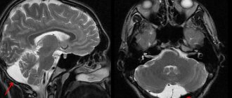
Vascular changes in the brain on MRI
MRI: ischemic zone during stroke (highlighted with a red oval)
If cerebrovascular pathology is suspected, special attention is paid to the condition of the arteries during MRI of the brain. The study always involves the introduction of contrast, and it is called magnetic resonance angiography. In urgent situations in case of vascular accidents, CT is performed, since X-ray diagnostics takes less time and clearly demonstrates the area of damage during hemorrhages, but after stabilization of health, magnetic resonance angiography is quite justified. The study shows atherosclerotic plaques, blood clots and aneurysms (protrusions), their localization, and wall deformation.
With an ischemic stroke, darkened and blurred areas of irregular shape are visible almost immediately on magnetic tomograms, appearing on a computer scan only at the end of the first day. The lesion is often unilateral. The outpouring of blood from a ruptured vessel gives an intense light color, but only in the first hour and a half from the moment of the disaster, and then becomes invisible on MRI, although it is clearly visualized using CT. As a consequence of a stroke, a fluid-filled pseudocyst is formed, and deformation of the nerve tissue appears. MRA is indispensable in the diagnosis of tumor angiogenesis. Increased vascularization of a pathological focus is always suspicious of a malignant neoplasm, which grows and feeds due to increased blood flow. If the vessels do not keep up with the growth of the tumor, areas of ischemia and necrosis appear.
Diet
In the early stages of this disease, it is enough to reconsider your lifestyle and diet, choosing a more gentle regimen and diet. In the diet, it is advised to reduce the consumption of animal fats, and it is better to completely replace them with vegetable ones. You should eat fish and seafood instead of fatty meat, and reduce the amount of salt in your diet. Fresh vegetables and fruits will be of great benefit.
Treatment
There are a huge number of focal anomalies, so each has its own cause. Treatment of brain pathologies is based on the destruction of those factors that led to the appearance of lesions in brain tissue. In addition to eliminating the underlying disease, the doctor may also prescribe vitamins and medications to help combat the deterioration of cerebral blood flow.
The treatment process depends directly on what somatic problems in the body led to the occurrence of lesions in the brain. For infections, for example, antibiotics are taken; for injuries, diuretics, decongestants, and anticonvulsants are taken. If the damage to the brain tissue was caused by a circulatory disorder, then nootropics and coagulants are prescribed.
Source
Symptoms
Demyelination always manifests itself as a neurological deficit. This sign indicates the beginning of the process of myelin destruction. The immune system is also involved. Brain tissue - spinal and brain - atrophies, and expansion of the ventricles is observed.
Manifestations of demyelination depend on the type of disease, causative factors and location of the lesion. Symptoms may be absent when the damage to the brain substance is minor, up to 20%. This is due to a compensatory function: healthy brain tissue performs the tasks of the affected areas. Neurological symptoms rarely appear - only when more than 50% of the nervous tissue is damaged.
The following are common signs of demyelinating brain diseases:
- paralysis;
- limited muscle mobility;
- tonic spasms of the limbs;
- dysfunction of the intestines and bladder;
- pseudobulbar syndrome (impaired pronunciation of sounds, difficulty swallowing, change in voice);
- impaired fine motor skills of the hands;
- skin numbness and tingling;
- visual dysfunction (decreased visual acuity, blurred images, fluctuations in the eyeballs, color distortions).
Neuropsychological disorders characteristic of the pathology in question are caused by memory deterioration and a decrease in mental activity, as well as changes in behavior and personal qualities. This is manifested by the development of neuroses, depression, dementia of organic origin, emotional swings, severe weakness and decreased performance.