Calcium is an essential trace element necessary for the functioning of the body. But its excess, caused by metabolic disorders, can provoke a disease such as calcification of the aortic walls. Excess calcium enters the blood, and then begins to be deposited on the vascular walls, growing and gradually clogging the vessels. It is especially dangerous if it occurs in the largest and most important vessel, the aorta and aortic valve. In this case, they speak of aortic calcification, and the rapid development of the disease requires immediate diagnosis and treatment.
The formation of calcium deposits can damage the valve.
Development and classification of aortic calcification
Calcium deposits can occur in various tissues. According to the type of process, there is the following classification of calcification:
- Metastatic – with massive accumulation of calcium in tissues. It is accompanied by increased leaching of the element from bone tissue during osteoporosis, injuries, and oncological bone diseases.
- Dystrophic - accumulation is limited, occurs due to focal areas where absorption is impaired, resulting in calcification.
- Metabolic - due to metabolic disorders, the element is not retained in the blood, but accumulates in tissues where it should not be.
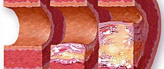
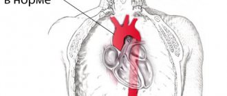
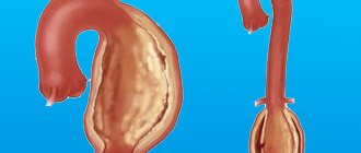
Calcification or calcification of the aortic arch consists of the deposition of calcium salts on its walls or valve. In this case, the elasticity of the walls is lost, they become fragile, and the valve loses its ability to function normally. The entire blood flow from the heart passes through the aorta, which is distributed through various branches to all organs. The condition of this vessel determines the nutrition of the brain, the functioning of the abdominal and pelvic organs, and the quality of human life in general.
Along with deposits and narrowing of the lumen of the vessel, degenerative processes occur in the vessel itself, creating an increased load on its walls. Lesions may spread further throughout the system, involving the ventricles and mitral valve. Sometimes cholesterol accumulations are added to calcified plaques, in which case atherocalcinosis is diagnosed. Aortic calcification is the cause of the development of aortic valve stenosis and subsequent heart valve defects. The fusion of the leaflets interferes with normal blood flow, pressure differences occur between the ventricle and the aorta, which leads to a weakening of the ventricle’s ability to contract and overload of the left atrium.
Aortic stenosis
Aortic stenosis is a fusion of the aortic valve leaflets. Since this pathology leads to a narrowing of the valve opening, the release of blood flow into the aorta is difficult, resulting in a significant increase in the load on the left ventricle, which leads to myocardial hypertrophy.
There are three forms of aortic stenosis: subvalvular, valvular and supravalvular. Among patients with heart defects, aortic stenosis occurs in 84% of cases, and in men it is three times more common. Of all the forms of aortic stenosis, valvular stenosis is the most common. Aortic stenosis can also be congenital or acquired (90-97%). The supravalvular form is a congenital pathology.
Calcification of the aortic walls: causes
The main cause of vascular calcification is disturbances in metabolic systems. They can be caused by:
- Diseases of the parathyroid glands, which regulate the processes of calcium removal from bones into the blood. These pathologies include malignant tumors.
- Diseases of the endocrine system, disturbances in the normal production of hormones.
- Problems with digestion, intestinal diseases, lack of enzymes, which leads to disturbances in the absorption of calcium.
- Diseases of the kidneys and urinary system, resulting in delayed removal of the element from the body.
- Infectious diseases suffered with complications.
Factors contributing to the occurrence of this pathology include:
- injuries, including fractures;
- oncological diseases;
- excess vitamin D;
- heart defects;
- blood diseases;
- vascular pathologies;
- operations on the heart and blood vessels;
- osteoporosis;
- elderly age;
- heredity;
- diabetes;
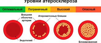
- atherosclerosis;
- smoking and excess weight, unhealthy lifestyle;
- lack of magnesium.
The presence of risk factors in combination with metabolic disorders creates a high probability of rapid development of the disease. The danger, as with many other vascular pathologies, is that in the first stages the symptoms practically do not appear. And only with significant disturbances in blood flow can signs of decreased functionality of some organ be noticed.
Causes of aortic stenosis
The causes of acquired valvular or subvalvular stenosis can be aortic atherosclerosis, rheumatic valve disease, aortic calcification, and infective endocarditis. Sometimes the cause is diabetes mellitus, chronic kidney disease, Paget's disease, systemic lupus erythematosus or carcenoid syndrome.
Symptoms of aortic stenosis may not appear for a long time, especially at an early age. However, over time, the patient begins to complain of squeezing, aching pain in the chest (in the heart), shortness of breath, fainting, increased heart rate, nausea, and fatigue. Since cardiac output is reduced, this can lead to loss of consciousness. In later stages, aortic stenosis can cause a sudden attack of suffocation (cardiac asthma).
Also, in patients with aortic stenosis, the likelihood of sudden death increases, especially during physical activity. Hypertrophied myocardium causes acute coronary insufficiency and arrhythmias that threaten the patient’s life.
Symptoms of aortic calcification
Calcification of the aortic walls in its pure form is rare, since the process quickly spreads to the heart valves. Until then, there may be a complete absence of symptoms, so people go to the doctor with already pronounced heart problems. Further general symptoms appear:
- fatigue and fatigue;
- pain in the chest and heart;
- heart rhythm disturbances;
- dizziness, clouding of consciousness;
- shortness of breath, which in severe stages can be observed at rest and sleep.
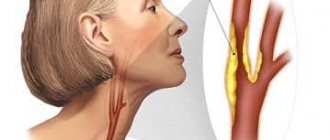
The aorta has two sections, thoracic and abdominal. Through the first, the brain is nourished; it is responsible for the functioning of the upper body. The abdominal region provides blood supply to the abdominal organs, pelvis, and lower extremities. Depending on the location of the main lesion, symptoms will vary.
Calcification of the upper aorta has the following characteristic features:
- headache;
- fainting;
- tingling in the limbs, chills;
- sore throat, difficulty swallowing;
- hoarse voice;
- increased blood pressure;
- chest pain radiating to the neck and shoulder blades, having a paroxysmal nature and lasting for several days.
Symptoms of abdominal aortic calcification:
- abdominal pain that gets worse after eating;
- digestive problems;
- bloating and constipation;
- weight loss and appetite;
- numbness and tingling in the legs, especially the feet;
- dry skin on the legs, ulcerative lesions;
- impaired coordination in the lower extremities, lameness, heaviness and pain, especially during exercise.
The consequence of progressive calcification is heart rhythm disturbances, ischemia, heart failure, difficulty breathing, cardiac asthma, and heart attack. One of the most dangerous consequences is the occurrence of an aneurysm, the rupture of which leads to profuse hemorrhage and death.
Symptoms and degrees of calcification
An increase in the concentration of free calcium in the blood serves as a provoking factor not only for calcification of the heart valves, but also for the development of pathologies such as heart failure, heart attack, and stroke. In this case, the symptoms of calcification are combined with signs of concomitant diseases.
Degree of development of calcification of heart valves
| Degree of disease | Signs of mitral valve pathology | Signs of aortic valve pathology |
| Compensated | An increase in the level of calcium in the blood, calcification of the valves. The tissues are compacted, there is a slight deterioration in hemodynamics | Difficulties arise with the opening of the valves, but the compensation mechanisms of the left ventricle keep the situation under control. During physical activity, minor heart pain is possible, which increases as the disease progresses. |
| Subcompensated | Shortness of breath during physical activity, tachycardia, arrhythmia, wet cough (sometimes with blood clots), hoarseness | Shortness of breath during physical activity, heart pain. Over time, signs of respiratory failure and aortic narrowing occur at rest |
| Decompensated | It develops rarely - with long-term ignoring of symptoms or an avalanche-like progression of the pathology. Pronounced weakness, shortness of breath with little physical activity. Cough with scarlet sputum, pale skin. Requires emergency medical attention | Signs of increased volume of both ventricles. Severe heart failure. The condition is extremely dangerous |
Aortic valve calcification
Patients do not feel any noticeable discomfort for a long time. Signs of pathology appear with stenosis of the aortic valve opening up to half of its passage. The progression of heart failure is accompanied by:
- dizziness;
- angina attacks;
- fainting with a sudden change in body position;
- shortness of breath at night;
- attacks of cardiac asthma and pulmonary edema (in severe cases).
- Aortic valve insufficiency: symptoms, diagnosis, treatment
The combination of heart pain and asthmatic attacks is very unfavorable. If left untreated, aortic valve calcification can cause bleeding in the lower digestive tract. If the valve opening is severely narrowed, sudden cardiac arrest may occur in elderly patients.
Mitral valve calcification
When blood stagnates in the pulmonary circulation, caused by calcification of the mitral valve, respiratory failure occurs, accompanied by characteristic symptoms. In case of serious damage, wheezing and shortness of breath at rest (to alleviate this, you need to sit down with your legs down) are observed, as well as pulmonary edema, characterized by:
- suffocation;
- wet wheezing when breathing;
- chest pain;
- fear of death.
Sometimes pinkish foam appears when exhaling. In such a case, the patient should be seated comfortably, with all the windows in the room opened, and tourniquets applied to his legs. You can hold a cotton swab soaked in alcohol to your nose and offer diuretics. Further therapy should be carried out exclusively in a hospital. Mitral valve calcification is similar in symptoms to signs of cardiosclerosis, rheumatism, and hypertension, so calcification often remains untreated, provoking the development of mitral valve insufficiency and stenosis.
Calcification of the aortic walls: diagnosis
Calcification can be easily diagnosed using modern research methods. But the problem of making a diagnosis is that patients for a long time do not suspect the presence of any abnormalities in their health, and even the doctor has no reason to issue a referral for examination. The reason to diagnose aortic calcification is the presence of the following pathologies:
- heart disease with unknown causes and no abnormalities in tests or ultrasound;
- heart murmurs in the absence of heart disease;
- attacks of tachycardia;
- cardiac ischemia.
Any problems with blood circulation or cardiac activity, for which it is difficult to reliably identify the cause, should be alarming; they are grounds for prescribing additional examinations. Among them:
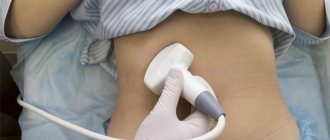
- Ultrasonography - allows you to identify areas with increased density of the aortic walls, but does not give an idea of the condition of the valve.
- Ultrasound diagnostics using echo signals helps detect calcium deposits.
- X-ray is more effective than previous methods in showing a picture of the vascular system and calcareous deposits, including on the valve.
- Computed tomography visually and clearly determines calcification and the extent of its spread in the aorta and heart.
- Ultrasound densitometry, which allows using a special scale to determine the severity of calcification.
Laboratory tests, especially in the early stages, do not have specific abnormalities, therefore they are not very informative in the diagnosis of calcification. Particular attention should be paid to the slightest suspicion of the disease in pregnant women. Their cardiovascular system is under increased stress, in addition, calcium deposits can accumulate in the placenta, which can be detrimental to the development of the fetus.
Diagnosis of aortic stenosis
If symptoms of aortic stenosis appear, you should contact a cardiologist or begin an examination with a general practitioner. Using a stethoscope, the doctor will listen for a murmur over the aorta as the left ventricle contracts. An electrocardiogram can reveal ventricular hypertrophy. Ventricular enlargement can also be detected using x-ray. An echocardiogram uses ultrasound to evaluate the condition of the valves and heart muscle.
Also, an information research method is cardiac catheterization (insertion of a catheter into an artery). Provides information about intracardiac pressure, the location of the narrowing, the concentration of oxygen in the blood, the condition of the valve and the presence of a defect.
The most informative diagnostic method for making a decision on surgical intervention is an angiocardiographic study. It takes place in two stages: ventriculography and oorthography.
Author of the article:
Mochalov Pavel Alexandrovich |
Doctor of Medical Sciences therapist Education: Moscow Medical Institute named after. I. M. Sechenov, specialty - “General Medicine” in 1991, in 1993 “Occupational diseases”, in 1996 “Therapy”. Our authors
Treatment of aortic calcification
Important for the success of treatment are determining the root cause of metabolic disorders and correcting the impact of negative factors. First of all, this is a change in diet and lifestyle. Drug therapy is aimed at improving blood circulation to prevent disruptions in the functioning of vital organs. Often the initial cause of the accumulation of calcium salts is increased cholesterol levels and the formation of atherosclerotic plaques. In this case, all means of treating atherosclerosis are used.
With calcinosis, complications may arise and symptoms of cardiac dysfunction may appear, then appropriate medications are prescribed:
- reducing blood pressure;
- improving blood circulation and relieving congestion;
- drugs that reduce blood thickness;
- remedies for ischemia and arrhythmia.
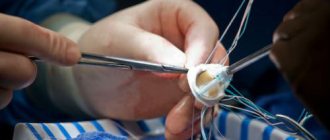
In advanced conditions, when treating aortic calcification with medications is ineffective, surgical intervention is used. This can be an open operation with cleaning of the vascular walls or manipulation using a catheter, as well as bypass surgery and prosthetics.
Calcification of the aortic walls: therapeutic diet
Conservative treatment will not be effective without following a special diet aimed at normalizing metabolic processes. Specific recommendations should be developed by the attending physician, taking into account the medical history and the reasons that caused the pathology. But there are a number of general principles for developing a diet for aortic calcification:
- Avoid foods rich in calcium.
- Consumption of foods containing magnesium.
- Limit salt.
- Reduce calories.
- Avoid fatty and fried foods.
Prohibited foods for aortic calcification include dairy products, fatty meats, spices, yeast baked goods, desserts, herbs, and cocoa. It is recommended to consume bran, nuts, buckwheat and barley. It is also necessary to strictly monitor the amount of food eaten, avoid excess weight and lead an active and healthy lifestyle.
Varieties
In medical practice, calcification is divided not only by complexity. There are its varieties depending on the manifestations and causes of occurrence. The 4 types of this disease are different:
- Lock.
- Universal.
- Dystrophic.
- Idiopathic.
Metastatic
This type is caused by high levels of calcium and vitamin D. The disease gains momentum against the abnormal functioning of internal organs:
- heart;
- liver;
- intestines;
- Bud.
The risk group includes both adults and children. For a long period of time, the disease does not have any special symptoms.
Universal
This type of disease occurs in people with hypersensitivity to calcium. The disease passes with clear symptoms. Continue very quickly.
Dystrophic
The limestone shell is formed in this variant. It involves the heart and other internal organs, preventing them from functioning properly. As a result, various other diseases develop in the patient.
Idiopathic
Newborns are at risk. Hence, this type is also called congenital. The cause of idiopathic calcinosis is a pathology in the development of the cardiovascular system.
Folk remedies for calcinosis
In addition to diet and traditional therapy for aortic calcification, treatment with folk remedies can also be used. With their help it is impossible to stop the pathological process, but the benefits in improving the functioning of the body are significant. When choosing a folk remedy, you need to take into account the presence of any chronic diseases and coordinate your intentions to take a folk remedy with your doctor.
Herbs with medicinal effects include:
- pharmaceutical camomile;
- Birch buds;
- motherwort.
There are a number of useful recipes that prevent the development of aortic calcification:
- 300 grams of crushed garlic is infused into 200 ml. vodka for 10 days, after that for up to 5 days taken three times a day before meals in a dosage of up to 15 drops. The first dose should start with 1 – 2 drops. Next, drink for another 5 days according to the same scheme, but with a gradual decrease to 1 drop. The tincture can be diluted in 30 ml before use. milk.
- Herbal mixture of St. John's wort, birch buds, immortelle, chamomile and motherwort. To prepare, mix the herbs in equal parts and brew at the rate of 400 ml. water per tablespoon. Herbs must be chopped before brewing. Drink warm in two parts throughout the day.
- Mix chopped garlic, lemon juice and honey in equal proportions, take a teaspoon three times a day for a month.
- Mix freshly squeezed carrot, beet and pineapple juice and drink 100 ml each. twice a day.
Aortic calcification is difficult to detect in the early stages and a very dangerous disease in its advanced form. Therefore, you need to take all measures to prevent it, lead a healthy lifestyle and monitor your diet. If there are risk factors, periodic preventive examinations and monitoring of the course of chronic diseases are necessary.