Tells Candidate of Medical Sciences, general practitioner, cardiologist at the Atlas biomedical holding Maria Kirillova.
Thrombosis is a condition in which blood clots (thrombi) form in blood vessels (veins and arteries). Most often, blood clots form in the veins or venous thrombosis occurs (in which a blood clot forms in a section of the venous vascular bed, mainly in the legs). Any blood clot interferes with normal blood flow, but a disaster occurs at the moment when the clot reaches a size comparable to the size of the vessel and completely blocks the blood flow, or when the clot (or a small part of it) breaks off and, along with the blood flow, enters the vessels of the lungs (this condition called pulmonary embolism.
Mechanism of formation and causes
The uniqueness of the human body is that any damage to it causes a protective reaction. So, when a blood vessel ruptures, platelets stick together and stick to the walls of the vessels. The blood clot formed in this way prevents bleeding and prevents pathogenic microorganisms from penetrating the damaged tissue. Over time, blood clots dissolve and tissues are restored. This mechanism does not relate to diseases and is natural.
However, there is pathological thrombus formation, which occurs due to various reasons.
Changes in the walls of blood vessels
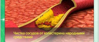
As a rule, vascular walls are subject to changes during atherosclerosis. According to medical studies, 85% of the 350 patients who died from this disease had blood clots.
The appearance of blood clots is caused by biochemical disorders inherent in atherosclerosis, in which excess cholesterol contributes to the formation of plaques in the vessels and the deposition of calcium in them.
As a result of this process, the vessels lose their softness and elasticity, become fragile, brittle and ulcerated. It is the damaged areas that become the site of dislocation of blood clots, which are designed to close the wounds.
In addition, some chronic pathologies contribute to the deformation and loss of elasticity of blood vessels, and the formation of growths in them.
Increased blood viscosity
Blood clots that form due to increased blood viscosity are its most dangerous consequence.
Many factors can lead to a decrease in the rheological properties of blood:
- genetic defects of the coagulation system;
- some diseases (oncology, autoimmune pathologies, diabetes, hepatitis, liver cirrhosis and others);
- dehydration;
- severe blood loss;
- sugar, carbohydrates, etc. consumed in large quantities.
In this case, preventing the formation of blood clots is possible only by getting rid of the underlying pathology.
Blood flow disorders
Thrombosis directly depends on the disruption of blood flow. Thus, when a person’s mobility is insufficient, it slows down, and high blood pressure is accompanied by vortex blood flow.
The occurrence of thrombosis is directly dependent on:
- Patient's gender. According to medical research, men are more likely to develop blood clots.
- Age. Due to loss of vascular elasticity and slowing blood flow.
- The central nervous system, which has a direct effect on metabolic processes. When the balance in the central nervous system is disturbed, temporary or permanent circulatory problems are often observed.
- Presence of oncology. As a result of cancer pathology, the coagulation and anticoagulation systems malfunction, an imbalance between them occurs, which provokes thrombosis.
- Hereditary predisposition to clot formation and cardiovascular disease.
- Blood characteristics. Changes in the properties or composition of the fluid may contribute to the appearance of undesirable formations.
- Infections that negatively affect blood properties.
- Cardiac disorders. Patients with heart defects, atherosclerosis, hypertension, and mitral stenosis are susceptible to the formation of blood clots.
- Pregnancy. The period of waiting for a baby is characterized by an increase in proteins in a woman’s body, which helps to increase blood clotting. Vessels can also rupture during childbirth.
- Climate, especially its changes. In case of changes in weather conditions, a negative reaction of the nervous system is observed, usually with cardiovascular pathologies and slow blood flow.
- Food. Poor quality food, as well as eating it in large quantities, contributes to the formation of blood clots in blood vessels.
- Activity, the absence of which leads to slow blood circulation and venous stagnation.
- The use of certain medications, including hormonal drugs, which negatively affect the properties of the blood.
- The presence of bad habits: smoking and alcohol abuse.
- Surgical interventions, the use of general anesthesia, as well as reduced mobility in the postoperative period.
- Injury to any organs.
- Physical activity, which may be excessive or, conversely, insufficient for the normal functioning of the body.
- Varicose veins. Increased lumen in the veins caused by varicose veins leads to impaired blood flow and the appearance of pathological clots.
The occurrence of blood clots can be observed in blood vessels located anywhere in the circulatory system, mainly in the presence of atherosclerotic plaques, insufficient elasticity and inflammatory processes in the walls.
Then you will find out...
2/3 of venous thrombosis are asymptomatic or with mild symptoms. A person is bothered by moderate pain in the leg and slight swelling, but not so severe as to force him to see a doctor. Of the three cases of thrombosis, one progresses, and two resolve spontaneously.
However, this does not always mean that resolved thrombosis has passed without a trace. Even in this case, irreversible changes occur on the wall and valve of the affected vein, which lead to the development of chronic venous insufficiency. With duplex scanning of blood vessels, doctors often see post-thrombotic changes in the deep veins of the leg in patients who have never been diagnosed with venous thrombosis.
Blood control and nutrition without “greens”. A doctor on how to live with thrombosis Read more
Kinds
According to their structure, blood clots are:
- white (platelet), affecting arteries and small vessels and characterized by slow formation with increased blood flow;
- red (blood fibrin). As a rule, they affect the veins and appear when blood clotting increases and blood flow slows;
- mixed (layered), the appearance of which is possible in the aortic aneurysm, veins;
- hyaline, which affect small vessels in various organs. The mechanism of their formation is not fully understood. Causes of occurrence: shock, burns, electrical injuries, etc.
In addition, pathological formations can be:
- arrowroot, occurring in old age when the water-electrolyte balance is disturbed with partial dehydration. They affect the superficial veins in the extremities and the sinuses of the lining of the brain;
- tumors that appear during metastases of malignant tumors;
- septic, which are the result of inflammation and infection.
There is also a classification of blood clots depending on their size and location in the vessels:
- Parietal. The most common type. They are localized in large vessels, as well as on the chambers and valves of the heart. Cover the vessel in diameter by no more than 50%. Initially they are not dangerous, but their layering one on top of another leads to group accumulations of blood clots and complete blockage of the blood vessel.
- Clogging. They are capable of blocking the lumen in blood vessels by more than 50%, disrupting blood circulation. As a rule, small blood vessels are affected. Appear when wall clots grow.
- Progressive. A rapidly growing formation that initially involves the venous wall, and then the collecting venous vessels.
- Globular. Located in the left atrium, they have the ability to quickly increase and separate from the place of their formation.
- Dilation. The aneurysm cavities are greatly stretched and grow greatly, causing the risk of their separation and complete blocking of blood flow.
The type of blood clot and its danger can only be determined by a specialist during the diagnostic process.
Consequences
Untimely detection and elimination of thrombosis is fraught with incurable consequences and death.
The presence of pathological clots in the body can provoke:
- thrombophlebitis, in which thrombosis is accompanied by inflammation of the venous walls, leading to complete closure of the lumen of the vessel;
- thromboembolism – separation of a blood clot from the site of its formation and its entry into the bloodstream, which causes acute blockage of the vessel (embolism).
With thrombophlebitis, blood clots can be located in both deep and superficial veins.
Complete or partial blockage of blood vessels reduces or stops the blood supply to organs and tissues, leading to their hypoxia.
If clots are not removed in a timely manner, even if the outcome is favorable, the vein valves are destroyed and post-thrombophlebic disease develops, which makes the patient’s full recovery impossible.
Modern medicine does not know why blood clots break off, but in any case, this process occurs under the following conditions:
- free placement of the thrombus in the vessel (does not completely close its lumen). Typically, such clots are observed in the veins of the legs and heart;
- sufficient blood flow to separate the mass from the vessel.
Migrating blood clots are very dangerous. Clots are capable of traveling long distances and fragmenting, which causes blockage of many vessels.
An example is pulmonary embolism, a blood clot formed in the lower extremities. People often do not attach much importance to varicose veins and thrombophlebitis, not even suspecting the threat of sudden death.
Symptoms
The symptoms of thrombosis are influenced not only by the location of the blood clot in the body, but also by the degree of vascular occlusion.
Clinical manifestations of the disease are directly dependent on the location of the thrombus
Venous thrombosis
Thrombus damage to the iliac, femoral, popliteal veins (deep great vessels) leads to the occurrence of:
- swelling;
- pastosity of the affected tissues,
- sensitivity disorder (numbness, tingling, goosebumps);
- nagging pain;
- trophic changes in the skin.
Portal vein thrombosis is characterized by:
- intense abdominal pain and bloating;
- constipation;
- black, tarry stool;
- bleeding from the anus;
- enlarged spleen;
- ascites;
- purulent peritonitis.
A renal vein affected by a thrombus does not cause renal colic, but the patient experiences severe lower back pain and decreased diuresis. Blood may appear in the urine.
With jugular vein thrombosis, septic complications and papilledema develop. Drug addicts and patients with malignant neoplasms are usually susceptible to pathology.
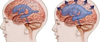
Thrombosis of the cerebral sinuses is rare and manifests itself:
- feeling of nausea;
- low-grade fever;
- meningeal signs;
- aching joints;
- increased intracranial pressure;
- cerebral edema;
- impaired blood circulation in the eyes.
The presence of a blood clot in the veins can contribute to the rapid proliferation of microorganisms, as a result of which the surrounding tissues undergo an inflammatory process, and then the patient begins to develop sepsis.
Arterial thrombosis
Arterial thrombosis can affect large vessels in various parts of the human body. The most common pathologies are:
- Abdominal aorta. The clot provokes circulatory problems in the abdominal and pelvic organs. The patient experiences a feeling of weakness in the lower extremities, pain, and intermittent claudication. Men are subject to impotence. The pulse in the legs weakens and then cannot be felt completely. The skin turns pale and becomes cold.
- Kidney vessels. A person’s blood pressure increases, which cannot be reduced. There are traces of blood in the urine. With rapid blockage, the kidney dies.
- Brachial artery. Thrombosis may have no symptoms. When a large part of the lumen closes, the hand begins to go numb, becomes cold, and a weakening of the pulse is observed. In case of complete occlusion, the limb may be amputated.
- Lower limbs. The patient is susceptible to swelling, muscle weakness and pain in the lower limb. Hair growth on the legs is significantly reduced or stops altogether. The skin turns red, and in advanced cases becomes purple-bluish. Pathological processes are fraught with gangrene.
- Brain. The patient's body temperature rises, headaches, drowsiness and nausea appear. Convulsive attacks, paralysis, impaired consciousness and other symptoms indicating a stroke may occur.
- Coronary artery. Lack of oxygen and nutrients in the heart causes the risk of angina or heart attack. Pain in the heart area can be of different types: pressing, squeezing, stabbing, etc.
- Pulmonary artery. The lightning-fast course of the disease leads to instant death. Otherwise, the onset of pathology is characterized by pain in the chest area, cough, hemoptysis, blue fingers, elevated body temperature, etc.
Blood clots in blood vessels provoke dangerous consequences, which can be avoided with timely diagnosis and effective treatment.
Advantages of thrombosis treatment at the Swiss University Hospital
- In our Center for Cardiovascular Surgery, treatment of deep venous thrombosis is carried out not only by conservative methods. We have widely introduced our own minimally invasive surgical interventions into clinical practice, thanks to which we were able to reduce not only the trauma of surgical intervention, but also save the patient from complications, which leads to rapid healing and a reduction in the duration of the rehabilitation period, improving the quality of life.
- To prevent venous thromboembolic complications, our clinic widely uses complex methods of preoperative, intraoperative, postoperative prevention of venous thromboembolic complications in accordance with the degree of risk of their occurrence.
- Vascular surgeons (phlebologists) at our clinic have many years of experience in performing surgical interventions, each of our specialists is fluent in all the techniques used in our clinic. Each surgeon has performed more than 2,500 successful operations for varicose veins, including those complicated by varicothrombophlebitis and deep vein thrombosis, with analysis of long-term results.
- Ultrasound diagnosis of thrombosis of the main veins of the lower extremities, its nature and the exclusion of local contraindications before surgery is carried out by vascular surgeons (phlebologists) using expert-class equipment at the highest professional level. Doctors of the Center for Vascular Surgery are certified specialists of the highest qualification category with an academic degree.
- We have developed and put into practice a protocol for ultrasound duplex scanning of the veins of the lower extremities, which allows not only to map in detail varicose veins and their sizes, but also to accurately determine the variants of pathological reflux with the location of sources of reflux, routes of spread of reflux, channels of blood return to the deep vein system, variant of thrombotic lesions of various venous segments of the limb. This protocol allows you to adequately select the method and scope of minimally invasive surgical intervention. Thus, in our practice, the number of tactical errors associated with the choice of method and volume of surgical intervention for venous thrombosis has significantly decreased, which has a qualitative impact on the results.
- The Vascular Surgery Center is equipped with expert-class equipment from world-famous manufacturers (such as Karl Storz, Covidien, ACUSON-Siemens, Valleylab, etc.). Cooperation with leaders in equipment production makes it possible to be several years ahead of other clinics and be one of the first to apply innovative technologies, scientific and medical developments in practice. To perform surgical intervention for deep venous veins, we use, in addition to proprietary low-traumatic surgical instruments, certified disposable surgical instruments.
- We provide treatment in accordance with international standards, the quality of the services provided is assessed by international organizations that regularly visit our clinic to verify compliance with international standards.
- For each patient, we select a set of surgical intervention techniques, based on the individual characteristics of the course and severity of varicose veins, the presence and severity of concomitant pathology, physical and social activity, attitude towards one’s disease, and motivation for a good long-term result.
- We strictly and unswervingly adhere to the principles of continuity of treatment and family medicine, the principle of “open doors”, when the patient has the right to contact the operating surgeon if necessary at any time.
- In addition to vascular surgeons (phlebologists), patients in our clinic, if necessary, can count on the help of other specialists, which makes it possible to undergo a comprehensive examination and treatment.
- Considering the chronic nature of the course of chronic venous diseases, the tendency to relapse, and thrombotic complications, we have moved away from the impersonal “operate and forget” system to a lifelong monitoring system, including annual ultrasound examination.
- We will determine the frequency of further follow-up examinations and examinations for the prevention and timely detection of relapse of venous thrombosis, correction of conservative treatment.
- When determining whether a patient is suitable for surgical intervention for venous thrombosis, we do not follow his wishes, observing, first of all, the principle of “do no harm.”
- Together with you, we will try not only to save you from severe manifestations of chronic venous diseases and possible relapse of varicose veins, but also to significantly improve the quality of your life in the future.
Diagnostics
The presence of one or more symptoms of thrombosis should prompt an immediate visit to the doctor. Diagnosis is made using:
- Questioning the patient and visually examining him.
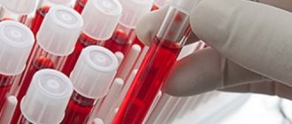
- Enzyme immunoassay blood test, which allows you to identify infectious diseases and hormonal disorders.
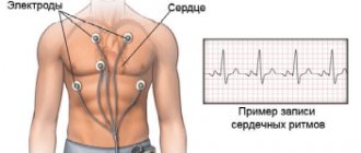
- Coagulograms to determine blood clotting parameters.
- Molecular genetic analysis if a hereditary factor for the appearance of blood clots is suspected.
- Functional tests to evaluate the saphenous veins, but not the localization of blood clots (Brodie-Troyanov-Trendelenburg and Hackenbruch tests).
- Angiography uses a radiopaque contrast agent injected into a vein to produce X-ray images.
- Ultrasound.
- Duplex scanning, the use of which allows a specialist to determine the condition of the vessels and blood flow in them using ultrasound.
- Phlebography. Refers to x-ray diagnostic methods, during which a contrast agent is injected into the vessel.
Only a specialist can accurately determine the location of blood clots in the vessels.
Modern medicine has a sufficient number of diagnostic methods for identifying blood clots in blood vessels in order to further help the patient get rid of them.
Wait, who's coming?
How to detect thrombosis at an early stage? More often it “starts” in the veins of the leg and is accompanied by quite tolerable local pain and swelling. In an unfavorable scenario, the blood clot increases in size, involving the veins of the thigh and pelvis. Gradually, massive swelling, redness of the skin, bursting pain, and swelling of the saphenous veins appear. From the moment the first symptoms appear until you see a doctor, it usually takes 5-10 days. The exception is iliofemoral thrombosis, which makes itself felt immediately - with pronounced swelling and noticeable pain.
“Silent” thrombosis (with a complete absence of symptoms) can occur in bedridden patients (after surgery or paralysis) or if a blood clot forms in the pelvic veins (usually this occurs during pregnancy and childbirth, in the postpartum period, when taking oral contraceptives). In such situations, the first sign of disease may be pulmonary thromboembolism “without a clear source.”
Article on the topic
There is a cork in the vessel. Where did the child get thrombosis?
Treatment
You can get rid of blood clots at home or in a hospital, following the instructions of your doctor.
Thrombosis therapy involves the use of the following methods:
- Drug therapy using: anticoagulants and antiplatelet agents; painkillers; thrombolytics.
- Non-surgical implantation of a vena cava filter to prevent thromboembolism.
- Non-drug therapy (elastic bandages, compression stockings).
- Surgical intervention in the form of: bypass surgery. In which a new path for blood flow is created that does not come into contact with the affected area; stenting. A special stent is installed at the site of narrowing of blood vessels; rhombectomy. Removal of a thrombus from a vein by traditional excision or endovascular method.
- Supportive therapy (massage, exercise therapy, traditional methods).
- Low cholesterol diet.
Self-treatment is unacceptable. Only a doctor will help remove a blood clot from the vessels and prevent irreversible consequences.
It is within the power of the patient to prevent the formation of pathology. To do this, you need to lead a healthy lifestyle, give up bad habits, eat healthy food and systematically undergo preventive examinations of the body.