Possible complications after cardiac cryoablation
Complications are rare, but no procedure is guaranteed to be risk-free. Before you undergo the procedure, you need to be aware of possible complications, which may include:
- Bleeding;
- Infection;
- Pain at the catheter insertion site;
- Blood clots;
- Injuries to blood vessels and heart;
- Abnormal heart rhythm;
- Heart attack;
- Heart puncture.
Factors that may increase the risk of complications include:
- Smoking
The level of risk may be related to the specific type of arrhythmia.
Efficacy of cryoablation pulmonary vein isolation in patients with atrial fibrillation
September 25, 2015 0
Purpose: To evaluate the effectiveness, long-term results and quality of life after performing balloon cryoablation to isolate the pulmonary vein ostia in patients with atrial fibrillation.
Methods: The study included 37 patients, of which 24 (64.8%) were men. The average age of patients was 53.0±10.1 (from 44 to 72 years). The average duration of arrhythmic history was 6.4±4.7 years. All patients had paroxysmal drug-resistant AF. Before surgery, all patients underwent a general clinical examination: general and biochemical blood tests, coagulogram, transthoracic and transesophageal echocardiography to determine the mechanical function of the atria and the presence of thrombus formation, computed tomography of the left atrium with its reconstruction.. All patients used a 28-mm balloon catheter (ArcticFront, MedtronicCryoCath LP, Pointe-Claire, Quebec, Canada), under the control of intracardiac echocardiography with an AcuNav catheter (BiosenseWebster). Cryoablation was performed at temperatures up to – 44 ± 7 C. In order to control the isolated area, stimulation was used with an amplitude of 20 mA and a pulse duration of 2 ms from an isolated vein with potential recording from the coronary sinus. Isolation of the right PVs was controlled against the background of sinus rhythm. This technique is well known and widely described in the world literature. All patients underwent isolation of the mouths of the left and right pulmonary veins. All patients were discharged from the hospital with a recommendation to take warfarin and a class IC or III antiarrhythmic drug. Daily ECG monitoring was carried out during the first month after surgery, then 3, 6, 12 months. The antiarrhythmic drug was discontinued after 3 months. The quality of life of patients was assessed before surgery, 3 and 12 months after surgery using the SF-36 questionnaire.
Results: In the early postoperative period, 9 (24.3%) patients experienced attacks of atrial fibrillation, which were easily controlled by the administration of antiarrhythmics (propanorm, cordarone). At 1-year follow-up, 35 (94.5%) remained in sinus rhythm; of these, 21 (56.7%) patients receive antiarrhythmic drugs. In 2 patients, an asymptomatic course of AF was noted in the form of short paroxysms, recorded during daily ECG monitoring. Patients in both groups answered the SF-36 questionnaire before surgery, three months and one year after surgery. Moreover, in the first three months of observation, most of them showed mainly an improvement in physical functioning criteria due to improved hemodynamics. Subsequently, over the course of 12 months, all indicators are observed to approach the indicators of the quality of life of the main population of Russian residents.
Conclusions: Thus, the results of this study showed that the balloon cryoablation technique for isolating the ostia of the pulmonary veins is highly effective in maintaining sinus rhythm. These circumstances lead to an increase in the average quality of life of patients, mainly due to improved hemodynamics and the absence of AF paroxysms.
Read the publication in full
How is cryoablation performed?
Preparation for the procedure
The doctor will likely do the following:
- Electrophysiological examination (cardiac EPS) to accurately determine the location of abnormal rhythms;
- Ask you to stop taking medications that were previously used to treat arrhythmia.
Before the procedure:
- You should not eat or drink anything for eight hours before the procedure;
- Follow your doctor's instructions.
Anesthesia
A local anesthetic will be administered. It will numb the area where the catheter will be inserted. You will also receive a mild sedative through an IV into your arm. This will help you relax during the procedure.
Description of the procedure
A special ablation catheter will be inserted into a blood vessel in the groin, upper thigh, arm, or wrist. The area where the catheter will be inserted will be cleaned and numbed using anesthesia.
The catheter will be inserted into the artery and connected to the blood vessels in the heart. The doctor will be able to see the catheter on the screen of a special X-ray machine.
The doctor finds the source of the arrhythmia. This will be done by identifying the location of the arrhythmia with a special catheter tip. Once the problem area is found, it will be cooled using the tip of the catheter. If the source of the arrhythmia is found correctly, the problem will disappear. If the site is chosen incorrectly, the cold end of the catheter is removed and the tissue will not be damaged in any way.
When the location of the arrhythmia has been correctly identified, the tip of the catheter will be cooled to -70°C. This will freeze the heart tissue and eliminate the arrhythmia. The search procedure is repeated until all sources of arrhythmia are eliminated.
Immediately after the procedure
You will be moved to the recovery room. Staff will monitor vital signs for several hours to look for symptoms of tenderness, heart rhythm problems, or bleeding from the catheter site.
You need to lie on your back for a period of time. A bandage may be placed over the site where the catheter was inserted to prevent bleeding. It is important to follow the directions of your nurse and doctor.
Heart rhythm disturbances
At the National Medical Research Center of Sports Sciences named after. A.N. Bakulev diagnoses heart rhythm disturbances, as well as their effective elimination using modern minimally invasive treatment methods. The center uses modern high-tech equipment and methods for treating arrhythmias, including life-threatening ones.
Invasive arrhythmology does not involve open heart surgery, but interventional endovascular (through blood vessels) intervention. This method of treatment allows us to minimize the volume of intervention, reduce the operation time and, most importantly, the patient’s stay in the center’s hospital to 3-5 days.
The Department of X-ray Surgical Interventional Diagnostics and Treatment of Arrhythmias treats the entire spectrum of heart rhythm disturbances: NVT - supraventricular tachyarrhythmias (junctional tachycardia, pre-excitation syndrome (Wolf-Parkinson-White syndrome), atrial tachycardia, atrial extrasystoles, atrial flutter, atrial fibrillation; ventricular arrhythmias (ventricular extrasystoles of various locations, ventricular tachycardia), as well as disorders that arose after open heart surgery.
To eliminate areas of cardiac arrhythmia, not only radiofrequency and cryoablation are used, but also non-fluoroscopic navigation mapping with the construction of a three-dimensional model of the heart and obtaining an image of the source of arrhythmia in real time. Our center has unique experience in using all types of navigation systems from all leading manufacturers of electrophysiological equipment. The use of such equipment makes it possible to help patients with complex multi-stage correction of rhythm disturbances with concomitant organic heart pathologies with maximum efficiency.
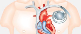
For bradyarrhythmias and life-threatening cardiac arrhythmias, the center uses implantation of an artificial pacemaker (Electrocardiac pacemaker), thanks to which an adequate heart rate is normalized.
The list of bradyarrhythmias requiring pacemaker implantation includes:
- Atrioventricular (atrioventricular) blocks, both congenital and acquired;
- Sinoatrial blocks;
- Permanent form of atrial fibrillation with impaired atrioventricular conduction;
- Various manifestations of sick sinus syndrome.
In addition, implantation of a cardioverter-defibrillator is used to prevent sudden cardiac death and life-threatening conditions.
Arrhythmias requiring implantation of a cardioverter-defibrillator:
- Life-threatening ventricular tachycardia and ventricular fibrillation;
- Risk of sudden death in patients with coronary heart disease;
- Genetic ventricular arrhythmias.
Resynchronization devices and cardiac contractility modulation devices (Optimizer) are also widely used to treat chronic heart failure.
In the clinic of the National Medical Research Center for Cardiovascular Surgery named after. A.N. Bakulev regularly tests all types of implantable devices for electrical pacing.
Patient care after cardiac cryoablation
Hospital care
- There may be bruising and pain at the site where the catheter was inserted;
- If the catheter was inserted in the groin area, you need to lie in bed for a while with your legs straight;
- If the catheter was inserted into the wrist or arm, you do not need to stay in bed;
- The catheter insertion site will be monitored for signs of bleeding, swelling, or inflammation;
- Vital parameters will be monitored.
Home care
When you return home after the procedure, follow these steps to ensure a normal recovery:
- Take aspirin as directed for 2 to 4 weeks. This will help reduce the risk of blood clots at the catheter insertion site;
- You can return to normal activities, walking or climbing stairs is allowed. Avoid heavy or physical activity for 24 hours. In most cases, you will be able to return to your normal activity level within a few days;
- It is necessary to make visits to the doctor in a timely manner. Catheter insertion sites should be checked for complications.
This procedure has an extremely high success rate and low risk of complications. However:
- If atrial fibrillation or ventricular tachycardia is present, antiarrhythmic therapy may need to be continued;
- After AV node cryoablation, you will need a pacemaker.
RFA or cryoablation?
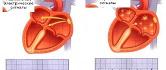
Isolation of the pulmonary veins, which produce the chaotic electrical signals that cause atrial fibrillation, is known to be a standard treatment for patients with atrial fibrillation. Cryoballoon ablation uses a coolant to create circular contiguous lesions and thereby isolate the pulmonary veins, while radiofrequency ablation uses heat (radiofrequency energy) but also requires 3D mapping and step-by-step treatment.
FIRE AND ICE is the largest randomized international clinical trial to compare two ablation techniques used to treat atrial fibrillation, cryoablation (“Ice”), which uses Arctic Front cryoballoons, and radiofrequency. ablation (“Flame”) using ThermoCool radiofrequency ablation catheters
The study results were presented at the Sixty-fifth Annual Scientific Session of the American College of Cardiology April 2-4, 2021, and simultaneously published in The New England Journal of Medicine.
The study was led by the famous Professor Karl-Heinz Kuck, Director of the Cardiology Department of the Asklepios Klinik St. Georg in Hamburg, Germany.
The study involved 769 patients from 16 medical centers across Europe. All study subjects had a diagnosis of paroxysmal atrial fibrillation, had failed at least one antiarrhythmic drug, and had been seen for no longer than 33 months (mean = 1.54 years) after the first ablation procedure. The main objective of this type of research is to prove that the new technology is comparable to the generally accepted existing technology.
The study met its primary efficacy endpoint of demonstrating that cryoballoon ablation was noninferior to radiofrequency ablation (p=0.0004) in terms of reducing the incidence of arrhythmia recurrences or the need for antiarrhythmic drug therapy and/or repeat ablation. The primary safety endpoint of time to first death from any cause, stroke or TIA (transient ischemic attack) from any cause, or treatment-related serious adverse events was also met (p = 0.24).
Both technologies showed comparable low complication rates. According to the study, cryoballoon ablation technology provided shorter procedure times (mean = 124 minutes) compared with radiofrequency ablation group (mean = 141 minutes; p = 0.0001), but the use of a radiofrequency catheter allowed shorter fluoroscopy time. time (mean = 17 minutes) compared with cryoballoon ablation catheter (mean = 22 minutes; p = 0.0001).
The study authors said they believe the findings will help move interventional arrhythmology interventions away from highly specialized clinics and toward less-trained physicians. If atrial fibrillation ablation can be performed easily and safely, it is likely that more patients will be treated, and perhaps at an earlier stage of the disease. Perhaps someday they will deal with fibrillation in the same way as is now recommended to do with WPW syndrome - perform intervention after the first paroxysm.
Although the study discussed was conducted on a small sample of patients, it is expected that the findings may change current clinical guidelines. In particular, it may be taken into account by experts currently working on the third update of the Heart Rhythm Society (HRS) consensus statement.
In June 2021, results regarding secondary endpoints were presented at the latest CARDIOSTIM European Congress. The study demonstrated significantly fewer need for repeat ablations and lower hospitalization rates for patients with paroxysmal atrial fibrillation in the cryoballoon pulmonary vein isolation group. Quality of life was assessed at baseline and 6 months after the procedure; the data were comparable in the study groups.
Thus, cryoballoon ablation is a safe and effective method that can reduce the duration of the procedure, make the treatment schedule more predictable in time, reduce the need for repeated procedures, reduce the number of hospitalizations and improve the quality of life of patients.
Contacting your doctor after cardiac cryoablation
After returning home, you should consult a doctor if the following symptoms appear:
- Signs of infection, including fever and chills;
- Redness, swelling, increased pain, bleeding, or discharge at the catheter site;
- The leg into which the catheter was inserted becomes cold, white or blue, numb, or tingling;
- cough, shortness of breath, chest pain, or severe nausea or vomiting;
- Discomfort in the jaw, chest, neck, arm, or upper back;
- Dizziness and weakness.

