Etiology of AF and cardiovascular risks
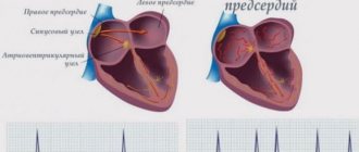
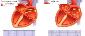
Atrial fibrillation is defined as a supraventricular tachyarrhythmia characterized by chaotic electrical activity of the atria at a high rate (350-700 per minute) and an irregular ventricular rhythm1. Previously, the term “atrial fibrillation” was widely used, which quite accurately conveys the essence of atrial fibrillation.
According to foreign studies, AF is the most common form of tachyarrhythmia in the world and occurs in 2% of the population2. There are valvular and non-valvular AF (NVAF). In the first case, AF is associated with damage to the heart valves and overload of the left atrium with pressure or volume, due to which fibrillation begins. The most common cause of valvular AF is rheumatic mitral valve stenosis or replacement1. All other cases of atrial fibrillation are defined as non-valvular.
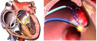
NFP occurs against the background of a number of diseases and conditions, such as coronary heart disease, hypertension, primary myocardial diseases, diabetes mellitus, pheochromocytoma, as well as alcohol abuse, excess body weight and hypokalemia3. Due to the chaotic movement of blood in the fibrillating left atrium, the prerequisites are created for the formation of blood clots, which can spread through the left ventricle throughout the systemic circulation. Thromboembolic complications are the most dangerous complications of AF. These include:
- ischemic cardioembolic stroke;
- heart attacks of internal organs;
- thromboembolism of the vessels of the extremities.
In a cardioembolic stroke, an embolus (thrombus) located in the left atrium breaks off and enters the arterial system of the brain.
According to a number of epidemiological studies, NFP increases the risk of stroke by 2.6-4.5 times, depending on the clinical parameters of patients4. Moreover, for women, these risks, other things being equal, are higher than for men5.
A stroke that occurs during NAF is severe and more often leads to disability than a stroke in patients without AF6. The basis for the prevention of thromboembolism in NFP is oral anticoagulants. They reduce the activity of blood clotting and thus prevent the formation of emboli (blood clots). 10 years ago, the drug of choice was the vitamin K antagonist warfarin, but direct oral anticoagulants (DOACs) appeared, in particular apixaban, which have a number of advantages over warfarin, which allowed DOACs to become the standard for stroke prevention in NAF.
Classification by duration of arrhythmia existence
Newly diagnosed arrhythmia . It is diagnosed in patients who have not previously had episodes of AF, regardless of the severity of symptoms associated with rhythm disturbances, complications, and other factors.
Paroxysmal form. Arrhythmia spontaneously arises and stops. Many patients experience frequent paroxysms of atrial fibrillation with characteristic symptoms. The duration of one episode of AF is more than 30 seconds. With this form of arrhythmia, paroxysms usually last from 48 hours, but not more than 7 days.
Persistent form. Atrial fibrillation is observed for at least 7 days (according to medical experts) or is stopped by electrical or drug cardioversion. This form of arrhythmia involves the desire to maintain normal sinus rhythm. This is what distinguishes persistent AF from permanent AF. Paroxysms can be observed throughout the year, but sinus rhythm is always restored.
Permanent form. Arrhythmia exists for a long time, usually the establishment of this form is preceded by a period of repetition of paroxysms. In this case, a rhythm frequency control strategy is used. Catheter ablation, cardioversion, antiarrhythmic drugs, and other interventions are not used to restore sinus rhythm.
The same patient with a long-term form of AF at different stages of arrhythmia may experience different forms of the disease, as well as their combinations.
Eliquis® (apixaban) for non-valvular atrial fibrillation
To date, the only drug with the INN apixaban registered in Russia is Eliquis®. This drug is a direct selective inhibitor of coagulation factor Xa, due to which it interrupts the coagulation cascade and prevents the formation of blood clots.
The superiority of apixaban over warfarin was demonstrated in the double-blind, randomized international clinical trial ARISTOTLE, the results of which were published in 20117. We analyzed data from 18,201 patients with atrial fibrillation and risk of stroke, divided into groups of patients taking INR-guided warfarin or apixaban.
The study results showed that apixaban, compared with warfarin, reduced the risk of stroke by 21%, the risk of major bleeding by 31%, and overall mortality by 11%.
Thus, to date, Eliquis® is the only direct oral anticoagulant that has shown superiority over warfarin in patients with NAF according to three main clinical indicators based on CT results.
In addition, the advantage of apixaban is its favorable safety profile. The drug has low renal excretion (renal excretion 27%), which allows its use in mild to moderate renal impairment8.
Also, Eliquis® at the full therapeutic dose (5 mg twice daily) is suitable for most patients with NFP and does not increase the risk of gastrointestinal bleeding7.
The treatment regimen for the drug on an outpatient basis looks very simple and is easy for the patient to remember: one 5 mg tablet twice a day, regardless of meals. (2.5 mg twice daily if two or more of the following are present: age 80 years or older, body weight 60 kg or less, or plasma creatinine concentration ≥1.5 mg/dL (133 µmol/L) ; or with creatinine clearance 15–29 ml/min).
6.6. Atrial fibrillation (AF)
DEFINITION, ETIOLOGY AND PATHOGENESIS to top
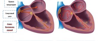
The most common supraventricular tachyarrhythmia is characterized by rapid (350–700/min), uncoordinated activation of the atria, leading to a loss of hemodynamic efficiency of their contractility, and is accompanied by an irregular ventricular rhythm. The frequency of the ventricular rhythm depends on the electrophysiological properties of the AV node, the state of the autonomic system and the action of drugs and can be normal (at rest 70–90/min), accelerated (tachyarrhythmia) or slowed (bradyarrhythmia).
Classification of AF →Fig. 2.6-9. If a patient experiences arrhythmia episodes in different categories, consider (first) the most common form of AF.
Figure 2.6-9.
Classification of atrial fibrillation
Causes:
1) cardiac - arterial hypertension, acquired valve defects (primarily mitral), ischemic heart disease, cardiomyopathies (especially dilated and hypertrophic), congenital heart defects (mainly with interatrial shunt), myocarditis and pericarditis, history of cardiac surgery, weakness syndrome sinus node (tachycardia-bradycardia syndrome), premature excitation syndrome, systemic diseases with heart damage - sarcoidosis, amyloidosis, hemochromatosis, cardiac tumors - primary and metastatic. AF is very common in patients with heart failure, regardless of its cause.
2) extracardiac - hyperthyroidism (most often), hypothyroidism, acute infection, general anesthesia, lung diseases, pheochromocytoma, obesity, diabetes mellitus, metabolic syndrome, chronic kidney disease, sports (disciplines that develop endurance), various substances (alcohol, oxide carbon, caffeine, some drugs [eg, β2-mimetics]).
Paroxysmal AF occurs in ≈50% of cases in individuals without organic heart disease; most often it is a focal type arrhythmia induced by excitation arising in the pulmonary veins, less often in the superior vena cava, vein of Marshall or coronary sinus. In persistent or constant AF, organic heart disease is detected in >90% of patients.
CLINICAL PICTURE AND NATURAL COURSE to top
Subjective symptoms: palpitations, bouts of sweating, weakness and decreased exercise tolerance, fainting or dizziness.
With persistent AF, patients often do not experience symptoms. Classification of symptom intensity (EHRA scale) → table. 2.6-5. Objective symptoms: irregular heart activity (complete irregularity), irregular pulse, pulse deficiency. In patients with focal AF, numerous extrasystoles or attacks of tachycardia (atrial tachycardia, atrial flutter) may occur. Table 2.6-5. Classification of complaints related to atrial fibrillation according to the modified EHRA scale
| Class | Description |
| 1 | without symptoms |
| 2a | mild symptoms - do not interfere with normal daily activities |
| 2b | moderate symptoms - unpleasant for the patient, but do not interfere with normal daily activities |
| 3 | severe symptoms - normal daily activities are limited |
| 4 | symptoms that make functioning impossible - normal daily activities are impossible |
A newly diagnosed episode of AF may be a single attack in a lifetime or another relapse of paroxysmal AF or even long-lasting persistent AF. A thorough history taking and analysis of the patient’s available medical documentation is important. Paroxysmal AF is self-limiting and most often resolves within 24 hours. Persistent AF does not resolve on its own and lasts >7 days; It may be either the first clinical manifestation of arrhythmia or a consequence of recurrent episodes of paroxysmal AF. Long-term persistent AF is recognized when AF persists for >1 year and a decision is made to restore sinus rhythm.
DIAGNOSTICS up
Screening studies
Screening studies for AF should be performed:
1) in persons >65 years old - palpation of the pulse or ECG;
2) in patients with a transient ischemic attack (TIA) or a history of ischemic cerebral stroke, continuous ECG monitoring for ≥72 hours (consider the need for long-term ECG monitoring using non-invasive devices or implantable loop recorders).
Additional research methods
1. ECG: complete irregularity of the ventricular complexes, absence of P waves (they are replaced by f waves) →Fig. 2.6-10A.
2. Holter ECG monitoring (sometimes >24 hours, up to 7 days), long-term (eg 2-4 weeks) and continuous telemetric ECG recording: in the case of paroxysmal AF, if diagnostic doubts arise.
3. Stress ECG test: if myocardial ischemia is suspected and before starting the use of class Ic drugs.
4. Echocardiography: transthoracic examination should be performed in all patients with AF in order to identify possible. organic heart disease or thrombus in the left atrium and its appendage (for this, a transesophageal study is required).
Differential diagnosis →Fig. 2.6-1
TREATMENT to the top
Emergency treatment
Tactics for paroxysmal AF depend on the accompanying symptoms and hemodynamic disorders:
1. If subjective symptoms are not expressed:
1) correct possible electrolyte disorders (potassium and magnesium levels) and wait for the attack to stop;
2) Monitor ventricular rate (target <110/min) using, e.g., verapamil or diltiazem (do not IV if heart failure), a β-blocker (e.g. metoprolol; if heart failure, or LVEF < 40% use at the minimum effective dose; if heart rate does not decrease <110/min, digoxin or amiodarone) or digoxin (do not use as monotherapy) can be added;
3) if AF is prolonged, especially >24 hours → cardioversion is indicated, most often pharmacological (most effective when AF lasts <7 days); use propafenone (in persons without significant organic heart disease) or amiodarone (in other cases); If an attack of AF lasts <48 hours, pretreatment with anticoagulant treatment is not necessary (except for patients at high risk of thromboembolic complications, such as patients with diabetes mellitus or heart failure).
2. If AF causes significant hemodynamic impairment or coronary pain → perform emergency electrical cardioversion → section. 24.18.
3. The next relapse → can be recommended to the patient (without organic heart damage, with significant symptoms of rare ari tablets - a single dose of 600 mg (450 mg for body weight <70 kg) of propafenone, if the effectiveness and safety of such treatment in hospital has been previously confirmed in this patient conditions: Apply a β-blocker or verapamil 30 min earlier to avoid 1:1 AV conduction if AF progresses to atrial flutter.
Long-term treatment
General principles
1. Paroxysmal AF: exclude factors that induce arrhythmia, such as alcohol, caffeine, nicotine; identify and treat the possible cause. Modify cardiovascular risk factors. After stopping the first paroxysm of AF in life, antiarrhythmic drugs are not used prophylactically. In case of rare relapses with good tolerance → possible. taking a “handy” pill (→see above).
2. Persistent AF: choose treatment strategy:
1) restoration of sinus rhythm (most often using electrical cardioversion) and its retention (most often pharmacological)
or
2) AF is treated as permanent and optimal ventricular rate control is used.
Rhythm control algorithm in patients with newly diagnosed AF →Fig. 2.6-11. Both strategies have demonstrated similar effects on the risk of death and stroke. Consider these approaches not to be mutually exclusive, but to be complementary. Even after adopting a strategy to maintain sinus rhythm, continue to use drugs to control the ventricular rate in order to protect the patient from developing tachyarrhythmia in the event of recurrent AF. Ventricular rate monitoring is recommended in elderly patients with asymptomatic AF or mild arrhythmia-related symptoms (EHRA 1 or 2a). Whereas in patients with at least moderate symptoms (EHRA ≥2b), in addition to controlling the ventricular rate, strive to maintain sinus rhythm. Consider maintaining sinus rhythm in these patients:
Figure 2.6-11.
Algorithm for monitoring heart rate in patients with newly diagnosed atrial fibrillation (based on ESC 2021, modified)
1) young people with symptoms of arrhythmia, in whom ablation cannot be excluded;
2) with AF and heart failure caused by arrhythmia;
3) with AF, when its cause has been eliminated (eg thyrotoxicosis).
3. Persistent AF: the goal of treatment is to control the ventricular rate (mild in asymptomatic patients or with well-tolerated symptoms - <110/min at rest; more strict in patients with symptoms due to AF - at rest <80/min, during moderate load <110/min); in the case of strict control, it is important to evaluate it using an exercise test (for symptoms during exercise) and Holter ECG monitoring. Do not use antiarrhythmic drugs of either class I or III, as well as amiodarone, for the purpose of long-term control of the ventricular rate.
Pharmacological treatment
1. Maintaining sinus rhythm: the choice of drugs is determined primarily by the safety of therapy, depending on the risk of proarrhythmia. The choice of drug depends on the manifestation of organic heart disease (attention: patients taking sotalol should be monitored for proarrhythmia; amiodarone is a 2nd line drug for a large number of patients due to non-cardiac side effects):
1) patients without significant organic heart disease → dronedarone, flecainide, propafenone, sotalol;
2) coronary heart disease, clinically significant valvular heart disease, pathological left ventricular hypertrophy → dronedarone, sotalol (patients with clinically significant left ventricular hypertrophy have a higher risk of proarrhythmia), amiodarone;
3) heart failure → amiodarone.
2. Control of ventricular rate: β-blockers (the most effective, preferable for ischemic heart disease, arterial hypertension, heart failure or hyperthyroidism), calcium channel blockers (verapamil, diltiazem; in patients with LVEF ≥40%, especially when there are contraindications to β -blockers; do not use if you have WPW syndrome). Digoxin is less effective, especially in active individuals, may be preferable in older patients, with less activity, in heart failure, quite often in combination with a beta-blocker or calcium channel blocker; do not use in WPW syndrome or hypertrophic cardiomyopathy. In patients with heart failure, without additional conduction pathways, or if other treatments are ineffective or contraindicated → IV amiodarone can be used.
Invasive treatment
1. Percutaneous ablation (pulmonary vein isolation - basic technique, linear ablations, destruction of fractionated electrogram zones, ablation of autonomic ganglia): recommend it to patients (refer them to a center with experience in using these methods) with paroxysmal AF, occurring with subjective symptoms, if ≥ 1 class I or III antiarrhythmic drug has been ineffective, and the patient wishes to achieve rhythm control. Consider ablation:
1) as a first-choice treatment in selected patients with severe symptoms of AF (as an alternative to antiarrhythmic drugs) after assessing the potential benefits and risks associated with the intervention, and if patients consciously choose this method of treatment;
2) in symptomatic patients:
a) with persistent or long-term persistent AF, resistant to pharmacological treatment;
b) with AF and heart failure with reduced LVEF, especially if the development of tachyarrhythmic cardiomyopathy is suspected;
3) in patients with bradycardia dependent on AF.
2. Surgical ablation: in those undergoing cardiac surgery, e.g. about mitral valve disease or coronary artery disease.
3. Percutaneous AV node ablation with pacemaker implantation: Consider in patients with persistent AF if pharmacologic rate control has failed. Stimulator implantation is also indicated for symptomatic patients with brady-tachycardia syndrome or persistent AF associated with symptomatic bradyarrhythmia.
COMPLICATIONS to the top
The most serious are thromboembolic complications, primarily ischemic stroke. They are associated with the occurrence of a blood clot in the left atrium (most often in its appendage).
1. Long-term prevention: for each patient with AF, assess the risk of thromboembolic complications based on the CHA2DS2-VASc scale → table.
2.6-6, as well as the risk of bleeding (see below). Recommendations for prevention →Fig. 2.6-12: Table 2.6-7. CHA2DS2‑VASc scale for assessing the risk of ischemic stroke in patients with non-valvular atrial fibrillation
| Risk factor | Points |
| symptoms of heart failure or decreased left ventricular ejection fraction | 1 |
| arterial hypertensiona | 1 |
| age ≥75 years | 2 |
| diabetesb | 1 |
| history of stroke or TIA or other thromboembolic episodea | 2 |
| vascular disease | 1 |
| age 65–74 years | 1 |
| female half | 1 |
| and blood pressure at rest >140/90 mm Hg. Art. with ≥2 measurements taken in a different setting or ongoing antihypertensive treatment b fasting blood glucose >125 mg/dL (7 mmol/L) or use of oral antidiabetic agents and/or insulin in previous myocardial infarction, atherosclerosis of peripheral arteries, atherosclerotic plaque in the aorta d increases risk if ≥1 other risk factor is present | |
Figure 2.6-12.
Prevention of stroke in patients with atrial fibrillation (based on ESC 2021, modified)
1) if the risk of stroke is assessed at 0 points (men) or 1 point (women) on the CHA2DS2-VASc scale → do not prescribe either anticoagulants or antiplatelet agents;
2) in other cases, if there are no contraindications to treatment with anticoagulants → constantly use an oral anticoagulant - dabigatran (110 or 150 mg 2 times a day depending on age, concomitant therapy, renal function and the risk of bleeding), rivaroxaban (15 or 20 mg 1 × per day) and apixaban (2.5 or 5 mg 2 × per day); possibly a vitamin K antagonist (VKA - acenocoumarol or warfarin) at a dose that maintains the INR in the range of 2-3 (after stabilizing the dose, monitor the INR monthly); in patients with AF associated with moderate or severe mitral valve stenosis or mechanical valve prosthesis, use a VKA;
3) assess the risk of bleeding - based on the presence of risk factors for bleeding. Modifiable factors: hypertension (especially systolic pressure >160 mm Hg), labile INR values in those receiving a vitamin K antagonist, with TTR <60% (Time in Therapeutic Range - percentage of time in which INR is kept in the therapeutic range ), taking medications that predispose to bleeding [eg, antiplatelet agents, nonsteroidal anti-inflammatory drugs], excessive alcohol consumption [≥8 drinks/week]). Potentially modifiable factors: anemia, worsening renal function, decreased platelet count or impaired platelet function. Non-modifiable factors: age [>65 years or ≥75 years depending on the scale], previous significant bleeding, previous cerebral stroke, dialysis therapy or condition after kidney transplantation, liver cirrhosis, cancer, genetic factors [CYP2C9 polymorphism]. Factors associated with biomarker levels: increased troponin concentration as determined by a high-sensitivity test, decreased estimated GFR. Aim to correct reversible risk factors for bleeding.
4) the use of ASA (or other antiplatelet drug) for the purpose of preventing stroke in patients with AF is not recommended;
5) in patients at high risk of stroke and with contraindications to chronic oral anticoagulants → you can consider the procedure of percutaneous closure of the left atrial appendage;
6) with a high risk of bleeding → the patient needs increased attention and regular monitoring after prescribing an anticoagulant.
2. Prevention during cardioversion
1) In patients with AF lasting ≥48 hours or unknown duration, use a VKA (INR 2–3) before attempting to restore sinus rhythm (electrical or pharmacologic cardioversion); dabigatran, rivaroxaban, or apixaban may be used instead of VKA; if prescribed, conversation with the patient to confirm regular drug intake; if in doubt, transesophageal echocardiography is necessary) for ≥3 weeks. before cardioversion and 4 weeks. after it (attention: longer treatment with anticoagulants may be indicated; consider risk factors for thromboembolic complications).
2) If emergency cardioversion is required and AF duration is ≥48 hours or unknown → rule out thrombus on transesophageal echocardiography, use unfractionated IV heparin (possibly low molecular weight heparin) before cardioversion and oral anticoagulant after the procedure.
3) In patients with AF lasting exactly <48 hours, you can perform cardioversion immediately after heparin administration, but if there is a risk of stroke, begin long-term use of an oral anticoagulant.
3. Prevention in patients undergoing PCI → table. 2.34-7.