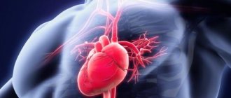
Cardiosclerosis is a pathological replacement of the working muscle tissue of the heart with rigid connective tissue that does not bear a functional load. An inelastic replacement is unable to contract and conduct impulses.
In cardiology, the disease is regarded as one of the manifestations of myocardial damage caused by a deficiency or cessation of blood supply, otherwise known as coronary heart disease (CHD). Cardiosclerosis is classified into four types:
- myocardial cardiosclerosis – a complication of inflammation of the muscular lining of the heart (myocarditis);
- post-infarction cardiosclerosis – consequences of necrosis (death) of a portion of the myocardium;
- primary cardiosclerosis is the result of congenital pathologies of the inner layer of the membrane of the myocardial walls and connective tissue;
- atherosclerotic cardiosclerosis.
According to ICD-10, the disease was assigned code I25.1.
General information. Cardiosclerosis - what is it and how to treat it
Cardiosclerosis is understood as a chronic heart disease that develops due to excessive growth of connective tissue in the thickness of the myocardium .
The number of muscle cells themselves also decreases noticeably. Cardiosclerosis is not an independent disease, because formed as a result of other pathologies. It would be more correct to consider cardiosclerosis a complication that seriously disrupts the functioning of the heart.
The disease is chronic and has no acute symptoms. Cardiosclerosis is provoked by a large number of causes and factors, so it is quite difficult to determine its prevalence. The main signs of the disease are found in most cardiac patients. Diagnosed cardiosclerosis always worsens the patient's prognosis, because replacement of muscle fibers with connective tissue is an irreversible process.
Prevention of post-myocardial cardiosclerosis
To prevent the development of myocardiosclerosis, a patient who has had myocarditis must systematically visit a cardiologist, as well as a therapist, and undergo appropriate heart-supportive therapy.
Preventive examinations should be carried out at least 2 times a year.
If the first and minor symptoms of post-myocardial cardiosclerosis appear, it is necessary to immediately visit a cardiologist in order to conduct a full instrumental examination and promptly begin therapy for sclerosis of the heart muscle.
Self-medication, or simply ignoring the developing pathology of the heart, can lead to very sad consequences, which will end in a sudden heart attack with a fatal outcome.
It is possible to stop the progression of the pathology of postmyocardial cardiosclerosis by following the doctor’s instructions, as well as a properly selected diet.
The diet should include the maximum amount of vitamins and fiber found in fresh vegetables and fruits, as well as polyunsaturated fatty acids found in sea fish and vegetable oils.
The diet should not include fatty meats and eggs, full-fat dairy products and trans fats. Limiting sweet foods and the amount of salt consumed.
Also, the diet should not contain fried and smoked foods, as well as canned food and marinades.
It is necessary to completely give up smoking, drugs and alcoholic beverages.
Constantly monitor the blood pressure index, as well as the index of cholesterol and glucose in the blood.
Avoid stress and overstrain of the nervous system. Do not overload the body physically, but also do not lead a sedentary lifestyle. Constantly fight obesity.
Pathogenesis
The development of cardiosclerosis is based on 3 mechanisms:
- Dystrophic changes. They are formed as a result of disturbances in the trophism and nutrition of the myocardium due to developed cardiovascular disease ( cardiomyopathy , atherosclerosis , chronic ischemia or myocardial dystrophy ). , diffuse cardiosclerosis develops .
- Necrotic processes . Develop after heart attacks , injuries and damage that occurred during heart surgery. , focal cardiosclerosis develops .
- Myocardial inflammation. The process starts as a result of the development of infectious myocarditis , rheumatism and leads to the formation of diffuse or focal cardiosclerosis.
Results
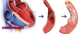
Atherosclerotic cardiosclerosis is one of the types of coronary heart disease. During the disease process, living, functional myocardial tissue is replaced by non-functional connective tissue.
Scars are formed, the free movement of blood, the saturation of the heart with oxygen, the conductivity of impulses, and the contractility of the myocardium are disrupted. Pathological changes develop against the background of progressive atherosclerosis of the aorta and coronary arteries. The consequences of unstable cardiac activity can be myocardial infarction, CHF, and coronary death.
Classification
Cardiosclerosis is classified according to the reasons that will be listed and described below in the appropriate section, by the intensity of the process and by localization. Depending on the classification, the course of the disease changes, different functions of the heart are affected.
Based on intensity and localization, they are distinguished:
- focal cardiosclerosis;
- diffuse cardiosclerosis (total);
- with damage to the valvular apparatus of the heart.
Focal cardiosclerosis
Focal damage to the heart muscle is observed after a myocardial infarction . Less commonly, focal cardiosclerosis forms after localized myocarditis. Characterized by a clear limitation of the lesion in the form of scar tissue, which is surrounded by healthy cardiomyocytes that are capable of fully performing all their functions.
Factors influencing the severity of the disease:
- Depth of damage . Determined by the type of myocardial infarction suffered. With a superficial lesion, only the outer layers of the wall are damaged, and after the formation of a scar, a fully functioning muscle layer remains underneath. With transmural lesions, necrosis affects the entire thickness of the muscle. The scar is formed from the pericardium to the cavity of the heart chamber. This option is considered the most dangerous, because... with it there is a high risk of developing such a serious complication as a cardiac aneurysm.
- Sizes of the hearth. The larger the area of myocardial damage, the more pronounced the symptoms and the worse the prognosis for the patient. There are small-focal and large-focal cardiosclerosis. Single small inclusions of scar tissue may not produce absolutely any symptoms and may not affect the functioning of the heart or the patient’s well-being. Large-focal cardiosclerosis is fraught with consequences and complications for the patient.
- Localization of the outbreak. Depending on the location of the source, dangerous and non-hazardous are determined. The location of a small area of connective tissue in the interventricular septum or in the atrium wall is considered harmless. Such scars do not affect the basic functioning of the heart. Damage to the left ventricle, which performs the main pumping function, is considered dangerous. Number of lesions. Sometimes several small foci of scar tissue are diagnosed at once. In this case, the risk of complications is directly proportional to their number.
- State of the conduction system. Connective tissue not only does not have the necessary elasticity, compared to muscle cells, but is also unable to conduct impulses at the required speed. If scar tissue has affected the conduction system of the heart, then this is fraught with the development of arrhythmias and various blockades. Even if only one wall of the heart chamber lags during the contraction process, the ejection fraction, the main indicator of the contractility of the heart, decreases.
From the above it follows that the presence of even small foci of cardiosclerosis can lead to negative consequences. Timely and competent diagnosis of myocardial damage is required to select appropriate treatment tactics.
Diffuse cardiosclerosis
Connective tissue accumulates in the heart muscle everywhere and evenly, which makes it difficult to isolate specific lesions. Diffuse cardiosclerosis most often occurs after toxic, allergic and infectious myocarditis, as well as with coronary heart disease.
Diffuse cardiosclerosis
The alternation of normal muscle fibers and connective tissue is characteristic, which prevents the heart muscle from fully contracting and performing its function. The walls of the heart lose their elasticity, do not relax well after contraction, and do not stretch well when filled with blood. Such disorders are often referred to as restrictive (compressive) cardiomyopathy .
Cardiosclerosis with damage to the valve apparatus
It is extremely rare that sclerosis affects the valve apparatus of the heart. Valves are involved in the process in rheumatological and systemic diseases.
Types of valve damage:
- Valve insufficiency. Characterized by incomplete closure and closure of the valves, which makes it difficult to eject blood in the desired direction. Through a poorly functioning valve, blood returns back, which reduces the volume of pumped blood and leads to the development of heart failure. In cardiosclerosis, valve insufficiency is formed due to deformation of the valve leaflets.
- Valve stenosis. Due to the proliferation of connective tissue, the lumen of the valve narrows. Blood does not flow in sufficient volume through the narrowed opening. The pressure in the heart cavity increases, which leads to serious structural changes. Thickening of the myocardium (hypertrophy) is observed as a compensatory reaction of the body.
In cardiosclerosis, the valvular apparatus of the heart is affected only in a diffuse process that involves the endocardium.
Symptoms
Atherosclerotic cardiosclerosis progresses slowly. Primary symptoms are not specific. Changes in the myocardium are manifested by slight shortness of breath, chronic drowsiness and fatigue, accelerated fatigue, tingling in the subclavian region (left).
Patients note increased heart rate, pale skin, and short-term numbness in the fingers of the left hand. As the sclerotic areas increase, sharp chest pain occurs (angina pectoris syndrome). Periodically, the heart “stabs”, and pain is felt not only in the heart area, but also on the left side of the body (under the shoulder blade, under the collarbone, in the arm).
No ads 3
Developing:
- dyspnea (shortness of breath);
- heart rate disturbance (tachycardia, bradycardia);
- painful sensations in the epigastric region, accompanied by nausea;
- dry chest cough;
- insomnia (insomnia).
At a later stage, the symptoms of coronary heart disease are added to the listed signs. Swelling of the face and lower limbs. Dizziness, periodic clouding of consciousness, fear of death, manic anxiety, and fainting occur. Angina attacks last up to 15 minutes.
One of the signs of hypercholesterolemia is fatty growths under the skin of the eyelids - xanthomas.
Causes
The transition of cardiomyocytes into connective tissue occurs due to the inflammatory process. In this case, the formation of connective tissue fibers is a kind of protective mechanism.
Depending on the reasons, several groups are distinguished:
- atherosclerotic form;
- post-infarction cardiosclerosis;
- myocardial form;
- other reasons.
Atherosclerotic cardiosclerosis
Includes diseases that lead to cardiosclerosis through long-term ischemia and ischemic heart disease. Atherosclerotic cardiosclerosis is not classified in a separate category according to ICD-10.
Coronary heart disease develops as a result of atherosclerosis of the coronary arteries. When the lumen of the vessel narrows, the myocardium ceases to receive normal blood supply. The narrowing occurs due to the deposition of cholesterol and the formation of an atherosclerotic plaque, or due to the presence of a muscular bridge over the coronary vessel.
With prolonged ischemia , connective tissue begins to grow between cardiomyocytes and cardiosclerosis forms. It is important to understand that this is a rather long process and most often the disease is asymptomatic. The first signs begin to appear only when a significant part of the heart muscle is filled with connective tissue. The cause of death is the rapid progression of the disease and the development of complications.
Myocardial form (Postmyocardial cardiosclerosis)
The mechanism of development of myocardial cardiosclerosis is completely different. A lesion forms at the site of former inflammation after myocarditis. This type of cardiosclerosis is characterized by:
- young age;
- history of allergic and infectious diseases;
- the presence of foci of chronic infection.
ICD-10 code for postmyocardial cardiosclerosis: I51.4.
The disease develops due to proliferative and exudative processes in the myocardial stroma, due to destructive changes in the myocytes themselves. With myocarditis, a huge amount of substances are released that have a damaging effect on the membranes of muscle cells. Some of them are destroyed. After recovery, the body, as a protective reaction, increases the production and volume of connective tissue. Myocardial cardiosclerosis develops much faster than atherosclerosis. The myocardial variant is characterized by damage to young people.
Post-infarction cardiosclerosis
Formed at the site of death of cardiomycytes after acute myocardial infarction. When blood access through the coronary artery to the heart muscle is stopped, necrosis of the corresponding area develops. The area may be of different locations depending on which vessel is blocked. Depending on the caliber of the vessel, the size of the affected area also changes. As a compensatory reaction, the body begins increased production of connective tissue at the site of the lesion. The code for post-infarction cardiosclerosis according to ICD-10 is I25.2.
The prognosis for survival after a heart attack depends on many factors. The cause of death after a heart attack lies in complications of the disease and lack of adequate therapy. Post-infarction syndrome is an autoimmune reaction that complicates myocardial infarction and is manifested by symptoms of inflammation of the pericardium, lungs and pleura.
Postpericardiotomy syndrome is an inflammatory autoimmune disease of the pericardium that develops after open heart surgery.
Other reasons
In addition to those described above, there are other reasons that are more rare.
- Radiation exposure. Under the influence of radiation exposure, changes occur in a variety of organs and tissues. After irradiation of the heart muscle, irreversible changes and complete restructuring occur in cardiomyocytes at the molecular level. Gradually, connective tissue begins to form, its proliferation and the formation of cardiosclerosis. The pathology can develop lightning fast (within several months after strong radiation) or slowly (several years after exposure to a low dose of radiation).
- Sarcoidosis of the heart. A systemic disease that can affect a wide variety of organs and tissues. In the cardiac form, inflammatory granulomas form in the myocardium. With proper therapy, these formations disappear, but in their place, pockets of scar tissue can form. Thus, focal cardiosclerosis is formed.
- Hemochromatosis . This disease is characterized by the deposition of iron in the tissues of the heart. Gradually, the toxic effect increases, an inflammatory process develops, which ends with the proliferation of connective tissues. With hemochromatosis, cardiosclerosis affects the entire thickness of the myocardium. In more severe cases, the endocardium is also damaged.
- Idiopathic cardiosclerosis. This concept includes cardiosclerosis, which has developed for no apparent reason. It is assumed that it is based on as yet unknown mechanisms. The likelihood of the influence of hereditary factors that provoke increased growth of connective tissue at a certain stage of the patient’s life is considered.
- Scleroderma. Damage to the heart muscle in scleroderma is one of the most dangerous complications of the disease. Connective tissue begins to grow from the capillaries that the heart muscle is so rich in. Gradually, the size of the heart increases against the background of constant thickening of the walls. Traditional signs of destruction of cardiomyocytes and the presence of an inflammatory process are not recorded.
There are many mechanisms and reasons for triggering the proliferation of connective tissue in the myocardium. It is quite difficult to reliably establish the true cause of the disease. However, identifying the root cause of the pathology is simply necessary to prescribe the correct treatment.
Development mechanism
Atherosclerotic cardiosclerosis develops against the background of coronary atherosclerosis and is a chain of disorders associated with damage to the left and right coronary arteries. Coronary (also known as coronary) arteries are the only source that ensures an uninterrupted supply of arterial blood to the heart.
In atherosclerotic cardiosclerosis, impaired hemodynamics (blood movement) and oxygen starvation of the heart muscle are a consequence of narrowing of the lumen of the coronary arteries. In turn, the cause of stenosis (narrowing) of the vascular bed is pathological deposits of cholesterol on the inner wall of the arteries (intima, endothelium) - atherosclerosis.
Endogenous cholesterol, produced by hepatocytes (liver cells), and exogenous cholesterol, coming from food, are necessary for the body:
- as a basis for sex and steroid hormones;
- as active participants in interneuronal connections of the spinal cord;
- for proper lipid metabolism, absorption of vitamin D and bile acids.
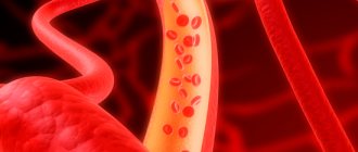
When cholesterol combines with blood proteins, lipoproteins (transport lipids) are formed. The composition of low and very low density lipoproteins (LDL and VLDL) is dominated by fat. Their task is to deliver lipids from the liver to the body’s cells to ensure the above functions.
High-density lipoproteins (HDL) are mostly composed of protein. HDL is responsible for promptly clearing the arteries of excess LDL and VLDL and moving them to the liver for processing and excretion. The well-functioning process of lipid metabolism fails with hypercholesterolemia (an increase in the concentration of cholesterol in the blood due to an increase in the LDL fraction).
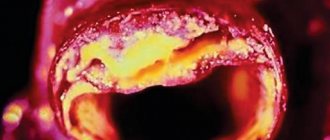
Excess low-density lipoproteins “get stuck” in microcracks in the endothelium. This intravascular deposit grows over time and transforms into a hard cholesterol plaque, which partially blocks arterial blood flow.
NC (circulatory failure) or ischemia causes organic and functional inferiority of the myocardium, replacement of functional tissue with connective fibers. Gradually, the replaced areas are deformed into scars, which further complicates the work of the myocardium. With occlusion (absolute obstruction of the vessel), cardiac activity stops. Lack of emergency resuscitation leads to the death of the patient.
Reference! Atherosclerotic plaques are a heterogeneous substance consisting of lipids (to a greater extent), connective fibers, calcium salts, and blood cells.
A type of ischemic heart disease - atherosclerotic cardiosclerosis - is diagnosed in men aged 45+, in women after 50-55 years. The gender difference is due to the cessation in the female body of the synthesis of sex hormones that protect the endothelium during the reproductive period.
No ads 1
Symptoms of cardiosclerosis of the heart
In the first stages of the disease, cardiosclerosis can be practically asymptomatic. The gradual proliferation of connective tissue negatively affects the elasticity of muscle tissue, the contractile force of the myocardium decreases, the cavities stretch, and the conduction system of the heart is damaged. Focal cardiosclerosis after a heart attack can proceed almost asymptomatically if the area of damage was small in area and located superficially. The main symptoms in the initial stages are not associated with cardiosclerosis, but with the underlying disease, which provokes the proliferation of connective tissue.
The main symptoms of cardiosclerosis:
- dyspnea;
- arrhythmia;
- cardiopalmus;
- dry cough;
- excessively rapid fatigue;
- dizziness;
- swelling of the limbs and body.
Dyspnea
Shortness of breath is one of the main manifestations of heart failure that accompanies cardiosclerosis. It does not manifest itself immediately, but years after the start of connective tissue growth. Shortness of breath increases most quickly after myocarditis or myocardial infarction, when the rate of progression of cardiosclerosis is maximum.
Shortness of breath with cardiosclerosis
Shortness of breath manifests itself in the form of respiratory failure. The patient has difficulty breathing normally. In some cases, shortness of breath is accompanied by chest pain, coughing and a feeling of rapid and irregular heartbeat. The mechanism for the appearance of shortness of breath is quite simple: with cardiosclerosis, the pumping function of the heart is impaired. With reduced elasticity, the chambers of the heart cannot absorb all the blood that flows to them, so fluid stagnation develops in the pulmonary circulation. There is a slowdown in gas exchange and, as a result, impaired respiratory function.
Shortness of breath most often occurs during physical activity, during stress, and while lying down. It is impossible to completely eliminate the main symptom of cardiosclerosis, because characteristic changes in the myocardium are irreversible. As the disease progresses, shortness of breath begins to bother patients even at rest.
Cough
Cough occurs due to stagnation in the pulmonary circulation. The walls of the bronchial tree swell, fill with fluid and thicken, irritating the cough receptors. In cardiosclerosis, stagnation is weakly expressed, so accumulation of water in the alveoli is observed quite rarely. A dry cough occurs for the same reasons as shortness of breath. With proper treatment, you can almost completely get rid of a dry, hacking and unproductive cough. Cough with cardiosclerosis is often called “cardiac”.
Arrhythmias and palpitations
Rhythm disturbances are recorded in cases where the connective tissue damages the conduction system of the heart. The conduction pathways that normally carry uniform rhythms are damaged. There is an inhibition of the contraction of certain areas of the myocardium, which negatively affects the blood flow as a whole. Sometimes contraction occurs before the chambers are filled with blood. All this leads to the fact that the required volume of blood does not reach the next section. With uneven contraction of muscle tissue, increased mixing of blood in the cavities of the heart is observed, which significantly increases the risk of thrombosis.
Most often, patients with cardiosclerosis experience:
- tachycardia;
- bradycardia;
- extrasystolic arrhythmia;
- atrial fibrillation.
Arrhythmias appear with severe cardiosclerosis. With small areas of cardiosclerosis or with moderate diffuse proliferation of connective tissue, the conducting fibers of the system are not affected. Arrhythmias worsen the life prognosis of a patient suffering from cardiosclerosis, because significantly increase the risk of developing serious complications.
With palpitations, the patient feels his heart beating at the level of the neck or in the abdomen. Upon careful examination, you can pay attention to visible pulsation near the lower point of the sternum (the area of the xiphoid process).
Fast fatiguability
When pumping function is impaired, the heart loses its ability to pump out a sufficient volume of blood with each contraction, and blood pressure becomes unstable. Patients complain of rapid fatigue not only during physical, but also during mental stress. When performing physical exercises or walking, the muscles cannot cope with the load due to insufficient oxygen supply. During mental activity, a negative factor is oxygen starvation of the brain, which leads to decreased concentration, attention and memory impairment.
Edema
Swelling appears in the later stages with severe cardiosclerosis. Edema is formed due to stagnation in the systemic circulation, with inadequate functioning of the right ventricle. It is into this part of the heart that venous blood enters and stagnates when the heart chamber is unable to pump the required volume of blood.
First of all, swelling appears in those areas where blood circulation is slow and blood pressure . Under the influence of gravity, edema most often forms in the lower extremities. First, there is expansion and swelling of the veins in the legs, then the fluid leaves the vascular bed and begins to accumulate in the soft tissues, forming edema. At first, swelling is observed only in the morning, because due to mechanical movements, blood flow accelerates and swelling goes away. In later stages, as heart failure progresses, edema is observed throughout the day and evening.
Dizziness
At later stages, not only mild dizziness is recorded, but also occasional fainting , which is a consequence of oxygen starvation of the brain. Fainting occurs due to a sharp drop in blood pressure or serious heart rhythm disturbances. The central nervous system does not receive enough nutrients. Fainting in this case is a defensive reaction - the body saves energy in order to function on the volume of oxygen that a sick heart can provide.
Complications
Atherosclerotic cardiosclerosis leads to disruption of the rhythm of heart contractions. Due to the fact that the conducted impulse “stumbles” over a healed area of the myocardium, the heart rate is lost. Types of failures:
- bradycardia – slowing of the rhythm. HR (heart rate) less than 60 beats/min.;
- tachycardia – heart rate more than 140 beats/min. without physical activity;
- extrasystole - uneven contraction of the heart (the most characteristic pathology for cardiosclerosis);
- atrial fibrillation is a chaotic contraction (in the atria the rhythm slows down, while the ventricles contract up to 140-150 times/min.).
Reference! The normal heart rate at rest for an adult ranges from 60 to 90 beats/min.
Unstable myocardial function leads to the development of:
- CHF (chronic heart failure);
- acute necrosis of a portion of the myocardium (infarction);
- congestion in the lungs and pulmonary edema;
- cardiomyopathy;
- sudden cardiac arrest (coronary death).
The main clinical syndrome of pathological changes in cardiosclerosis is angina pectoris (an imbalance in the myocardial oxygen demand and the supply of blood to the heart muscle).
Tests and diagnostics
At the initial stages of the disease, diagnosing cardiosclerosis causes certain difficulties. Most diagnostic examination techniques do not detect small accumulations of connective tissue among healthy cardiomyocytes. In addition, patients do not present any specific complaints. That is why cardiosclerosis is most often diagnosed in the later stages, when heart failure and other complications of the disease occur.
Only patients who have suffered myocarditis or myocardial infarction undergo targeted and timely examination. In this category of patients, myocardial sclerosis is a predictable and expected consequence.
Basic diagnostic methods:
- objective examination by a doctor;
- ECG;
- EchoCG;
- chest x-ray;
- scintigraphy;
- MRI or CT;
- specific laboratory tests.
Objective examination
It is the first step towards diagnosis. The examination is carried out by a therapist or cardiologist during communication with the patient. During examination, it is impossible to diagnose cardiosclerosis itself, but the disease can be suspected if there are signs of heart failure. The doctor examines the patient, performs palpation, auscultation, history taking and percussion.
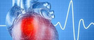
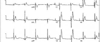
Electrocardiography
Allows you to evaluate the bioelectrical activity of the heart. Characteristic changes on the ECG in cardiosclerosis:
- reduced voltage of the teeth of the QRS complex (an indicator of impaired ventricular contractility);
- reduction of the “T” wave or its negative polarity;
- decrease in the ST segment below the isoline;
- rhythm disturbances;
- blockades
The ECG should be assessed by an experienced cardiologist who can, based on the nature of changes in electrical impulses, determine the location of the lesion, the form of cardiosclerosis and diagnose complications.
EchoCG
It is the most informative method in assessing heart function. Ultrasound of the heart is a painless and non-invasive procedure that allows you to determine the morphological state of the heart muscle, evaluate its pumping function, contractility, etc.
Characteristic changes in patients with cardiosclerosis:
- conduction disturbance;
- impaired contractility;
- thinning of the heart wall in the area of sclerosis;
- focus of fibrosis or sclerosis, its location;
- disturbances in the functioning of the valvular apparatus of the heart.
Radiography
X-ray is unable to clearly display all changes in the heart during cardiosclerosis, and therefore is an optional diagnostic method. Most often, R-graphy is used to make a preliminary diagnosis for the purpose of further examination. The method is painless, but is contraindicated for pregnant women due to the small dose of radiation. Pictures are taken in two projections to evaluate the heart from both sides. In the later stages of cardiosclerosis, the heart is noticeably enlarged. An experienced doctor can even see large aneurysms on X-rays.
Computed tomography and magnetic resonance imaging
They represent high-precision methods for studying the structures of the heart. The diagnostic significance of CT and MRI is equivalent, despite the different principles of image acquisition. The images allow you to see even small foci of connective tissue spread in the myocardium (most often after a heart attack). Diagnosis is difficult with diffuse process of damage to the heart muscle, because changes in myocardial density are uniform. The difficulty in examining the heart using CT and MRI methods is due to the fact that the heart is in constant motion, which does not allow obtaining a clear picture.
Scintigraphy
An instrumental examination method based on the introduction of a special substance into the bloodstream that marks certain types of cells. The target for the administered substance in case of cardiosclerosis is healthy cardiomyocyte cells. The contrast does not accumulate in damaged cells, or accumulates in smaller quantities. After the substance is administered, pictures of the heart are taken, which show how the contrast is distributed in the heart muscle.
Myocardial scintigraphy
In healthy myocardium, the injected substance accumulates evenly. Areas of damage in focal cardiosclerosis are very clearly visible - there will be no accumulation of contrast in them. The examination is informative and practically safe (with the exception of allergic responses to a contrast agent). The disadvantage of scintigraphy is the low prevalence of the method due to the high cost of the equipment.
Laboratory research methods
In the TAM and OAK, no specific changes are usually observed. Laboratory examination methods allow us to find the cause of the development of cardiosclerosis. For example, with atherosclerosis, the patient will have increased cholesterol; with myocarditis, there will be signs of an inflammatory process in the CBC. The data obtained during a laboratory examination of the patient only allows one to suspect the disease based on indirect signs. Drug therapy cannot be started without assessing the functioning of the renal and hepatic systems, for which a biochemical blood test, OBC, and OAM are performed.
Diagnostics
The diagnosis of “atherosclerotic cardiosclerosis” is based on the results of laboratory and instrumental studies. The patient receives an appointment for tests and diagnostic procedures upon presentation of symptomatic complaints. Characteristic changes in the biochemical blood test indicate myocardial damage.
An increase in enzyme concentration is determined:
- aspartate aminotransferase (ALT);
- creatine phosphokinase (CPK);
- lactate dehydrogenase (LLH).
The lipidogram (the patient's lipid profile, which determines the quality of fat metabolism) shows a significant excess of the norm for total cholesterol, triglycerides, low and very low density lipoproteins. A general clinical blood test determines the hanging platelet count, the discrepancy between thrombocrit and platelet indices from normal values.
A number of pathological changes are recorded during a coagulogram (blood clotting test). Primary examination of the heart - ECG (electrocardiogram) reveals areas of the myocardium that have lost the ability to conduct impulses.
Depending on the nature and intensity of the violations, the following may be prescribed:
- transthoracic or transesophageal echocardiography - ultrasound of the heart with an external probe or a tubular probe through the esophagus;
- echocardiography with Doppler – examination of the aorta and coronary arteries;
- stress echocardiography – ultrasound of the heart in a calm state and after a dynamic load (physical exercise or administration of medications);
- coronography - x-ray of the coronary arteries of the heart with the introduction of a radiopaque substance.
If possible, magnetic resonance imaging or computed tomography of the cardiovascular system is performed.
Treatment of myocardial dystrophy with folk remedies
Alternative medicine can only be used as an addition to the main therapy in consultation with the treating doctor. Traditional medicine recommends herbal decoctions and tinctures. A decoction of cumin and hawthorn gives good results.
Below are simple folk remedies to combat cardiosclerosis.
- A mixture of sour cream, honey and fresh protein. Beat 2 fresh chicken whites with 1 teaspoon of honey and 2 teaspoons of sour cream. The resulting mass should be taken on an empty stomach.
- Healing collection. Pour 300 ml of boiling water over dry raw materials (1 teaspoon each of hawthorn, yarrow, mistletoe, periwinkle). Let it brew for 1 hour. Should be taken in small portions throughout the day.
- Elecampane root. Pour 300 g of dry raw material into 500 ml of vodka and keep in a dark place for a week. The filtered decoction should be taken three times a day, 30 ml.
- Lemon and pine. Peel the lemon and divide in half. Pour half a lemon with pine decoction. Let it brew. You need to take it for a week, then take a two-week break and repeat the course.
- Onion juice and honey. For myocardial diseases, drinking onion juice mixed with honey is beneficial. The mixture should be stored in the refrigerator.
Prevention of atherosclerosis of the aorta and coronary arteries
How to influence the process of calcification, leading to hardening of the plaque, how to prevent the development of this process?
The main factor influencing the development of atherosclerosis of the coronary arteries and thoracic aorta is a person’s lifestyle. Prevention of the disease will be:
- lack of excess weight;
- balanced quality nutrition;
- giving up alcohol and cigarettes;
- sufficient physical activity during the day;
- elimination of stressful situations and overwork;
- normalization of metabolism (sufficient levels of calcium, vitamins and other microelements).
People with a hereditary predisposition to atherosclerosis must adhere to the rules of a healthy lifestyle to prevent the development of this pathology.
The article described in detail the process of development of the disease, from which it is clear that plaques can form due to excess calcium in the arteries. This happens when the body lacks certain nutrients that would direct calcium into the bones and teeth.
Numerous studies indicate that vitamins such as D3 and K2 help calcium to be absorbed. A person must obtain them from animal foods; vitamin D is also synthesized in the skin under the influence of sunlight. Calcitriol, a derivative of D3, reduces inflammation in the plaque area, and vitamin K2 activates the protein osteocalcin, which redistributes calcium from plaques to where it is most needed - to bone tissue. In addition, K2 is needed for the synthesis of another protein (MGP protein), which is a vascular orderly and removes excess calcium.
Unfortunately, today a person cannot get the required amount of these substances from food, so scientists have learned to create special supplements that, when taken, will compensate for the deficiency of vitamin D3 and K2 in the body. One of these drugs is Osteo K2; you can learn more about it here.
Prevention
Prevention of the development of cardiosclerosis consists of timely treatment of the underlying trigger disease, which provokes the proliferation of connective tissue.
There are basic rules, the observance of which allows you to slow down the progression of cardiosclerosis and avoid the development of complications:
- Complete smoking cessation. When using tobacco, spasm of the coronary vessels is observed, which increases myocardial hypoxia;
- Limiting physical activity. Jerks, sudden lifting of weights, and physical overload are unacceptable. Moderate cardio exercises are encouraged, which keep the muscles toned, but do not overload the heart;
- Constant monitoring by a cardiologist. Regular visits to the doctor, even without deterioration in health, every 3 months will allow you to diagnose complications in a timely manner and promptly correct the treatment regimen;
- Timely treatment of hormonal disorders and acute infectious diseases. This measure will avoid the negative impact of concomitant pathology on the functioning of the heart;
- Avoid stress.
Ignoring these simple rules can lead to the development of complications, despite taking prescribed medications.
Treatment
Treatment of post-myocardial cardiosclerosis is based on drug therapy, and compliance with nutritional and dietary measures, as well as lifestyle changes. If a diagnosis of post-myocardial cardiosclerosis is made, it is necessary to reduce physical activity on the body for the next six months.
After 6 months, with normal restitution of cardiac function, physical activity is carried out slowly and gradually.
Once the functionality of the heart has been restored, you can begin to exercise, but only if the systolic function of the left ventricle is completely restored.
Smoking and alcohol are strictly prohibited.
Salt restriction is only for those patients who have been diagnosed with heart failure.
The main therapy is aimed at:
- Treatment of left ventricular dysfunction and treatment of left ventricular failure,
- Decrease in blood pressure index,
- Elimination of abnormal heart rhythm,
- Elimination of impaired electrical conductivity of cardiac impulses.
It is also necessary to stop heart failure and restore all metabolic processes in the body.
Drug therapy consists of the following groups of medications:
- ACE inhibitors,
- Beta blockers in acute forms of pathology development, but only during the period of stabilization of the condition,
- Mineralocorticoid receptor antagonists drug Spironolactone,
- For swelling, diuretics are prescribed,
- Many patients are prescribed Digoxin, or the drug Ivabradine.
If drug therapy does not have a positive result, then surgical treatment of post-myocardial cardiosclerosis is used.
Drug therapy
Immunosuppressive drug therapy for cardiosclerosis
This therapy is prescribed to patients in whom an inflammatory process in the myocardial infiltration has been confirmed by biopsy, but if viral and infectious agents are not detected in the cardiac myocardium.
Immunosuppressive therapy with Prednisolone and Azathioprine significantly improves left ventricular systole and reduces severe symptoms of left ventricular failure.
Immunosuppressive therapy lasts from 90 days to 180 days.
During pregnancy
When diagnosing certain dangerous conditions, pregnancy is called into question:
- myocarditis in the acute phase;
- severe coronary pathology;
- heart failure IIA;
- cardiosclerosis complicated by arrhythmia.
In such cases, in order to save the child, it is necessary to undergo a set of therapeutic measures that have a positive effect on the course of pregnancy. The first thing that needs to be done is the sanitation of foci of infection (pharyngitis, sore throat, caries). For this purpose, antibiotics are prescribed. Additionally, beta blockers, metabolic drugs and glucocorticosteroids are prescribed. Pregnant women with myocardial cardiosclerosis are under special supervision, because This category of patients has a high risk of developing complications from the cardiovascular system and kidneys.
Methods for treating atherosclerosis of the coronary vessels of the heart
The patient's priority problem with atherosclerosis of the coronary arteries is the formation of plaques, which can lead to blockage of vital arteries. Therefore, treatment is aimed both at eliminating existing calcifications and at preventing the formation of new ones.
According to clinical recommendations, for atherosclerosis of the coronary arteries, drugs that reduce the level of harmful fats are prescribed; they are successfully used, influencing the first link of the mechanism of plaque formation: statins, fibrates, bile acid sequesters.
In particularly difficult cases, an operation can be performed in which the affected artery is cut and the growths are removed, sometimes part of the artery is removed.
Treatment can only be prescribed by a doctor after appropriate diagnosis. The patient can only follow the rules of prevention.
Diet for cardiosclerosis
Diet for vascular atherosclerosis
- Efficacy: therapeutic effect after 2 months
- Dates: no data
- Cost of products: 1700-1800 rubles. in Week
Diet for heart failure
- Efficacy: therapeutic effect after 20 days
- Timing: constantly
- Cost of products: 1700-1800 rubles. in Week
Diet for cardiac arrhythmia
- Efficacy: therapeutic effect after 30 days
- Timing: constantly
- Cost of products: 1700-1800 rubles. in Week
Diet for coronary heart disease
- Efficacy: therapeutic effect after 30 days
- Timing: constantly
- Cost of products: 1700-1800 rubles. in Week
Diet after a heart attack
- Efficacy: therapeutic effect after 2-6 months
- Terms: 2-12 months
- Cost of products: 1800-1900 rubles. in Week
Diet for cardiosclerosis is important. The essence of dietary nutrition is to reduce cholesterol in the blood. Correction of the blood lipid profile helps prevent the development of atherosclerosis in the coronary vessels. Additionally, it is necessary to limit salt intake (up to 5 g per day, and in severe cases - up to 3 g). A salt-free diet can reduce the volume of circulating blood, reducing the load on the heart.
Prohibited foods for cardiosclerosis:
- animal fats (butter, fatty meat);
- smoked meats, pickles;
- chocolate;
- coffee and strong tea;
- flour products;
- alcoholic drinks, energy drinks.
The emphasis in nutrition should be on eating lean boiled meat, various cereals, fresh fruits and vegetables. It is better to give preference to foods rich in potassium. The effect of cardiac glycosides is better when there is sufficient potassium in the body.
It is useful to eat raisins, bananas, dried fruits. It is necessary to monitor the total daily caloric intake (1800-2600 kcal). A well-designed menu will cover all the body’s needs in conditions of limited physical activity. Calorie content can change by 20-30% only if the patient suffers from obesity or cachexia (exhaustion). Poor nutrition worsens the prognosis for the patient. A qualified nutritionist and attending physician will help create a specific menu for the patient.
Causes of hypercholesterolemia and atherosclerosis
Atherosclerosis develops under the influence of correlation factors: hypercholesterolemia and the presence of microdamage to the endothelium. The normal level of total cholesterol in the blood for adults is 3.2-5.2 mmol/l. Analysis results > 7.7 mmol/l are considered pathologically overestimated.
An increase in LDL and VLDL in the blood is caused by:
- unhealthy eating habits (excessive fatty and sweet foods in the diet);
- low physical activity (hypodynamia);
- long-term psycho-emotional instability (stressful state);
- chronic diseases of the gallbladder and liver;
- malabsorption is a violation of the resorption (absorption) of nutrients in the intestine.
Hypercholesterolemia accompanies diabetes mellitus and other metabolic disorders. Microcracks in the endothelium appear as a result of the aggressive influence of nicotine, alcohol, and certain medications. Intimal damage is caused by:
- diseases associated with blood disorders (including metastatic cancer and diabetes mellitus);
- high pressure on the walls of blood vessels - arterial hypertension.
Fragility of arteries and cracks can be a consequence of hypovitaminosis.
The inner layer of the coronary and all other arteries is protected by flat blood cells - platelets. They are also responsible for blood clotting. When a microcrack appears in the intima, platelets (in large quantities) try to “seal” the damage.
Due to the identical charge of LDL molecules and platelets, excess fats are attracted to the crack, which is the beginning of the formation of cholesterol deposits. At first, a spot forms, then it increases in volume and area, turning into a plaque - an obstacle to blood flow. Then the plaque calcifies and transforms into a hard growth - the cause of NK and atherosclerotic coronary artery sclerosis.
Consequences and complications
It is important to understand that cardiosclerosis is an irreversible process, which in any case worsens the functioning of the heart and increases the risk of developing various complications. Even if the causes that worsen the course of the disease are eliminated, the presence of connective tissue in the thickness of the heart muscle can lead to adverse consequences. All complications are corrected with medication or treated surgically.
The most common consequences and complications:
- formation of CHF;
- development of cardiac aneurysm ;
- development of arrhythmias ;
- acquisition of heart defects;
- development of thromboembolism ;
- diagnosing chronic fatigue syndrome.
Kinds
The diffuse type of cardiosclerosis is characterized by damage to the cardiac myocardium of a uniform shape, and is distributed diffusely throughout the heart muscle. A diffuse type of post-myocardial cardiosclerosis most often occurs with ischemic heart disease and in the pre-infarction state.
The focal type of cardiosclerosis is a partial lesion of the heart muscle with a specific localization of sclerosis. The localization of foci of cardiosclerosis can be in different places.
Prognosis for cardiosclerosis
The prognosis is assessed after a thorough examination of the patient. If the patient has no pronounced complaints, and the disease is amenable to drug therapy, then the prognosis is considered favorable. Most often, positive dynamics are observed with small focal cardiosclerosis.
In patients with large-focal lesions, as well as with diffuse proliferation of connective tissue, the prognosis is considered unfavorable. After a transmural infarction, an aneurysm forms in the affected area, which entails very serious complications and consequences. Often, after large-focal infarctions, arrhythmias , which are also considered prognostically unfavorable. The prognosis for survival in post-infarction cardiosclerosis depends on the area of the lesion, treatment regimen, concomitant pathology, age, and the presence of complications.
List of sources
- Shopin A.N. Prediction of recurrent myocardial infarction in patients with post-infarction cardiosclerosis / A.N. Shopin, Ya.B. Khovaeva, E.N. Burdina, B.V. and others // Practical medicine, 2011
- Samorodskaya I.V. Cardiovascular morbidity and risk factors for cardiovascular events in the Russian Federation / I.V. Samorodskaya // Cardiovascular therapy and prevention, 2005. No. 3
- Tokareva E.A., Khatuntseva Yu.A., Makeeva A.V. ETIOLOGY, PATHOGENESIS AND DYNAMICS OF THE INCIDENCE OF CARDIOSCLEROSIS IN THE VORONEZH REGION // International Student Scientific Bulletin. – 2016
How does atherosclerosis of the coronary vessels manifest?
The atherosclerotic process can begin at a young age and last for decades, only with a combination of predisposing factors and a narrowing of the lumen of the vessel by half or more, can it make itself felt with the following conditions:
- stable and unstable angina. Characterized by cardiac and chest pain, shortness of breath when moving, and in the advanced stage and at rest, the heart works intermittently;
- myocardial infarction. Gives unbearable chest pain, pronounced fear of death, a sharp drop in blood pressure, reaching shock. The severity of the consequences depends on the location and size of the dead zone. The damaged part of the heart will no longer be able to recover.
The destruction of the arteries supplying the heart can be asymptomatic for a long time and the person will not complain of any abnormalities, which is why it is so important to undergo annual examinations and tests.