Information about other types of diseases starting with the letter “V”: Vegetative state, Ventricularity, Vestibular ataxia, Vestibular neuronitis, Vibration disease, Viral meningitis, Viral encephalitis, Temporal lobe epilepsy, Intracerebral hematoma, Intracranial tumors of the cerebral hemispheres, Intracranial hypertension, Inflammatory myopathy, Inflammatory polyneuropathy , Congenital myopathy, Congenital paramyotonia, Secondary parkinsonism
In the medical community, the syndrome of increased intracranial pressure is called intracranial hypertension or hypertension. This disease develops against the background of damage to the brain tissue or appears autonomously, due to factors independent of the body.
Hypertension is a polysyndromic disease, and its manifestations are diverse: severe headaches with a feeling of pressure on the eyes, sometimes with temporary distortion of visual fields, a feeling of lightheadedness and vomiting. When blood pressure rises to high levels, loss of consciousness is possible.
The diagnosis is based on clinical manifestations, head tomography data, and echoEG results. An analysis of spinal fluid and ultrasound of brain vessels may also be necessary. Due to the large number of syndromes, treatment consists of eliminating or alleviating them. Neurosurgical operations are indicated in special circumstances.
Types of disease
Intracranial hypertension (ICP) is a disease that often occurs not only in adults, but also in children. The measure of intracranial pressure corresponds to the height of pressure in the spinal cord fluid, which is why this disease is also called cerebrospinal fluid hypertension syndrome. After head injuries or during brain diseases, hypertension is of a secondary nature. In a larger number of patients, hypertension is primary and manifests itself without visible motives - idiopathic hypertension, which is defined according to the ICD-10 classifier as benign. This diagnosis is established by a differential method of eliminating all possible causes of increased pressure inside the head.
ICP is divided into acute and chronic. The acute form occurs against the background of traumatic brain injury or infection, the chronic form occurs when vascular problems or intracerebral formations (tumors, cysts) occur. Chronic hypertension can be a side effect after brain surgery or after strokes, infections, or injuries.
Pathogenesis
The pathogenesis of increased ICP in adults may be based on various mechanisms - edema/swelling of the brain, an increase in the mass of the contents of the cranium ( tumor , hematoma , abscess ), difficulty in the outflow of cerebrospinal fluid, disturbances in the venous outflow of the cerebral blood fraction). Within one article, it is not possible to consider the pathogenesis of high-frequency hypertension in a particular pathology, so we will only consider the pathogenesis of the development of high-frequency hypertension with cytotoxic cerebral edema .
Understanding the development of increased intracranial pressure (ICP) is based on a model of progressive cerebral edema, based on the theoretical Monroe-Kelly model, which is based on the thesis of a close relationship between the rigid cranium of an adult and components such as the brain, blood, and cerebrospinal fluid. The basis of such a connection is the interaction of any of the components with the others, manifested by a reaction to an increase in one of them by a corresponding (proportional) decrease in the volume of the other, due to which the constancy of intracranial pressure is maintained.
cytotoxic edema due to disruption of the transmembrane transport of electrolytes . Edema cells, due to their increased volume, exert a compressive effect (pressure) on neighboring cells, thereby promoting the spread of edema to intact cells (mass effect).
As the pathological volume of cells with cytotoxic edema increases, compression develops in the capillary-pial bed system, which leads to disruption of microcirculation and the development of hypoxemia / ischemia in areas of the brain that are not directly related to the primary mass effect, that is, this leads to pathological separation of different sections of the contents of the cranium. As a result, the pressure created by the pulse oscillations of the arteries and the cerebrospinal fluid loses the ability to spread freely along the tissues and spaces of the cerebrospinal fluid located inside the skull/spinal canal. This leads to the emergence of a difference in parenchymal pressure between intact and those involved in edema of the brain structures, which initiates its dislocation in the direction of relatively low pressure.
As a result of this process, diffuse edema of the entire brain develops and its gradual dislocation (movement in the direction of the foramen magnum (the only open exit from the cranium). As a result, various types of dislocation occur. More often, this is a hernial protrusion into the tentorial notch of the middle-basal parts of the temporal lobes and compression of the mesencephalic structures of the brain stem with inhibition of the primary circulatory/respiratory centers and a sharp disruption of brain function, up to the cessation of its vital activity. Neurological symptoms manifest themselves at the stages of cerebrovascular accident. The figure below shows a diagram of the progression of ICP and the stages of the cerebral protection mechanism.
Etiology and pathogenesis of intracranial hypertension
The sources of the disease are divided into groups:
The first is brain formations - primary or metastatic tumors, cysts, cerebral aneurysms, hematomas.- The second progresses against the background of a head injury, oxygen deficiency, ischemic stroke, toxic effects, encephalitis, edema of the brain or its membranes.
A separate group includes vascular causes that cause increased filling of the brain vessels with blood. This is possible when the temperature rises, when there is an excess of carbon dioxide in the blood, or when venous circulation is impaired.
Liquorodynamic disorders were identified as a separate category of causes of cerebral hypertension. It is caused by an increase in the production of cerebrospinal fluid. In this case, the volume of cerebrospinal fluid becomes excessive and leads to hydrocephalus.
Benign ICP occurs for unclear reasons. It develops more often in women than in men due to weight gain. This pattern gave rise to the hypothesis of an endocrine cause of the disease. Hypertension is caused by excessive intake of vitamin A, taking certain medications, and withdrawal of corticosteroid hormones after long-term use.
The skull is a confined cavity, so growing parts of the brain causes increased pressure. The result of this is compression of the brain of varying degrees. This condition leads to dysmetabolic changes in brain neurons. The more pressure increases, the more significant the shifting of brain structures, which can lead to wedging of the cerebellar tonsils into the foramen magnum. The cardiovascular and respiratory nerve centers are located in the brain stem, and when compression of these areas occurs, the vital functions of the body are disrupted.
The occurrence and development of the disease in children is facilitated by malformations of the brain (hydrocephalus, microcephaly, defects in the development of arteries and veins of the brain), birth injuries to the head, intrauterine infections, asphyxia and hypoxia of newborns. Due to the mobility of cranial sutures and their elasticity in infancy, the manifestations of high ICP contribute to long-term compensation.
General information
Intracranial hypertension is a pathological condition (syndrome) manifested by increased intracranial (intracranial) pressure.
Intracranial hypertension syndrome (syn. liquor-hypertension syndrome) is quite common in adult and pediatric neurology and can be either idiopathic or develop with a variety of brain lesions and skull injuries. The most common cerebral hypertension of unknown origin is idiopathic (primary) intracranial hypertension (ICH), which is classified as benign intracranial hypertension (ICD-10 code: G 93.2). As a rule, this diagnosis is made only after there is no confirmation of the specific causes of hypertension (the presence of a space-occupying formation in the cranial cavity, vein thrombosis, infectious lesions of the brain, etc.).
Determination of intracranial pressure (ICP) and its physiological norm
Intracranial pressure is the difference between atmospheric pressure and pressure in the cranial cavity (epidural/subarachnoid spaces, meningeal sinuses, ventricles of the brain). The level of intracranial pressure is formed by cerebrospinal fluid (CSF) circulating in the structures of the central nervous system and arterial/venous blood entering the brain.
These fluid environments are in constant motion (spinal fluid circulates through the ventricles of the brain/spinal canal, and blood through the vascular bed). Normally, the physiology of the circulation of fluid media in the brain is determined by:
- Mean arterial pressure, i.e. the average value (difference) between the systolic/diastolic blood pressure of arterial blood entering the skull, which is normally 80 mm Hg. Art.
- The average venous pressure at the exit of the skull, which is normally 0 mm Hg. Art., that is, there is no resistance to blood flow.
- The average cerebrospinal fluid pressure in the skull, which is external in relation to the brain and is equal to 10 mm Hg. Art. It is the cerebrospinal fluid pressure that has a constant compressive effect on the brain (creates constant ICP). The biomechanical balance normally present in the skull maintains the average cerebral tissue pressure at a level of 10 mmHg. Art. In a healthy adult, the total volume of circulating cerebrospinal fluid averages 150 ml, while ICP remains normal. Its slight increase is compensated by its resorption by brain tissue and outflow from the cranial cavity to the spinal cord along the liquor subarachnoid ducts through all parts of the spine, up to the sacral outlet openings.
The constant pressure inside the cranium is maintained by the formation of reserve spaces due to a decrease in the volume of CSF, as well as the cerebral blood fraction. In cases of an increase in any of the components occurring against the background of various pathologies (excessive accumulation of CSF, cerebral edema , cerebral hyperemia , impaired venous outflow), as well as with the development of pathological volumes (tumors, hematomas, parenchymal hemorrhage, abscess), a conflict occurs between the intracranial components and hypertension develops .
An increase/decrease in ICP can be observed both during natural physiological processes in the human body ( coughing , loud screaming, sneezing , crying, straining, physical/nervous strain, sudden bending forward), and indicate pathology. Normally, in an adult, the pressure inside the skull should not exceed 10–15 mmHg. ICH refers to a persistent increase in ICP to a level of 20 mm or higher. rt. Art. The severity of intracranial hypertension is presented in the table below.
How can you measure VD?
In specialized medical institutions, invasive techniques are used to measure the pressure inside the ventricles of the brain using specialized pressure sensors that are inserted into the cerebral ventricles (a catheter is inserted and connected to the sensor). Sensors can also be installed subarachnoidally, subdurally, or epidurally. This procedure has a low risk of brain injury. In practice, intracranial pressure is in most cases measured indirectly using a spinal puncture, measuring it at the level of the lumbar spine in the spinal subarachnoid space.
Why is intracranial hypertension dangerous?
The severity and specificity of disorders in ICH are determined by the degree of increase in ICP, its nature (diffuse/local) and localization, as well as the duration of the impact of increased ICP on brain structures. And if, with a weak and unstable degree of increase in ICP, no significant changes are observed, then in patients with a stable increase in ICP of moderate and severe degrees it can cause serious disorders - headaches (tension, ischemic), disturbances in fine motor skills of the hands, vision, hearing, hypertonicity of the upper muscles / lower extremities, skeletal muscle rigidity, convulsions , paresis , disturbances in emotional manifestations (sleep, behavior), fatigue, delayed speech development, neurogenic disorders of the cardiovascular and respiratory systems (vegetative-vascular dystonia , pain in the heart, bradycardia / tachycardia , arrhythmias , low-grade body temperature , breathing rhythm disturbances - shortness of breath , apnea ) and others.
Benign intracranial hypertension
First of all, what is it? As already noted, benign ICH is a condition characterized by a persistent increase in cerebrospinal fluid pressure in the absence of intracranial mass formation, vein thrombosis and abnormalities in the composition of the cerebrospinal fluid. Since intracranial hypertension of a specific origin can be considered within the framework of a particular pathology/disease, we will consider only idiopathic (benign) ICH.
The primary syndrome of idiopathic intracranial hypertension (IIH) today means a condition that is accompanied by an increase in ICP without identified etiological factors (possibly due to obesity ). The incidence rate of IIH is 0.7–2 cases/100,000 population.
This type of hypertension most often occurs in young women who are overweight. Much less common in children and men. The most typical symptoms of idiopathic liquor hypertension are headache and transient (transient) visual disturbances in the form of deterioration in image sharpness, blurring, double vision, and in 30-35% of patients there is a decrease in visual acuity.
Clinical signs of pathology
The main symptom of the disease is headache. A growing, intense headache is a sign of an acute attack of hypertension. Intermittent headaches are a sign of a chronic form of the disease. The pain is symmetrical, with a feeling of pressure on the eyes, localized in the frontal and parietal parts of the head.
Patients report nausea, vomiting, pain when moving their eyes, and a feeling of fullness in the head. An acute, growing headache leads to loss of consciousness, and in severe cases to coma. Long-term hypertension worsens the general nervous condition of the patient, causing physical fatigue, nervousness, sleep disturbances, and mental instability.
Chronic intracranial hypertension lasts with crises - periods of sudden surges in pressure with severe headaches, nausea, vomiting, and fainting. Most often, patients experience darkening of the eyes, deterioration of sharpness, or double vision. 30% of patients note a decrease in vision clarity.
Intracranial hypertension in children under one year of age is accompanied by refusal of mother's milk, crying and restlessness, frequent regurgitation, and tension of the fontanel. Chronic hypertension in an infant can cause mental retardation, even mental retardation.
Causes
Intracranial hypertension in adults can be caused by many different causes, the diversity of which can be grouped according to the mechanism of development of the pathology:
- Intracranial space-occupying formations causing an increase in ICP ( benign/malignant tumor intracranial hematoma , parasitic cyst , abscess );
- Cytotoxic cerebral edema caused by hypoxic damage to cells of brain structures (severe respiratory failure after cardiac arrest), early stage cerebral ischemia hepatic / renal encephalopathy , hyponatremia , Reye's syndrome , syndrome of inadequate production of andidiuretic hormone ).
- Vasogenic cerebral edema caused by damage to the blood-brain barrier (infectious diseases - meningitis / encephalitis , intracranial trauma - bruises, concussions, birth injuries), hematomas , ischemic / hemorrhagic stroke .
- Interstitial edema caused by impaired outflow of cerebrospinal fluid ( occlusive hydrocephalus ).
Establishing diagnosis
Only a neurologist, after thorough examinations, can make a diagnosis of high intracranial pressure. The main difficulty is that there is no generally accepted standard for ICP indicators. The experience of many years of research allows us to conclude that pressure values from 70 to 220 mm of water can be considered normal. pillar
Papilledema detected by an ophthalmologist may be a sign of ICP. X-rays of his skull show areas of thinning of his bones. A successful diagnostic method was lumbar puncture. When a needle is inserted into the cerebrospinal fluid space, cerebrospinal fluid flows out in strong jerks.
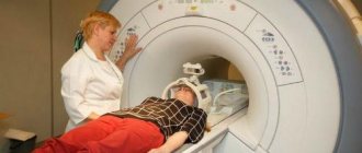
The use of electronic sensors requires craniotomy and therefore becomes an extremely undesirable way to make a diagnosis. This method is useful in severe cases of ICP to monitor pressure dynamics in the brain. Among the instrumental methods, MRI and CT of the head, ultrasound of the vessels of the head, biopsy of intracranial tumors are most often used; in children, neurosonography is performed through the fontanel.
Diagnostics
Because intracranial pressure can fluctuate, there is no consensus on what is normal. It is quite difficult to make a diagnosis. The patient is prescribed a special examination, during which he undergoes:
- echo-encephalography;
- examination by an ophthalmologist, who reveals swelling of the optic nerves during ophthalmoscopy;
- X-ray of the skull, where “finger impressions” that arise during chronic conditions are visible.
However, the most reliable methods for diagnosing intracranial hypertension are lumbar puncture and puncture of the cerebral ventricles.
To identify the causes of increased intracranial pressure, MRI, CT, MSCT of the brain are prescribed, as well as vascular ultrasound and stereotactic biopsy of identified tumors.
Treatment methods
Regular use of medications is necessary for the chronic course of the disease or during crises in the absence of deep disorders of consciousness. Depending on the level of ICP, one or another diuretic pharmaceutical is prescribed. In mild cases - furosemide, hydrochlorothiazide, spironolactone, in acute form - mannitol. Taking diuretics should be combined with potassium preparations - potassium chloride, potassium aspartate.
In the presence of concomitant infectious diseases, antiviral and antibacterial drugs are prescribed. When the body is intoxicated, detoxification measures are carried out. For venous circulation - venotonics. In conditions of ICP, nerve cells need special nutrition; for this purpose, piracetam, gamma-aminobutyric acid, and glycine are prescribed. It is advisable to undergo courses of cranio-manual therapy, which improves the outflow of venous blood. During crises, excessive strain on the eyes and emotional overload should be avoided.
Neurosurgical surgical interventions can be performed urgently or plannedly in severe forms of the disease. In acute prolonged crises, external ventricular drainage or craniotomy is indicated to reduce intracranial pressure. Removal of brain tumors, elimination of hydrocephalus, and cerebral shunting are routinely performed.
Symptoms
- Classification of arterial hypertension
An increase in intracranial pressure leads to compression of nerve cells, which affects their function. Regardless of the cause, intracranial hypertension syndrome manifests itself:
- bursting diffuse headache. The headache is more pronounced in the second half of the night and in the morning (since the outflow of fluid from the cranial cavity worsens at night), is dull in nature, and is accompanied by a feeling of pressure on the eyes from the inside. The pain intensifies with coughing, sneezing, straining, physical stress, and may be accompanied by noise in the head and dizziness. With a slight increase in intracranial pressure, you may simply feel a heaviness in the head;
- sudden nausea and vomiting. “Sudden” means that neither nausea nor vomiting is provoked by any external factors. Most often, vomiting occurs at the height of the headache, during its peak. Of course, such nausea and vomiting are completely unrelated to food intake. Sometimes vomiting occurs on an empty stomach immediately after waking up. In some cases, vomiting is very strong, fountain-like. After vomiting, a person may experience relief, and the intensity of the headache decreases;
- increased fatigue, rapid exhaustion both during mental and physical exertion. All this may be accompanied by unmotivated nervousness, emotional instability, irritability and tearfulness;
- weather sensitivity. Patients with intracranial hypertension do not tolerate changes in atmospheric pressure (especially its decrease, which happens before rainy weather). Most symptoms of intracranial hypertension intensify at these moments;
- disturbances in the functioning of the autonomic nervous system. This is manifested by increased sweating, changes in blood pressure, and a feeling of palpitations;
- visual impairment. Changes develop gradually, being transitory at first. Patients note the appearance of periodic blurriness, as if blurred vision, sometimes double images of objects. Movement of the eyeballs is often painful in all directions.
The duration of existence of the symptoms described above, their variability, and the tendency to decrease or increase are largely determined by the main cause of intracranial hypertension. The increase in the phenomena of intracranial hypertension is accompanied by an increase in all signs. In particular, this may manifest itself:
- persistent daily morning vomiting against a background of severe headache for the whole day (and not just at night and in the morning). Vomiting may be accompanied by persistent hiccups, which is a very unfavorable symptom (it may indicate the presence of a tumor in the posterior cranial fossa and signal the need for immediate medical attention);
- increasing depression of mental functions (appearance of lethargy, up to disturbance of consciousness such as stupor, stupor and even coma);
- increased blood pressure along with depression (decreased breathing) and slowed heart rate to less than 60 beats per minute;
- the appearance of generalized seizures.
If such symptoms appear, you must immediately seek medical help, since all of them pose an immediate threat to the patient’s life. They indicate an increase in the phenomena of cerebral edema, in which it can be pinched, which can lead to death.
With the long-term existence of the phenomena of intracranial hypertension, with the gradual progression of the process, visual impairment becomes not episodic, but permanent. A great diagnostic aid in such cases is an examination of the fundus by an ophthalmologist. In the fundus of the eye, ophthalmoscopy reveals congestive optic discs (essentially their swelling); small hemorrhages in their area are possible. If the phenomena of intracranial hypertension are quite significant and exist for a long time, then gradually the congestive discs of the optic nerves are replaced by their secondary atrophy. In this case, visual acuity is impaired, and it becomes impossible to correct it with lenses. Optic nerve atrophy can result in complete blindness.
With the long-term existence of persistent intracranial hypertension, distension from the inside leads to the formation of even bone changes. The plates of the skull bones become thinner, the back of the sella turcica is destroyed. The convolutions of the brain seem to be imprinted on the inner surface of the bones of the cranial vault (this is usually described as increased digital impressions). All these signs are detected during a simple x-ray of the skull.
A neurological examination in the presence of symptoms of increased intracranial pressure may not reveal any abnormalities at all. Occasionally (and even when the process persists for a long time), one can detect a limitation in the abduction of the eyeballs to the sides, changes in reflexes, a pathological Babinski sign, and impaired cognitive functions. However, all these changes are nonspecific, that is, they cannot indicate the presence of intracranial hypertension.
Prevention and prognosis
The outcome of liquor-hypertensive syndrome depends on the underlying disease, pressure level and adequate surgical therapy. In severe cases, the patient's death is possible. ICP of unknown origin responds well to therapy. If a child remains in this state for a long time, the most severe complication may be mental retardation.
Preventive measures that prevent the development of intracranial hypertension include: current diagnosis and treatment of intracranial neoplasms, dyscirculatory disorders, regular use of blood pressure-normalizing drugs in the chronic course of the disease, adherence to a daily routine, proper management of pregnancy and obstetric care.
Consequences and complications
A long-term (chronic) increase in ICP, even with minor deviations from the norm, negatively affects the function of the central nervous system and is manifested by signs of nervous exhaustion: irritability , frequent headaches, sleep disturbances, increased fatigue, decreased physical/intellectual performance, decreased cognitive functions (decreased concentration, memory , weakening of intelligence). With a significant increase in ICP, there is a high risk of developing visual impairment and epileptic seizures.
Benign form of pathology
Benign intracranial hypertension is a separate form of the disease that occurs with the mildest manifestations, without symptoms of damage to the nervous system. It is also called idiopathic or false brain tumor. Women who are young and obese most often suffer from the pathology. The main symptoms are the urge to vomit, unsteadiness of gait, pain in the head and eyes. Why such a syndrome is formed is still not completely clear to doctors.
Initially, headaches are mild, which is why people do not seek medical help, especially since attacks are quickly eliminated after taking analgesic tablets. Then the problem becomes constant, accompanied by nausea and vomiting. Vision problems appear, in women the regularity of the menstrual cycle is disrupted, in men - weakening libido.
As a rule, there are no signs of damage to the nervous system, and in the initial stages the disease goes away on its own without treatment. Doctors have identified a connection between the development of symptoms in women and the use of oral contraceptives, after which the pathology disappears without a trace. If the clinical picture is pronounced, the same drugs are used in conservative treatment as in the treatment of the usual form of intracranial hypertension.