Author Alexandra Balan-Senchuk
08/17/2019 18:07 (Updated: 10/02/2021 18:07)
Health
Diastolic dysfunction refers to the mechanical failure of the heart chambers to fill properly with blood during the diastolic phase of the cardiac cycle. This is caused by inadequate relaxation of the ventricles during diastole.
What it is?
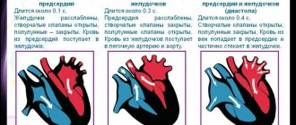
The heart operates in systole (contraction) and diastole (relaxation).
Dysfunction is said to exist if there are malfunctions in the functioning of an organ. When the diastolic function of the left ventricle is impaired, the myocardial muscle tissue loses the ability to relax during diastole. As a result, the ventricle does not receive the required amount of blood. To compensate for its deficiency, the left atrium is forced to intensify its work, trying to absorb more blood.
All this negatively affects the condition of the atrium, gradually leading to overload and its increase in size. Against the background of systolic dysfunction, stagnation can occur in the venous system and lungs, which entails disruptions in the blood supply to all organs of the human body. The transition of this pathological condition to a more severe form can lead to chronic heart failure.
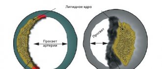
Diastole is important because thanks to it the heart muscle receives the necessary oxygen, which is distributed throughout the circulatory system through the coronary arteries.
If it is not able to fully perform its tasks, the left ventricle suffers from oxygen deficiency. This leads to metabolic disturbances in myocardial tissue and ischemia.
Prolonged ischemia is detrimental to the cells, instead of which connective tissue is formed. This process is called sclerosis or fibrosis. The altered tissue structure causes difficulty in contracting the left ventricle. Ultimately, systole fails.
Possible complications
Possible consequences:
- Cardiac arrest as a result of insufficient nutrition and a decrease in myocardial contractility.
- Heart attack. Necrosis of active, functional tissues. Usually extensive, associated with almost certain mortality.
- Cardiogenic shock. As a result of a catastrophic drop in basic vital signs. It is almost impossible to get out of this state. The risks are maximum.
- Stroke. Weakening of the trophism of nerve tissue. Accompanied by neurological deficit of varying severity. Possible disturbances in thinking, speech, vision, hearing, mnestic, cognitive abilities, behavior and other aspects.
- Vascular dementia. The symptoms are similar to Alzheimer's disease. Taking into account the persistence of disorders of the cardiovascular system, it has a poor prognosis and is difficult to reverse.
- Respiratory failure, pulmonary edema.
- Thromboembolism.
Death or disability as a result of all the consequences described above.
Complications occur as a result of insufficient or absent treatment. Particularly resistant forms, unfortunately, cannot be treated at all or the results have no clinical significance. Such situations are minimal, but they do exist.
Classification
The first type of disease is the most common. It is fraught with serious danger, since at the initial stage of development it occurs practically without any symptoms. It is characterized by a decrease in the ability to drive blood into the ventricle from the paired blood vessel of the pulmonary trunk. The reason for this is insufficient elasticity of the myocardial walls.
The second type of disease manifests itself against the background of increased pressure from the left atrium, which leads to impaired diastole function. It is also called pseudonormal.
The most severe type of pathology is considered to be restrictive, when there is a threat to a person’s life due to serious disorders in the heart. In such situations, a heart transplant is usually performed.
If a person has type 1 left ventricular diastolic dysfunction, this may be indicated by edema, which is observed mainly in the evening. This condition is caused by stagnation of fluid in the body. Swelling is usually noted in the lower extremities.
In this case, the patient may complain of heart pain caused by myocardial ischemia. Shortness of breath often occurs after physical activity. Type 1 left ventricular diastolic dysfunction should not be ignored; it requires drug correction.
At the initial stage of development, the disease may not even manifest itself. If left untreated, it will progress, resulting in the following signs of left ventricular diastolic dysfunction:
shortness of breath at rest or after minor physical exertion;- increased heart rate;
- a feeling of tightness in the chest and lack of oxygen;
- swelling of the lower extremities;
- bluish color of the skin;
- fast fatiguability;
- heart pain.
Very rarely, patients may experience a cough that occurs in the evening. Its appearance indicates the presence of congestion in the lungs.
Blood flow in the heart goes through 3 stages:
- muscle relaxation (diastole);
- slow filling of the left ventricle with blood, ensured by the pressure difference inside the atria;
- filling the left ventricle with the remaining blood after the heart contracts.
We are talking about diastolic dysfunction when some kind of failure occurs in such a well-functioning system. Pathology of this type may occur due to the presence of the following factors:
- old age;
- previous myocardial infarction;
- disturbance of blood flow in the cardiovascular system;
- excess body weight;
- hypertension;
- myocardial dysfunction.
Deviations in the functioning of the heart provoke harmful habits such as smoking and drinking alcohol. Love for caffeine-containing drinks does not have the best effect on the condition of the heart muscle.
According to medical experts, the main provoking factor of this disease is the deterioration of the contractile and relaxing ability of the myocardium. This is usually due to poor elasticity of its muscle tissue. This condition can lead to a number of diseases, including heart attack, myocardial hypertrophy and arterial hypertension.
Diastolic dysfunction can also affect newborn babies. If a child has increased blood supply to the lungs, this may cause:
- the size of the heart will increase;
- atrial overload will occur;
- tachycardia will appear;
- heart contraction will worsen.
This condition is not considered pathological, and therefore does not require special treatment if it occurs in children immediately after birth. But if the child suffered hypoxia, or was born prematurely, this problem may persist for two weeks.
Factors provoking the development of pathology
First of all, it should be noted that the development of diastolic myocardial dysfunction is facilitated by its hypertrophy, i.e. thickening of the walls of the ventricles and interventricular septum.
The main cause of cardiac muscle hypertrophy is hypertension. In addition, the danger of its development is associated with excessive physical stress on the body (for example, intense sports, heavy physical labor).
How to arrive for treatment
Factors contributing to the development of the main cause – hypertrophy – are separately identified and these are:
- arterial hypertension;
- heart disease;
- diabetes;
- obesity;
- snoring (its effect is caused by involuntary cessation of breathing for a few seconds during sleep).
Treatment
A diagnosis of “left ventricular diastolic dysfunction” of type 1, 2 or 3 is possible only after the patient has undergone a series of examinations. To do this, you will need to undergo a general urine test and blood biochemistry. It may also be necessary to check the functioning of the thyroid gland, kidneys, and liver.
The most informative way to study if there is a heart abnormality is an ECG.
The duration of the procedure is only 10 minutes. During this procedure, electrodes are attached to the patient’s chest area and read the necessary information. It is important that the body is relaxed and breathing is calm. The study is recommended to be carried out 2-3 hours after eating.
Additionally, an ultrasound of the heart may be prescribed. This diagnostic method allows you to determine the condition of the organ and also check the blood flow. Ultrasound examination does not require any preparation.
Only after receiving the results of a comprehensive examination does the doctor make a diagnosis and determine further treatment tactics. The main goals of therapy are as follows:
- normalize heart rate;
- prevent the occurrence of arrhythmia;
- cure coronary heart disease;
- stabilize the pressure.
To normalize heart rate, beta blockers are used, represented by drugs such as Concor and Atenolol. Cardiac ischemia is treated with nitrates. Blood pressure can be normalized with diuretics such as Hypothiazide or Spironolactone.
For diastolic dysfunction, ACE inhibitors are also indicated. Their action is aimed at normalizing blood pressure. They are usually prescribed to hypertensive patients. Inhibitors, in addition to reducing blood pressure, protect the heart and help relax the myocardial walls. Drugs in this group include Captopril and Fosinopril.
For preventive purposes, your doctor may recommend taking Aspirin Cardio. With its help, the blood is thinned, thereby minimizing the risk of blockage of blood vessels.
Methods for detecting pathology
Diagnosis of the development of a pathology such as diastolic dysfunction in the myocardium includes the following types of examinations:
- echocardiography in combination with Dopplerography (the study makes it possible to obtain an accurate image of the myocardium and assess functionality in a given period of time);
- electrocardiogram;
- ventriculography (in this case, radioactive albumin is also used to determine the contractile function of the heart);
- X-ray examination of the lungs;
- laboratory blood tests.
Forecast
Type 1 left ventricular diastolic dysfunction, in the vast majority of cases, has a favorable prognosis, which cannot be said about the transition of the disease to a restrictive form. It is accompanied by high atrial pressure and is complicated by concomitant heart failure. The prognosis in this case is not always reassuring. To cope with the pathology, a heart transplant may be required.
The incidence of readmission for patients diagnosed with diastolic dysfunction is 50%. The mortality rate for this pathology is 3-7% per year.
To prevent the development of irreversible processes, increased attention should be paid to preventive measures. It is very important to eat right, limit salt intake, and control water intake. The diet should consist of fresh vegetables, lean meats, cereals and dairy products. Dishes will be healthier if they are steamed or baked in the oven. It is also necessary to completely avoid fried and spicy foods, alcohol, and smoking.
Causes
The main reasons for the deterioration of relaxation of the left ventricle are hypertrophy of its walls and loss of elasticity. Various factors lead to this condition:
- arterial hypertension;
- aortic stenosis;
- cardiomyopathy;
- heart rhythm disturbances;
- myocardial ischemia;
- age-related changes;
- gender factor (women are more susceptible);
- abnormal condition of the coronary arteries;
- inflammation of the pericardium of the constrictive type;
- overweight;
- diabetes;
- heart defects;
- heart attack
results
The groups were homogeneous in age, gender composition, height and body weight, number of smokers, and the number of people with underweight. Obese patients were more common in groups 2 and 3. In group 1 there were no patients with coronary artery disease, hypertension, type 2 diabetes mellitus, or signs of heart and respiratory failure.
Spirometry data in the groups of patients with lung pathology did not differ significantly from each other, but were naturally reduced compared to those in the comparison group (Table 2). When comparing the structural parameters of echocardiography, a statistically significant increase in the diameter of the pulmonary artery was revealed in the PH group compared with group 1 ( p
<0.0001).
Diameter P.P. in patients without signs of PH was 6% more than in group 1 ( p
= 0.00023), and in the PH group it was 21% more than in healthy people, and 12% more than in patients without signs of PH (
p
< 0.0001 in both cases).
In the PH group, there was an increase in the EDR of the pancreas by 14 and 3% compared with that of groups 1 and 2 ( p
<0.0001 and
p
= 0.001, respectively).
TAPSE in the PH group was 15% lower than in the comparison group, and 5% lower than in the group of patients without signs of PH ( p
<0.0001 in both cases).
In the same group, there was a decrease in RV EF to 51%, which is 9 and 6% lower than in the first two groups ( p
<0.0001 in both cases).
There was an increase in pancreatic wall thickness compared to that in group 1 by 11% in patients without signs of PH and by 33% in patients with PH ( p
<0.0001 in both cases).
Table 2. Spirometry data, structural parameters of the right heart and pulmonary arterial pressure in the examined groups Note.
Here and in the table. 3: the differences in indicators are significant (p<0.05) when comparing * with the 1st group, ** with the 2nd group. EDV - end diastolic size; RVd - thickness of the wall of the pancreas in diastole. When assessing the diastolic function of the RV (Table 3), patients in the PH group showed a decrease in the rate of early diastolic flow Et by 8% compared to that in group 1 ( p
=0.46).
The Et/At ratio also did not differ significantly between the groups. There were virtually no differences between groups in the atrial systole velocity At and the time of deceleration of early tricuspid flow. In the group of patients with lung pathology without PH, compared with group 1, there was a significant decrease in CPTP by 6% ( p
<0.0001).
In patients with PH, compared with groups 1 and 2, there was a decrease in CPTP by 19 and 14% ( p
<0.0001 and
p
= 0.021, respectively).
The Et/CPTP ratio in the PH group was 19% higher than in group 1 ( p
= 0.00023), but did not differ significantly from that in the group of patients without PH (
p
= 0.18).
Table 3. Indicators of RV diastolic function in the examined groups Note.
Et/SRTP is the ratio of the speed of early diastolic transtricuspid flow to the speed of its propagation. When analyzing the indicators of spectral tissue Doppler annulus of the tricuspid valve (Table 4), there was a tendency towards a decrease in the speed of its early diastolic movement e't in the PH group compared with group 1 ( p
=0.19).
In the same group, compared with the 1st group, there was a tendency towards a decrease in the speed of late diastolic movement of the tricuspid ring a't ( p
= 0.07).
The e't/a't ratios did not differ between groups. There were also no intergroup differences in the speed of systolic movement of the tricuspid valve ring (s't) and the Et/e't ratio. In group 2, compared to group 1, there was a non-significant increase in the Thea index by 14% ( p
= 0.54).
In the PH group, this indicator was significantly higher by 43% than in group 1 ( p
= 0.00078), and there was a tendency to increase by 25% compared to group 2 (
p
= 0.07).
Table 4. Indicators of spectral tissue Doppler in the groups examined Note.
e't, a't, e't/a't, - speeds of early and late diastolic movements of the tricuspid valve ring and their ratio; s't is the speed of systolic movement of the distal part of the pancreatic wall; Et/e't is the ratio of the flow velocity of early RV filling to the velocity of early diastolic movement of the tricuspid valve annulus. Correlation analysis revealed a statistically significant relationship between SRTP and LADsr values ( r
=–0.51;
p
<0.0001) with a gradient of tricuspid regurgitation - GTR (
r
=–0.41;
p
<0.0001), SPAP (
r
=–0.44;
p
<0.0001).
In univariate logistic regression analysis, the probability of SRTP below 32 cm/s (Table 5) increased with increasing age, in the presence of COPD, respiratory failure stage II, coronary artery disease, CHF III F.K. The likelihood of detecting a reduced SRTP also increased the increase in GTR above 20 mm Hg, LBP above 25 mm Hg. Art. and MPAP above 30 mm Hg.
Table 5. Analysis of factors influencing SRTP less than 32 cm/s in logistic regression analysis Note. OR—odds ratio; CI—confidence interval.
When constructing a multiple linear regression model, which included such features as age, the presence of COPD, stage II DN, ischemic heart disease, an increase in LBP above 25 mm Hg, an increase in GAD above 20 mm Hg. Art., and MPAP is above 30 mm Hg. Art., it was revealed that only two factors had an independent effect on the decrease in SRTP: the presence of II degree DN (the risk of detection increased by 2.1 times) and an increase in LBP above 25 mm Hg. (the risk of detection increased by 2 times).
Enhanced function of the left shank
In a patient with diastolic dysfunction, it is important to control the heart rate quickly and avoid tachycardia in order to maximize the period of diastolic activity. To achieve this goal, beta blockers are especially helpful, but they do not directly affect myocardial relaxation. By increasing the heart rate for beta blockers, it has been shown that they can effectively reduce arterial pressure, reverse myocardial ischemia, relieve regression of left sac hypertrophy, and prevent suprarenal adrenaline. intense stimulation, which occurs in heart failure. For beta blockers, it has been reported that odors are significantly associated with increased survival in patients with diastolic heart failure. This group of drugs needs to be used for the treatment of diastolic heart failure, especially in the presence of hypertension, chronic heart disease or arrhythmia.
Diagnosis of diastolic heart failure
Diagnosing the disease at an early stage will help avoid irreversible changes.
To establish a diagnosis, three conditions are usually necessary:
- manifestation of symptoms and signs of heart failure;
- PV systolic function is normal or slightly decreased;
- instrumental diagnostic methods reveal disturbances in the functioning of the LV and an increase in its stiffness.
Early diagnosis helps prevent irreversible changes in heart function
The main instrumental diagnostic methods include:
- two-dimensional echocardiography with Doppler ultrasound is one of the most effective methods for determining the diagnosis;
- radionuclide ventriculography will determine the failure of myocardial contractility;
- EchoCG reveals signs of myocardial ischemia;
- A chest x-ray will help determine pulmonary hypertension.
How to treat left ventricular hypertrophy of the heart
Many people are susceptible to cardiac hypertrophy. If the problem is severe, medical or surgical treatment of left ventricular hypertrophy is performed. In this case, depending on the extent of the damage, treatment can be aimed at preventing the progression of the disease or at returning the myocardium to its normal size. But it happens that this condition is reversible; if the disease cannot be cured completely, then regression can be achieved by correcting such things as:
- Lifestyle;
- food type;
- hormonal balance;
- excess weight;
- amount of physical activity.
Treatment of left ventricular hypertrophy with medication
Medicines for left ventricular hypertrophy of the heart can have an effective result if taken under the supervision of a doctor. It is impossible to completely eliminate the symptoms of hypertension, but taking antihypertensive drugs for this disease and following a diet will help fight the causes and prevent deterioration of health. To treat LVH, the following medications are prescribed:
- Verapamil is an angiarrhythmic drug from the group of calcium channel blockers. Reduces myocardial contractility, reduces heart rate. Can be used by both adults and children, doses are set individually.
- Beta blockers - reduce the pressure and volume load in the heart cavity, help to even out the rhythm and reduce the risk of defects.
- Sartans - effectively reduce the overall load on the heart and remodel the myocardium.
Myocardial hypertension of the left heart belongs to class 9 on the ICD-10 scale, along with other diseases of the circulatory system. Preference should be given exclusively to drugs whose quality has been tested and proven clinically; experimental drugs may not only not have the expected effect, but also negatively affect overall health.
Cardiomyopathy surgical treatment
Surgery for left ventricular hypertrophy may be necessary to remove the hypertrophied portion of the muscle in late and advanced stages of the disease. To do this, a transplant of the entire heart or its individual parts is performed. If the cause of LVMH is damage to a valve or septum, transplantation of these specific organs is first attempted, which is simpler than whole-heart surgery. In the case of such an intervention, the patient will have to be under the supervision of a cardiologist for the rest of his life and take medications to prevent coronary thrombosis.
Traditional treatment of left ventricular hypertrophy
Treatment of hypertrophy of the left ventricle of the heart with folk remedies cannot help in the later stages of lesions, but it can be effective with minor increases, to prevent their development, and reduce the risk of more serious consequences. You will not be able to completely cure the disease, but traditional medicine can relieve discomfort, chest pain, weakness and fainting. The following means are known:
- Herbal infusions as an auxiliary therapy during the main treatment (blueberry, motherwort, blasphemous hawthorn, horsetail, cornflower flowers, adonis)
- Infused milk: boil and pour into a thermos overnight, or put in the oven until it turns brown.
- Lily of the valley in the form of drops of tincture or gruel. For the tincture, pour vodka or alcohol into the lily of the valley, leave in a dark place for 2 weeks, take 10 drops 3 times a day for 2 months. Gruel: pour boiling water over lily of the valley flowers, leave for 10 minutes. Then drain the water, chop the plant and take a tablespoon 2 times a day. Recommended in combination with drops.
- Garlic honey: mix crushed garlic with honey in a 1:1 ratio, leave for a week in a dark place, take 1 tablespoon 3 times a day before meals.
- Dry red wine infused with dried rosemary. Pour wine over the leaves, leave for about a month in a dark place, strain and take before eating.
- Cranberries, mashed with sugar: a teaspoon 4 times a day.
Significant and diagnostic criteria
Diastolic heart failure is indicated as a result of increased resistance to the replacement of one or both cords; This should not lead to symptoms of stagnation due to pathological diarrhea, crooked diastolic pressure - volume. Although we would like to thoroughly describe the underlying pathophysiological mechanism of diastolic heart failure, it is impossible to state it clinically. More practical is the significance of what can be observed clinically: the condition in which the classic symptoms of CHF appear, rather than the appearance of a calm pathological diastolic, or normal systolic function. The Diastolic Cardiac Failure Study Group asked clinicians to combine clinical and echocardiographic information with data from patients with diastolic cardiac failure. This goes up to the stage of diagnostic singing.