Dextrocardia is a congenital anomaly that involves the location of the heart on the right side. This feature is extremely rare. With this disorder, the main vital functions of the cardiovascular system are not affected.
An ultrasound diagnostician can detect such a deviation even during the intrauterine development of the embryo. The anomaly occurs due to pathology of the heart tube in the fetus, which occurs in the first months of pregnancy. It bends to the right, but this in no way affects the overall development of the cardiovascular system. People with a mirror heart are not sick, and if dextrocardia is not accompanied by functional pathologies, then this anomaly remains only a feature that requires observation by a doctor.
The etiology of displacement of the heart tube to the right side during intrauterine development of the fetus is unclear, and therefore the disorder cannot be prevented. In very rare cases, a child is born with a mirror arrangement of all organs.
Types of violation
Cardiac dextrocardia is divided into 3 types:
- Simple. It occurs quite rarely and consists of a displacement to the right side of only the heart and its vessels.
- Mirror. Characterized by incorrect location of parts of the digestive and respiratory organs.
- Full transposition. With it, all the organs of the chest are positioned incorrectly.
There is also a type of dextrocardia called dextroversion. It is characterized by the fact that the apex of the heart is turned to the right side. In this case, the anatomical structure of the organ does not suffer.
What does ultrasound reveal?
Our clinic uses modern 4D equipment with Doppler mode. With its help, you can obtain an image of 3 main vessels - the superior vena cava, the pulmonary trunk and the ascending aorta. During the examination, not only the location of the vessels is revealed, but also their diameter and other parameters.
The following fetal pathologies will be visible on the monitor screen:
- A decrease in the diameter of the aorta with expansion of the pulmonary trunk can indicate hypoplasia (underdevelopment) of the left parts of the baby’s heart, which are responsible for the onset of blood circulation;
- A decrease in the size of the pulmonary artery trunk while maintaining normal diameters of the aorta and superior vena cava indicates stenosis (narrowing) of the pulmonary artery. In the fetus, only pronounced forms are detected;
- The small diameter of the aorta with a normal 4-chamber structure of the heart is a consequence of coarctation of the aorta (narrowing of the aorta of the heart in a certain segment);
- Visualization of 2 vessels instead of 3 may be a consequence of the connection of the vessels into a common arterial trunk;
- Displacement of the aorta forward or to the right of the pulmonary artery is observed during transposition of the great vessels;
- The diameter of the aorta is expanded, but the diameter of the pulmonary artery is narrowed, and the aorta is displaced forward. This may be tetralogy of Fallot (a very severe combined cardiac anomaly). The problem includes stenosis or hypertrophy of the right ventricular outflow tract, ventricular septal defect, and dextroposition of the aorta (outflow to the right side). Diagnosis of the fetus is extremely difficult, so the Doppler mode comes to the rescue, helping to visualize the flow of blood into the aorta from both ventricles;
- Hypoplasia (underdevelopment) of the right chambers of the heart is determined by a decrease in their size relative to the left chambers. This pathology is usually accompanied by dysplasia (sagging or bulging) of the mitral valve;
- The common atrioventricular canal is seen as a cardiac septal defect with splitting of the atrioventricular valve;
- Hypoplastic syndrome of the left heart is manifested in the form of underdevelopment of the ventricle and mitral and aortic valves;
- A single ventricle is also not normal, because there should be two of them and they are clearly visible in a four-chamber section;
- When the tricuspid valve is underdeveloped, blood from the right atrium does not enter the left atrium, which is clearly visible during Doppler examination;
- From the 2nd trimester, endocardial fibroelastosis is visualized as thickening of the myocardium and worsening of its contraction;
- Underdevelopment of the myocardium of one of the ventricles (Uhl's anomaly) is noticeable in the 2nd trimester.
Danger of incorrect positioning
Dextrocardia itself does not pose a threat to the patient. People also live with the right heart, their health is no worse than that of people with the normal location of the organ. Only untimely detection of this disorder can lead to a doctor’s mistake during surgical intervention in emergency cases, when the patient is not examined first.
Danger to humans arises when dextrocardia is accompanied by other heart diseases. The presence of additional congenital defects can lead to extremely adverse consequences.
Cardiologists identify several heart defects that can occur along with dextrocardia. These include the following diseases:
- Endocardial defect.
- Pulmonary artery stenosis.
- Hole in the interventricular septum.
Also, the most common pathology accompanying dextrocardia is Kartagener-Sievert syndrome. With it, there is always an incorrect arrangement of organs and a violation of the structure of the respiratory tract. Male patients with this syndrome often suffer from infertility.
In some cases, the development of abnormal location of the heart is observed along with diseases of the abdominal organs. Patients often experience a concomitant disease such as heterotaxy syndrome. In this case, patients do not have a spleen or it is underdeveloped.
What problems arise
A person whose organs are mirrored will feel the symptoms of common diseases differently, which naturally complicates the work of doctors. So, for example, when appendicitis develops, all the characteristic signs will be felt, but not on the right, but on the left, or even practically not felt at all, since the appendix is often located slightly posteriorly. If the spleen hurts, the doctor, without additional research, can attribute such manifestations to the liver or gall bladder. As a result, the wrong treatment will be prescribed.
The issues of transplantology in patients with mirror placement of organs stand apart. After all, donors, as a rule, are people with the usual variant of internal development. Replacing an organ in the presence of a reverse arrangement is a rather complex process and requires special qualifications and experience from the doctor, since it is necessary to correctly position the vessels so that they do not reject the organ and take root well.
Article on the topic
Surgery in the womb: doctors operated on the heart of an unborn child
Symptoms
If the heart on the right side is the only disorder that is not accompanied by any defects of this organ, then no clinical manifestations are observed. A person, just like everyone else, lives an ordinary life and does not even suspect that he has the wrong disposition of heart.
Most often, dextrocardia is accompanied by other heart defects. Some are detected immediately after birth, others after some time. Each heart pathology has its own symptoms, but a heart disorder can be suspected based on the following signs:
- Yellowish or bluish color of the skin.
- Difficulty breathing.
- Pale skin.
- Pain in the sternum.
- Failure of heart rhythm.
- Weakness, fatigue.
- Retarded physical development.
If such signs appear, you should definitely consult a doctor and undergo a heart examination.
When the heart is on the right. Doctors talk about Kazakhstanis with mirror arrangement of organs
We are accustomed to the fact that an important human organ, pumping up to 10 thousand liters of blood per day, should be located on the left. However, there are exceptions. TengriMIX met an amazing girl whose heart is on the right side, and learned from doctors how many such unusual people live in Kazakhstan. 27-year-old Almaty resident Assem Smagulova did not immediately find out about her peculiarity. However, I noticed something strange in myself - as if my heart was beating not like ordinary people, but to the right. When she was a student, the girl suddenly became ill. She ended up in the hospital. There she learned that she had organ transposition.
“I was 18 years old, then I was in my first year at university. After class, I felt unwell and had a stabbing pain in my left side in the lower abdomen. An ambulance was called. They took him to the hospital. There they wheeled me around on a gurney for three hours and couldn’t figure out what was wrong with me. Only the chief surgeon announced suspicion of appendicitis. They did an ultrasound. He told me that I have mirror organs. The most interesting thing is that I kept telling my mother that when I run, my heart beats on the right side. She laughed and didn’t believe it, saying that this doesn’t happen,” recalls Asem.
© Asem Smagulova
Then the doctor consoled the patient, explaining that this was a rare case, but with such an anomaly people can live a full life. The girl tried to independently study the reasons for the unusual phenomenon.
“I read many different articles on the Internet, but did not find specific explanations of the pathology. I wonder how many of us there are in the country? The location of the organs itself does not interfere with my life; I lead a normal lifestyle. Since childhood, I have been involved in dancing and sports and I feel quite healthy,” said the Almaty resident.
According to Sundet Sargelov, head of the radiology department of the Scientific Center for Pediatrics and Child Surgery of the Ministry of Health of the Republic of Kazakhstan, organ transposition has been quite well studied in medical science. For the first time, the Italian medical scientist Hieronymus Fabricius in 1606, during his anatomical studies, discovered the location of the heart on the right side in humans. And 37 years later, his colleague Marco Aurelio Severino described in detail the mirror location of organs.
Sundet Sargelov
Transposition of internal organs is a rare variant of biologically normal anatomy, in which the main internal organs have a mirror arrangement compared to the usual normal position: the heart is on the right, the liver is on the left, the stomach is on the right. This case occurs in one person out of 10 thousand. The main reason is a gene mutation, and the anomaly is inherited.
“The heart of the unborn child begins to form already in the third week of pregnancy, it is then that it is determined where it will be located: in the left or right side of the chest. The reason is a gene mutation, that is, some kind of gene breakdown occurs in either the mother or the father of the embryo. Under no circumstances should an anomaly be associated with a person’s nationality, place of residence, lifestyle, or ecology,” explains Sundet Shakirovich.
Transposition of internal organs. Photo courtesy of Sundet Sargelov
Unfortunately, there are no accurate statistics about all Kazakhstanis whose heart is located on the right side of the chest. However, according to the health worker, in our country the situation is similar to the world. Thus, if the average population of Kazakhstan is 18 million, then after deduction it turns out that there are about 1,800 right-hearted people living among us. Most people find out about the abnormality when they take an x-ray. In addition, a post-mortem autopsy is often not performed, since the relatives of the deceased are against this procedure.
“In the West, particularly in England, as well as the USA, after death the bodies are examined, and then an anomaly is discovered. Therefore, if our citizens do not get sick, are not examined, then they live without suspecting their uniqueness. This is the same as among people there are right-handers and there are left-handers. If a person does not have other congenital heart defects or intrauterine developmental anomalies, then he can live, study and work peacefully on an equal basis with everyone else. Many of them live to an old age,” explains the candidate of medical sciences.
Also, a mirror arrangement of internal organs occurs in Sievert-Kartagener syndrome, which is an autosomal recessive genetic disease. Its rate is one in 50 thousand people. Currently in Kazakhstan there are 16 children registered with this pathology.
“This is a gene disorder. It causes people to have unhealthy airways. Characteristic features: synclititis, bronchiectasis and mirror arrangement of organs. People with this diagnosis experience weakness, fever, ear pain, shortness of breath, and wheezing. Therefore, patients undergo treatment from an early age. They are registered with a pediatric pulmonologist. They receive hospital treatment about five times a year, are under the supervision of doctors, and take antibiotics,” the interlocutor says.
Sievert-Kartagener syndrome. Photo courtesy of Sundet Sargelov
As Sundet Sargelov notes, the anomaly is so rare that many medical workers simply do not encounter it. And when identified, they study it with interest.
“This pathology as a subject is very interesting to doctors. Some doctors may never meet such patients. In this regard, I can say that I was lucky. Two years after I started working in the pulmonology department, I first discovered an anomaly in a 9-year-old child. He was admitted with a cough. When they began to examine him with X-ray machines and took a picture, they realized that he had a reverse-positioned, right-formed heart, the right lung was on the left side. Later I learned the story of another patient. We diagnosed him at the age of 7. Mom worked well with him: breathing exercises and swimming pool. Thanks to this, at the age of 13-14 he had good respiratory muscles and could relieve bronchospasm with gymnastics. Once he scared his fellow students, they ran into the medical center of the educational institution and said that he was wheezing a lot. When the paramedics came to him, he was already breathing calmly. I would like to say to the parents of such children that yes, this is indeed a disability, but the main thing is that the child does not have frequent exacerbations. Need treatment. Remove phlegm, do rinsing. Physical activity is useful for such people, but it is difficult to be professional athletes, since the vital capacity of the lungs is small,” emphasized Sundet Shakirovich.
Similar anomalies are found in the fauna world. For example, among grape snails there are so-called “kings”, in which the heart, respiratory and reproductive tracts are located abnormally on the left side.
Author: Sahibam Sadyrova
Diagnosis of heart position
Thanks to modern diagnostic methods, it is possible to detect an abnormal location of the baby’s heart even during pregnancy. When the child is born, a more thorough examination is carried out to check for any defects associated with dextrocardia.
Pathologies can be detected using the following methods:
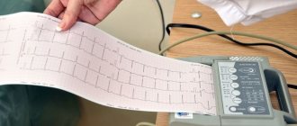
- Electrocardiogram. Using an ECG for dextrocardia, you can identify disturbances in the functioning of the heart and evaluate its rhythm.
- Echocardiography. Ultrasound makes it possible to examine the structure of an organ in detail and check the blood circulation in it.
- Ultrasound examination of the abdominal cavity. It is necessary to assess the location of organs.
- Magnetic resonance imaging. It is used as an additional diagnosis if there is a suspicion of the development of concomitant defects.
Even with normal listening to the heart with a stethoscope, the doctor may notice that the heartbeat is on the right side.
Reasons for originality
Until now, the reasons why “mirroring” of human organs occurs are not precisely known. But doctors have a number of theories about this. For example, some doctors are sure that twins first developed in the womb, but then they “merged” into one fetus. And it turns out that a person gets it for two. True, this version is often called unconvincing.
Article on the topic Congenital heart disease in children - are genetics and ecology to blame?
The second scenario is that changes occur under the influence of a woman’s hormonal fluctuations in the early stages of pregnancy. For example, the catalyst may be severe stress experienced by the expectant mother, as a result of which there was a rapid surge of hormones, and the organs were out of place.
There are also those experts who believe that the mirror arrangement of organs is caused by infectious diseases suffered by the mother. Ecology and heredity are also blamed.
Treatment methods
Treatment depends on whether dextrocardia is accompanied by other cardiac pathologies. If not, then therapy is not required. The patient can lead a full active life. Only when deciding to engage in sports is it recommended to undergo an ECG.
If the incorrect location of the heart occurs along with other diseases, treatment tactics will depend on the specific diagnosis. Typically, heart defects are eliminated through surgery, during which the existing defects are removed.
Before the operation, the patient must be prepared by prescribing medications. To maintain the patient's condition, diuretics, antihypertension agents, and drugs that support the myocardium are used.
Special attention is paid to antibacterial treatment. It is carried out both before surgery and during the period of rehabilitation of the body after it. But taking antibiotics for a long time is not recommended, as they negatively affect the condition of the organs.
Any medications are prescribed exclusively by the attending doctor, taking into account the results of the patient’s examination. The specific list of medications depends on the type of disease and the severity of its course.